May 2, 2016 - postmitotic (3, 4), cardiac growth is mediated by CM hypertrophy ... Patrick Brien,1,6 Kanar Alkass,7,8 Antonio Tomasso,7 Asmita Agrawal .... in EHMT1/2 expression was necessary and sufficient to induce the ...... acclimatization at a running velocity of 6 m/min for 30, 45, 60, 75, 90, ..... 2009;64(5):678â691.
RESEARCH ARTICLE
The Journal of Clinical Investigation
The H3K9 dimethyltransferases EHMT1/2 protect against pathological cardiac hypertrophy Bernard Thienpont,1,2 Jan Magnus Aronsen,3,4 Emma Louise Robinson,1,5,6 Hanneke Okkenhaug,1 Elena Loche,1,5 Arianna Ferrini,1 Patrick Brien,1,6 Kanar Alkass,7,8 Antonio Tomasso,7 Asmita Agrawal,1 Olaf Bergmann,7,9 Ivar Sjaastad,3 Wolf Reik,1,10,11 and Hywel Llewelyn Roderick1,6 Epigenetics Programme, Babraham Institute, Cambridge, United Kingdom. 2Laboratory of Translational Genetics, Vesalius Research Center, VIB and KU Leuven, Leuven, Belgium. 3Institute for Experimental
1
Medical Research, University of Oslo and Oslo University Hospital, Oslo, Norway. 4Bjørknes College, Oslo, Norway. 5Wellcome Trust – MRC Institute of Metabolic Science, University of Cambridge, Cambridge, United Kingdom. 6Laboratory of Experimental Cardiology, Department of Cardiovascular Sciences, KU Leuven, Leuven,
Belgium. 7Cell and Molecular Biology, Karolinska Institutet, Stockholm, Sweden. Department of Forensic Medicine, The National Board of Forensic Medicine, Stockholm, Sweden. 9DFG Research Center for Regenerative Therapies, Technische Universität Dresden, Dresden, Germany.
8
Centre for Trophoblast Research, University of Cambridge, Cambridge, United Kingdom. 11The Wellcome Trust Sanger Institute, Cambridge, United Kingdom.
10
Cardiac hypertrophic growth in response to pathological cues is associated with reexpression of fetal genes and decreased cardiac function and is often a precursor to heart failure. In contrast, physiologically induced hypertrophy is adaptive, resulting in improved cardiac function. The processes that selectively induce these hypertrophic states are poorly understood. Here, we have profiled 2 repressive epigenetic marks, H3K9me2 and H3K27me3, which are involved in stable cellular differentiation, specifically in cardiomyocytes from physiologically and pathologically hypertrophied rat hearts, and correlated these marks with their associated transcriptomes. This analysis revealed the pervasive loss of euchromatic H3K9me2 as a conserved feature of pathological hypertrophy that was associated with reexpression of fetal genes. In hypertrophy, H3K9me2 was reduced following a miR-217–mediated decrease in expression of the H3K9 dimethyltransferases EHMT1 and EHMT2 (EHMT1/2). miR-217–mediated, genetic, or pharmacological inactivation of EHMT1/2 was sufficient to promote pathological hypertrophy and fetal gene reexpression, while suppression of this pathway protected against pathological hypertrophy both in vitro and in mice. Thus, we have established a conserved mechanism involving a departure of the cardiomyocyte epigenome from its adult cellular identity to a reprogrammed state that is accompanied by reexpression of fetal genes and pathological hypertrophy. These results suggest that targeting miR-217 and EHMT1/2 to prevent H3K9 methylation loss is a viable therapeutic approach for the treatment of heart disease.
Introduction
The heart is a remarkable organ that adapts to the changing hemodynamic needs of the organism throughout life by modulating its output. Acute increases in cardiovascular demand are met by an increase in cardiac contractility, whereas chronic increases associated with development, pregnancy, or sustained exercise are accommodated by cardiac growth. Despite this apparent plasticity, cardiovascular diseases are a prime health burden worldwide and are anticipated to increase even further (1). Pathologies such as chronic hypertension and aortic stenosis similarly induce cardiac growth, but while these responses are initially adaptive, they ultimately compromise cardiac output, leading to heart failure and death (2). Since cardiomyocytes (CMs) in the adult heart are generally postmitotic (3, 4), cardiac growth is mediated by CM hypertrophy rather than proliferation. The defining characteristics of cardiac
Authorship note: B. Thienpont, J.M. Aronsen, and E.L. Robinson contributed equally to this work. I. Sjaastad and W. Reik contributed equally to this work. Conflict of interest: W. Reik is a consultant and shareholder of Cambridge Epigenetix. License: This work is licensed under the Creative Commons Attribution 4.0 International License. To view a copy of this license, visit http://creativecommons.org/licensesby/4.0/. Submitted: May 2, 2016; Accepted: October 17, 2016. Reference information: J Clin Invest. 2017;127(1):335–348. doi:10.1172/JCI88353.
growth (altered contractility, changed geometry, hypertrophy) reflect changes in the phenotype of CMs, making this cell type pivotal to the hypertrophic response. However, the multicellular composition of the heart represents a potent confounder for analyzing hypertrophy, as CMs are outnumbered up to 4-fold by non-CMs — a ratio that decreases further during pathological hypertrophy through a gain of fibroblasts and immune cells and death of CMs. Thus, analyses of heart tissue often only identify the composite of changes across its constituent cell types. To overcome this and allow the study of CM-specific responses in the intact heart, we used a powerful method involving fluorescence-assisted cell sorting to selectively isolate CM nuclei (4, 5). To bring about the extensive morphological, functional, and cellular changes associated with pathological or physiological hypertrophy, a wholesale reprogramming of the CM transcriptome is required. Notably, pathological, but not physiological, hypertrophy is characterized by a reexpression of the fetal gene program, with possible deleterious consequences for cardiac function (6). For example, reexpression of the fetal myosin heavy chain MYH7 diminishes cardiac contractility, and muscle pyruvate kinase (PKM) accompanies a return to an energetically less favorable glycolytic metabolism (7, 8). It is, however, unclear how CMs depart from their adult state. Importantly, a role for the epigenome in the heart jci.org Volume 127 Number 1 January 2017
335
RESEARCH ARTICLE
is increasingly being recognized; for example, histone 3 lysine 27 methylation is involved in heart development (9), and signalresponsive changes in histone deacetylation control gene expression in cardiac hypertrophy (10). Hypertrophy-associated changes in DNA methylation were also reported, although inactivation of the DNA methyltransferases Dnmt3a/3b did not affect the hypertrophic response (11). Despite this progress, it remains unknown whether or how the adult identity of the CM epigenome is lost during and contributes to disease. Two important repressive marks, methylation of lysines 9 and 27 of histone 3 (H3K9me2 and H3K27me3), mediate cellular memory by establishing gene programs during development that stabilize the differentiated state (12–14). Given that pathological hypertrophy in adult CMs is characterized by reexpression of fetal-associated transcripts, we hypothesized that this reprogramming arises through changes in these stable marks that determine and maintain the differentiated state. By analyzing purified CM nuclei from hypertrophied hearts, we identified loss of H3K9me2 as a distinguishing, conserved feature of pathological hypertrophy. Moreover, we show that the euchromatic histone methyltransferases EHMT1 and EHMT2 (also known as GLP and G9a or KMT1C and KMT1D) maintained these marks in adult myocytes but that pathological hypertrophic cues reduced EHMT1/2 expression posttranscriptionally by activating expression of microRNA 217 (miR-217). We found that a reduction in EHMT1/2 expression was necessary and sufficient to induce the expression of fetal-associated transcripts and pathological hypertrophy. Thus, we propose that the EHMT family of H3K9 dimethyltransferases “locks” adult CMs in their healthy adult state, protecting them against pathological hypertrophic remodeling.
Results
Pathological hypertrophy reduces H3K9me2 in cardiomyocytes. During development, cells become increasingly locked in their terminally differentiated state by stable epigenetic marks such as H3K9me2 and H3K27me3. To investigate how these marks are altered in different forms of hypertrophy, we compared hearts from well-established models of pathological and physiological hypertrophy in rats: pathological hypertrophy was triggered over a 6-week period through an ascending aortic banding–induced (AB-induced) pressure overload, and physiological hypertrophy was induced through a 6-week exercise regime of increasing intensity. Both treatments resulted in a similar 1.5-fold increase in the left ventricular (LV)/body weight (BW) ratio but yielded different functional and structural phenotypic outcomes. Specifically, AB-induced hypertrophy was associated with concentric cardiac growth, reexpression of the fetal gene program, expression of fibrosis-associated markers (both determined by quantitative realtime PCR [qRT-PCR]), and a moderate reduction in cardiac function (ejection fraction [EF], determined by echocardiography) (Figure 1, A–C, and Supplemental Figure 1, A and B; supplemental material available online with this article; doi:10.1172/JCI88353DS1). In contrast, exercise stimulated eccentric hypertrophy, without overt activation of the fetal gene program or fibrosis markers, and enhanced cardiac function (Figure 1, A–C, and Supplemental Figure 1, A and B). Pathological remodeling induced by AB was intentionally of a mild phenotype to allow the study of the mechanistic factors contributing to hypertrophy induction, without possible confounding sequelae associated with heart failure. 336
jci.org Volume 127 Number 1 January 2017
The Journal of Clinical Investigation Having validated these hypertrophy models, we next profiled H3K9me2 and H3K27me3 occupancy using ChIP, followed by high-throughput sequencing (ChIP-seq), and this was done specifically in flow-sorted pericentriolar material 1–positive (PCM1+) CM nuclei (n = 4 for each) (Supplemental Figure 1, C and D). Consistent with studies in other tissues, these histone modifications marked broad, homogeneous segments of the genome (Supplemental Figure 1, C and E) known as broad local enrichments (BLOCs) and large, organized chromatin K9 modifications (LOCKs) (15, 16). Of note, H3K9me2 and H3K27me3 levels were substantially altered in a subset of these genome segments during hypertrophy (Figure 1D). The changes were more pronounced in pathological hypertrophy than in physiological hypertrophy, however: 9% and 0.9%, respectively, of the genome showed more than 20% change in H3K9me2, and 3.5% and 0.9% showed more than 20% change in H3K27me3 (FDR = 10%; Figure 1D). Remarkably, hypertrophy induced by AB was almost exclusively associated with a loss of H3K9me2. This pervasive loss was also evident by immunofluorescence of heart sections and by immunoblotting, which revealed a decrease of 40% to 60% in H3K9me2 levels in CM nuclei upon AB (Figure 1E and Supplemental Figure 1F), whereas H3K9me2 levels remained unchanged with exercise (Figure 1E and Supplemental Figure 1G). Likewise, consistent with the lower number of BLOCs exhibiting alterations in H3K27me3, global levels of H3K27me3 were unaffected by either form of hypertrophy at this time point (Supplemental Figure 1, H and I). Changes at other time points and of other histone modifications cannot be excluded. Pathological hypertrophy reduces H3K9me2 at fetal heart genes. We next characterized the regions of the genome affected by loss of H3K9me2. A systematic ontology term analysis of genes in regions that were decreased in H3K9me2 upon AB revealed an enrichment of hypertrophy-associated processes including signaling, responses to hormone stimuli, and heart development (Figure 2A). Genes depleted in H3K9me2 included the fetal gene program canonical markers atrial natriuretic peptide (Nppa), brain natriuretic peptide (Nppb), and Myh7 (Figure 1D and Figure 2B, and Supplemental Figure 1J). These marks thus appear to repress the fetal gene program in the adult CM. To establish that H3K9me2 is indeed acquired during CM maturation, we profiled a time series of postnatal CM nuclei (P1 to adult) for H3K9me2 using ChIP-seq, which revealed a widespread gain in H3K9me2 during CM maturation that was particularly evident for LOCKs that showed a loss of H3K9me2 upon AB (Figure 2C), including Nppa, Nppb, and Myh7 (Supplemental Figure 1, K and L). Immunofluorescence analyses again underlined the pervasive nature of this H3K9me2 acquisition (Figure 2D). In conclusion, we observed a widespread and substantial loss of H3K9me2 upon hypertrophy induced by AB but not by exercise. Our data suggest that pathological hypertrophic cues strip CMs of this epigenetic constraint imposed during development, thereby enabling a reactivation of the fetal gene program and a pathological remodeling of gene expression. Loss of H3K9me2 correlates with increased transcription in hypertrophic cardiomyocytes. To assess whether the epigenetic changes observed underlie transcriptional regulation, we next used RNAseq to profile transcriptomes of sorted CM nuclei from hearts subjected to physiological and pathological hypertrophy. In addition to providing a CM-specific transcriptome, this nuclear RNA also pro-
The Journal of Clinical Investigation
RESEARCH ARTICLE
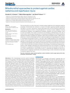
Figure 1. H3K9me2 is decreased in CMs upon AB in rats. (A) Graphs show LV/BW ratio, normalized to sham or control (mg/g); EF percentage; and ratio of LV lumen diameter to myocardial wall thickness at end-diastole. n = 7, 9, 6, and 8 animals for control, exercise, sham, and AB, respectively. (B) M-mode echocardiogram showing, at end-diastole, the LV diameter (LVDd) and thickness of the posterior wall (PWd) and of the interventricular septum (IVSd). Horizontal scale bars: 0.1 second; vertical scale bars: 2 mm. (C) qRT-PCR showing expression of Nppa, Nppb, Myh6, and Myh7 in the LV. (D) Volcano plot of differential markings by H3K9me2 and H3K27me3 of, respectively, LOCKs and BLOCs in PCM1+ nuclei upon pathological (left) and physiological (right) hypertrophy, as determined by ChIP-seq. Red and green dots represent regions enriched and depleted upon hypertrophy (FDR 20% change). (E) Plots show H3K9me2 signal in PCM1+ nuclei, determined by immunofluorescence analysis of the LV from AB, sham-operated, exercise, and control animals (n = 4, 5, 3, and 5, respectively). Representative confocal immunofluorescence images show staining for wheat germ agglutinin (WGA, red) and PCM1 (white), and H3K9me2 (green) in the LV. Scale bars: 50 μm. Error bars indicate the SEM. n = 5 (C) and 4 (D) replicates. *P < 0.05, **P < 0.01, and ***P < 0.001, by Student’s t test (A and C) and nested ANOVA (E).
vided the benefit of establishing levels of active transcription at the point of heart removal. Of note, our sorting protocol did not affect the nuclear transcriptome, as a comparison between samples before and after sorting revealed expression of the same genes as well as
highly similar intronic RNA transcript counts (Pearson’s r2 = 0.91), although, as expected, nuclear RNA was depleted of mitochondrial transcribed RNAs (Supplemental Figure 2, A–C). In addition, RNA from PCM1+ nuclei was highly enriched in marker genes and jci.org Volume 127 Number 1 January 2017
337
RESEARCH ARTICLE
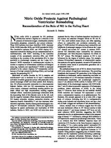
Figure 2. H3K9me2 is acquired during cardiomyocyte maturation and lost at fetal genes upon AB. (A) Selected ontology terms enriched among genes in regions altered in H3K9me2 upon AB. (B) H3K9me2 enrichment at loci on chromosomes (Chr) 5 and 15 in PCM1+ nuclei. Enrichment is displayed per 10-kb bin as log2(ratio of AB vs. sham). Green denotes significantly altered LOCKs. (C) Changes in H3K9me2 during CM maturation at LOCKs showed loss or no loss (green or gray) upon AB. (D) Quantification and representative images of immunofluorescence staining of H3K9me2 (green) in CMs (PCM1, red) in neonatal (3-day-old) and adult (3-month-old) mice (n = 4 each). Hypertrophy was induced for 6 weeks. Error bars indicate the SEM. n = 4 (B and D) and 2 (C) replicates. **P < 0.01, by nested ANOVA (D).
338
jci.org Volume 127 Number 1 January 2017
The Journal of Clinical Investigation processes specific to CMs, whereas PCM1– nuclei were enriched in markers of other cardiac cell types (fibroblasts and endothelial and immune cells), highlighting the purity (>98%) and proper identity of our CM nuclei (Supplemental Figure 2, D–G). Overlaying epigenomic and transcriptomic profiles revealed that genes and regions heavily covered by H3K9me2 or H3K27me3 were transcriptionally silent, as anticipated (Figure 3, A and B, Supplemental Figure 2, H and I) (17). Notably, however, whereas H3K9me2 levels decreased linearly with increasing levels of transcription, H3K27me3 levels were consistently low across the expressed genes (Figure 3, A and B and Supplemental Figure 2, J and K). This suggests that, in adult CMs, H3K9me2 is involved in modulating gene expression levels, while H3K27me3 marks an on/off state. In line with the more marked epigenetic changes observed upon AB, we also noted that transcriptomic changes were more pronounced in pathological hypertrophy than in physiological hypertrophy (Figure 3, C and D), with 705 and 331 proteincoding genes (3.1% and 1.4%), respectively, being differentially expressed (FDR 10%; Supplemental Tables 1 and 2). A comparison between differentially expressed genes here and those found in studies in which transcriptional changes were assessed in heart tissues confirmed that genes overexpressed in non-CMs were overrepresented in tissue-based profiles but not in our pure CM profiles, further validating our sorting approach (Supplemental Figure 2L). Genes that increased in expression upon AB included established pathological hypertrophy marker genes such as Nppa, Nppb, and Myh7 (Figure 3C). These genes were also depleted in H3K9me2 (Figure 1D and Figure 2B). A global comparison of changes in H3K9me2 and gene expression revealed that regions that were decreased in H3K9me2 upon AB were highly enriched in genes that were upregulated in response to this intervention (Figure 3E and Supplemental Figure 2M), suggesting that H3K9me2 alterations underlie gene expression changes. Notably, the few loci that exhibited gains in H3K9me2 levels upon AB (Figure 1D) were associated with 61 genes that were either not expressed in the heart (including genes involved in olfaction) or expressed at very low levels. Ontological analysis of genes upregulated upon AB revealed that they encode proteins involved in a diverse array of processes including heart contraction, cardiac morphogenesis, and signaling, processes that were similarly enriched among genes showing an AB-associated loss of H3K9me2 (Figure 3F). To further determine whether genes that lose H3K9me2 upon AB are part of the wider transcriptional program of the fetal heart, we also assessed their expression during in vitro differentiation of embryonic stem cells (ESCs) into CMs (18) and during maturation of the postnatal heart (19). This assessment revealed that genes losing H3K9me2 upon AB were strongly enriched among genes that were upregulated during in vitro differentiation into CMs, as well as among genes downregulated in the heart after birth (P < 10 –16 for both; Figure 3, G and H), which was in line with the observed gain of H3K9me2 upon CM maturation (Figure 2, C and D). Together, these data show that loss of H3K9me2 correlates with an overexpression of hypertrophy-induced genes, including the fetal gene program. Such transcriptional changes, although compensatory at the onset, are considered to reduce the efficiency of CM contraction and metabolism and thus to underlie pathological cardiac remodeling (6).
The Journal of Clinical Investigation
RESEARCH ARTICLE
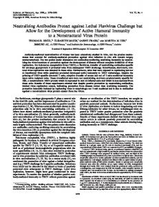
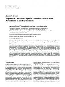
Figure 3. Hypertrophy-associated transcriptomic changes in flow-sorted rat CMs. (A and B) Violin plots illustrating the gene-wise relation between RNA expression and H3K9me2 (A) or H3K27me3 (B). Data were quantified per gene and are represented as log2((fragments + 1) per kb per million) (FPKM). (C and D) Gene expression, represented as fragments per million (FPM), in PCM1+ nuclei sorted from hypertrophied and control hearts. Highlighted are the genes that were significantly upregulated (green) and downregulated (red) (FDR