0013-7227/99/$03.00/0 Endocrinology Copyright © 1999 by The Endocrine Society
Vol. 140, No. 7 Printed in U.S.A.
3*,5* Cyclic Adenosine Monophosphate Mediates the Salmon Calcitonin-Induced Increase in Hypothalamic Tyrosine Hydroxylase Activity* LYDIA A. ARBOGAST, GIRISH V. SHAH,
AND
JAMES L. VOOGT
Department of Physiology (L.A.A.), Southern Illinois University School of Medicine, Carbondale, Illinois 62901-6512; Department of Pharmaceutical Sciences (G.V.S.), Texas Tech University Health Sciences Center, Amarillo, Texas 79106; and Department of Molecular and Integrative Physiology (J.L.V.), University of Kansas Medical Center, Kansas City, Kansas 66160-7401 ABSTRACT This study examined the effect of salmon calcitonin (sCT) on hypothalamic tyrosine hydroxylase (TH) activity and evaluated the cellular signaling mechanisms involved in the response. Fetal hypothalamic cells were cultured in a defined medium and treated with sCT and/or specific protein kinase inhibitors on day 14 in vitro. sCT (0.1–10 nM) increased both TH activity and cellular cAMP content in a concentration-dependent manner. sCT (10 nM) increased TH activity to 150 –175% of control values and resulted in a 10-fold increase in cellular cAMP content. Both the C1a and C1b CT receptor isoforms were present in the cultures, as assessed by RT-PCR. Rp-adenosine 39,59-cyclic monophosphothioate (Rp-cAMPS), a cAMP antagonist, and H-8, a cyclic nucleotide kinase inhibitor, blocked the sCT-induced increase in TH activity, with complete abolition of the response observed at concentrations of 1 mM and 5 mM, respectively. sCT (10 nM)
C
ALCITONIN (CT), a 32-amino acid peptide, is produced and released by the parafollicular cells of the thyroid gland and has a well established role in lowering extracellular calcium. More recently, CT-like peptides, CT receptors, and CT-induced biological responses have been identified in other tissues (1– 8). High affinity binding sites for CT are abundant in the central nervous system with the highest densities found in the various hypothalamic nuclei (1, 6). However, we still know relatively little about specific neuronal targets in the brain. Two isoforms of the CT receptor, C1a and C1b, have been identified in the brain (7, 8). The isoforms are identical except for a 37 amino acid insert in the second extracellular domain of the C1b isoform (7, 8). The C1b isoform appears to be present predominantly in the brain, whereas the C1a isoform is found in the brain and peripheral tissues (7, 8). The CT receptors are seven-transmembrane G protein-coupled receptors, coupled to activation of the adenylate cyclase/cAMP/protein kinase A pathway and the phospholipase C/calcium mobilization/protein kinase C pathway (3, 9 –12). Both of the cloned brain isoforms
Received November 9, 1998. Address all correspondence and requests for reprints to: Dr. Lydia A. Arbogast, Department of Physiology, School of Medicine, Southern Illinois University at Carbondale, Carbondale, Illinois 62901-6512. E-mail:
[email protected]. * This work was supported by NIH Grant HD-35332 (L.A.A.), University Women’s Professional Advancement/Women’s Studies Research Award from Southern Illinois University (L.A.A.), NIH Grant HD-24190 (J.L.V.) and NIH Grant DK-45044 (G.V.S.).
increased radiolabeled phosphate incorporation into TH protein to 169% of control values and 1 mM Rp-cAMPS completely blocked this effect. In contrast, neither Calphostin C, a protein kinase C inhibitor, nor U-73122, a phospholipase C inhibitor, significantly altered the ability of sCT to increase TH activity. Likewise, the sCT-induced increase in TH activity was observed after pretreating the cells with either BAPTA/AM, an intracellular calcium chelator, or thapsigargin, an inhibitor of the endoplasmic reticulum calcium pump. These data indicate that sCT has a profound stimulatory effect on TH activity in fetal hypothalamic cells and that enhanced phosphorylation of TH coincides with the sCT-induced increase in enzyme activity. Moreover, CT receptors, which are linked to cAMP production, are expressed in the hypothalamic cells and a cAMP-dependent mechanism mediates the sCT-induced activation and phosphorylation of TH. (Endocrinology 140: 3273–3281, 1999)
of the CT receptor are functionally linked to cAMP when transfected into COS cells (7, 8). In contrast, CT analogues do not cause adenylate cyclase activation in brain homogenates (13, 14). The reason for the differential effect on adenylate cyclase activation between the cloned receptors and brain membrane preparations is not clear. A likely role for the hypothalamic CT receptors is in the control of anterior pituitary function. CT or CT-like peptides can alter circulating PRL levels in vivo (15–17). The physiological significance of an inhibitory role is supported by the marked increase in circulating PRL levels of ovariectomized rats after administration of an anti-sCT serum (18). An sCTlike immunoreactive peptide, synthesized and secreted by the anterior pituitary, likely contributes to the inhibition of PRL secretion by a paracrine action within the anterior pituitary gland (3, 4, 12, 19). In addition, some evidence indicates that one or more sCT-like substances may also exert an indirect effect, possibly at the hypothalamic level, to control PRL release. First, the increase in PRL release induced by anti-sCT serum in ovariectomized rats is more profound in vivo than in vitro (18). Second, the sCT-induced decrease in PRL levels in male rats in prevented by median eminence lesions (15). Indeed, both the brain and pituitary are potential sources of sCT-like compounds (4, 5, 18). A possible site for sCT’s action is the tuberoinfundibular dopaminergic neurons. The dopamine released from these neurons is well established as a major PRL inhibiting hormone (20). Moreover, these dopaminergic neurons have their cell bodies in
3273
3274
CT INCREASES TYROSINE HYDROXYLASE ACTIVITY
Endo • 1999 Vol 140 • No 7
the arcuate nucleus of the hypothalamus (21), which has a high expression of CT receptor and high density of sCT binding sites (6, 22). The use of an in vitro model allows for isolation of brain area of interest and enhances the ability to explore the intracellular signaling mechanisms. Indeed, dopaminergic neurons in fetal hypothalamic cell cultures respond to many hormones and neuropeptides in a manner similar to the tuberoinfundibular dopaminergic neurons in an adult animal (23–27). The primary goal of this study was to evaluate the effect of sCT on dopaminergic neurons in fetal hypothalamic cell cultures and to determine the intracellular signaling pathway(s) involved. Tyrosine hydroxylase (TH) is the rate limiting enzyme in catecholamine biosynthesis (28), and this study focused on this key control point in the dopaminergic neurons. The specific aims were: 1) to determine the effect of different concentrations of sCT on TH activity and cellular cAMP content; 2) to assess the expression of sCT receptors; 3) to investigate the intracellular signaling pathway by which sCT alters TH activity by using specific protein kinase inhibitors; and 4) to examine the effect of sCT on the phosphorylation state of TH.
included in the preincubation medium. For experiments to reduce available intracellular calcium, the preincubation medium was phenol redfree and calcium-free Earle’s Balanced Salt Solution (Life Technologies, Inc.) containing 1 mm ethylene glycol-bis (b-aminoethylether)N,N,N9N9tetra-acetic acid (EGTA), 20 mm tyrosine and 20 mm potassium chloride. Where indicated, 1,2-Bis(2-aminophenoxy)ethane-N,N,N9,N9-tetraacetic acid tetraacetoxy-methylester (BAPTA/AM; Molecular Probes, Inc., Eugene, OR) or thapsigargin (Molecular Probes, Inc.) at the indicated concentration was included in the preincubation medium. The preincubation medium was removed and 120 ml medium containing 100 mm brocresine (4-bromo-hydroxybenzyloxyamine; gift from American Cyanamid Co., Pearl River, NY), an aromatic L-amino acid decarboxylase inhibitor, and the indicated concentration of rat CT, human CT, sCT (Bachem California, Inc., Torrance, CA), forskolin (Sigma Chemical Co., St. Louis, MO), phorbol 12-myristate 13-acetate (PMA; Research Biochemicals International), 4-a-phorbol 12-myristate 13-acetate (4-a-PMA; Research Biochemicals International) with or without the kinase inhibitors was added. For experiments to reduce available intracellular calcium, the medium was calcium-free with 1 mm EGTA and thapsigargin, but not BAPTA/AM, included in the incubation medium as indicated. The cells were incubated for 1 h at 37 C under an atmosphere of 10% CO2-90% air. The medium was the removed, acidified with 12 ml 1 n perchloric acid and frozen until samples were analyzed for L-dihydroxyphenylalanine (DOPA) accumulation in the media by HPLC with electrochemical detection (30).
Materials and Methods
The hypothalamic cells were preincubated for 15 min with 1 ml Earle’s Balanced Salt Solution containing 20 mm tyrosine and an additional 20 mm potassium chloride. The preincubation was removed and medium containing 1 mm isobutylmethylxanthine (IBMX; Sigma Chemical Co.), a phosphodiesterase inhibitor, and the appropriate concentrations of sCT was added. After 15 min, the medium was removed and assayed for cAMP levels. Immediately thereafter, 500 ml 3% perchloric acid was added to the cells. The cells were subjected to a freeze-thaw cycle, scraped from the plate, and homogenized by sonication. The pH was adjusted to pH 6.0 with 30% potassium bicarbonate. The samples were centrifuged at 10,000 3 g for 10 min and the supernatant transferred to the assay. The cAMP contents of the medium and cells were assayed separately according to the manufacturer’s directions using a kit from Diagnostic Products, Inc., (Los Angeles, CA) with 3H cAMP as the tracer. The lower limit of detection for the assay was 0.55 pmol/1 3 106 cells. Each sample was analyzed in duplicate and all samples from the same experiment were analyzed in a single assay to avoid intraassay variability.
Animals Adult female (200 –250 g) and male rats were obtained from Sasco (Omaha, NE) or Harlan Sprague Dawley, Inc. (Indianapolis, IN). Rats were maintained under controlled temperature and lighting conditions and food and water were available ad libitum. All procedures were approved by the institutional animal care and use committees at the University of Kansas Medical Center or Southern Illinois University. The estrous cycles of female rats were monitored by daily vaginal lavage, and proestrous rats were placed individually with male rats. If sperm were present in the vaginal lavage on the following day, it was designated as day 0 of pregnancy.
Fetal hypothalamic cell cultures Fetal hypothalamic cells were cultured as described previously (23, 27, 29). Briefly, the medioventral hypothalami of day 19 –20 rat fetuses were excised, and the hypothalamic cells were dispersed with trypsin. After dispersion, the cells were resuspended in a modified phenol redfree, high glucose Dulbecco’s medium containing 2.5% heat-inactivated FBS and 5% heat inactivated horse serum and were plated at a density of 150,000 cells/well in 96-well plates for determination of TH activity, or 1 3 106 cells/well in 24-well plates for radiolabeled phosphate incorporation, cellular cAMP content determinations or RNA isolation. After 18 –24 h, the serum containing medium was replaced with a serumfree, phenol red-free, chemically defined, high glucose modified DMEM (27). The cells were maintained under an atmosphere of 95% air-5% CO2. The medium was changed every 1–2 days as needed.
Determination of TH activity TH activity was determined on day 12–14 in vitro. The hypothalamic cells were preincubated for 30 – 45 min in phenol red-free Earles’ Balanced Salt Solution (Life Technologies, Inc., Grand Island, NY) with 20 mm tyrosine and 20 mm potassium chloride added. When indicated, Rp-adenosine 3959-cyclic monophosphothioate triethylamine (RpcAMPS; Research Biochemicals International, Natick, MA), N-[2-(methylamino) ethyl]-5-isoquinoline-sulfonamide dihydrochloride (H-8; BIOMOL Research Laboratories, Inc., Plymouth Meeting, PA), Calphostin C (Kamiya Biomedical Company, Thousand Oaks, CA; BIOMOL Research Laboratories, Inc.), 1-(6-((17b-3-methoxyestra-1,3,5 (10)trien-17-yl)amino)hexyl)-1H-pyrrole-2,5-dione (U-73122, BIOMOL) or 1-(6-((17b-3-methoxyestra-1,3,5 (10)-trien-17-yl)amino)hexyl-2,5-pyrrolidine-dione (U-73343; BIOMOL) at the indicated concentration was
Determination of cAMP content
RT-PCR and Southern blot for CT receptor The hypothalamic cells (2 3 106 cells) were lysed in 800 ml RNAzol B (Tel-Test, Inc., Friendswood, TX) and total RNA isolated according to the protocol provided by the manufacturer. RNA (5 mg) was used for first strand complementary DNA (cDNA) synthesis using the Superscript Preamplification System (Life Technologies, Inc.) with oligo dT primers. The cDNA was amplified through 30 cycles of PCR with Taq polymerase using 62.5 C as the annealing temperature. The 21 mer primers were described by Albrandt et al. (7) and were 59-GTT GAG GTT GTG CCC AAT GGA-39 and 59 CCC TGG AAA TGA ATC AGA GAG-39. These primers generate a 545-bp product for the C1a receptor and a 656-bp product for the C1b receptor. The PCR samples were run along with a 100-bp ladder (Life Technologies, Inc.) in an agarose gel containing ethidium bromide. After depurination of DNA fragments with 0.25 m HCl, denaturation with buffer containing 0.5 m sodium hydroxide and 1.5 m sodium chloride and neutralization with buffer containing 0.5 m Tris HCl pH 7.0 and 1.5 m sodium chloride, the DNA from the PCR reaction was transferred to a NYTRAN membrane using the Schleicher & Schuell Turboblotter downward transfer apparatus (Midwest Scientific, St. Louis, MO). The PCR product was verified by Southern blot hybridization. The partial pituitary cDNA probe for sCT receptor was generated by RT-PCR of a total RNA preparation from rat anterior pituitary glands using gene-specific sense and antisense amplimers as previously described (7). The amplified cDNA was subcloned in pGem-T vector (Promega Corp., Madison, WI) and the DNA sequence
CT INCREASES TYROSINE HYDROXYLASE ACTIVITY
3275
was determined. The partial pituitary CT receptor cDNA was 546 bp long and displayed 99% homology with rat C1a receptor (544/546) and C1b receptor (503/504) cDNA sequences (7).
Evaluation of radiolabeled phosphate incorporation into TH The procedure to determine the phosphorylation state of TH is adapted from the procedures described by Cahill and Perlman (31) and Porter (32). The basic incubation medium contained 116 mm sodium chloride, 5.3 mm potassium chloride, 2.5 mm calcium chloride, 0.8 mm magnesium sulfate, 5 mm glucose, and 25 mm HEPES buffer pH 7.4. Fetal hypothalamic cells (1 3 106 cells/well) were washed 2 3 5 min with medium and then incubated for 1 h in 0.25 ml medium containing 32P orthophosphoric acid (500 mC/ml). The radiolabeled phosphate was the sole source of phosphate in the medium. Hypothalamic cells were then incubated an additional 30 min with 1 mm Rp-cAMPS as indicated and subsequently for 15 min with 10 nm sCT and/or 1 mm Rp-cAMPS as indicated. At the end of the incubation time, cells were quickly rinsed four times with medium. Ice-cold phosphatase-inhibiting buffer (80 ml), consisting of 30 mm potassium phosphate, pH 7.5, 25 mm sodium fluoride, 5 mm sodium pyrophosphate, 0.5 mm okadaic acid, 1 mm EDTA, 0. 5 mm phenylmethylsulfonylfluoride, 10 mg/ml aprotinin and 0.5% IGEPAL CA-630, was added to each well. The cells were subjected to a freeze-thaw cycle and scraped from the plate. Individual wells were rinsed with an additional 40 ml of ice-cold phosphatase inhibiting buffer. Homogenates were centrifuged for 1 min and 100 ml of the homogenate was used for immunoprecipitation with 2 ml TH antibody (East Acre Biologicals, Southbridge, MA) at 4 C for 18 h. The antigen-antibody complex was then precipitated at 4 C for 30 min with 100 ml of a 10% solution of Staphylococcus aureus cells (Pansorbin; Calbiochem). The S. aureus-antibody-TH pellet was vigorously washed 4 times, sonicated in 20 ml of sample buffer, boiled for 2 min, and centrifuged at 10,000 3 g for 5 min. The sample (20 ml) was subject to electrophoresis on a 7.5% polyacrylamide gel slab using a buffer containing 0.375 m Tris buffer pH 8.8 and 0.1% SDS. The gels was calibrated with molecular weight standards between 14,000 and 100,000. After electrophoresis, the slab gel was dried and exposed to a phosphor screen (Molecular Dynamics, Inc., Sunnyvale, CA). The phosphor screen was scanned with a Molecular Dynamics, Inc. Phosphorimager 445SI. Individual TH bands on the gels were identified and the volume quantified with the associated FragmeNT analysis software. Total protein concentration was determined with 10 ml of the original homogenate by the method of Bradford (33), and the volume values obtained from the phosphorimager were normalized to the total protein content of the individual samples. Subsequently, the gels were exposed at 270 C for 3– 4 h to Kodak XAR-2 film (Eastman Kodak Co., Rochester, NY) using an intensifying screen, the film was developed, and the 32P-labeled TH bands on the gel localized.
Statistical analysis To normalize data, experimental values were adjusted to a percentage of control values for individual experiments. For TH activity, the control values were a mean of 9 –12 wells and each experimental value was the mean of triplicate wells for each hypothalamic culture. For radiolabeled phosphate incorporation experiments, control and experimental values were determined from duplicate wells. The results are expressed as the mean 6 se of determinations from five to eight different experiments. Data were evaluated by ANOVA, and multiple comparisons were made with Fisher’s least significant procedures (34, 35).
Results Effect of CT on TH activity and cAMP content in fetal hypothalamic cells
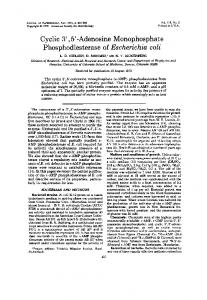
The first experiment examined the effect of sCT on TH activity in fetal hypothalamic cells. Different concentrations of sCT were included during the 1-h incubation period to determine TH activity by DOPA accumulation in the medium. sCT caused a concentration-dependent increase in TH activity in the fetal hypothalamic cells (Fig. 1; top panel). There was no significant increase in enzyme activity with 1 and 10
FIG. 1. Top panel, TH activity (DOPA accumulation) in fetal hypothalamic cell cultures after a 1-h treatment with different concentrations of sCT. Control values were normalized and are indicated by the dashed line. Experimental values are represented as percent of control and are indicated with the solid line. A significant increase was observed with 100 pM sCT and near maximal increase with 1–100 nM sCT. Bottom panel, Cellular cAMP content in fetal hypothalamic cells after 15 min treatment with 1–10 nM sCT. A significant increase in cAMP content was observed with 0.1 nM and continue to increase to 10 nM. Each value is a mean 6 SE of determinations from five to six different cell culture experiments. *, Significantly different from control (P , 0.05).
pm sCT, but 100 pm sCT increased TH activity to 130% of control values. The sCT-induced increase in TH activity reached near plateau levels between 1–100 nm sCT with values 145–157% of control levels. In a separate experiment, the hypothalamic cells were incubated with either rat CT or sCT to determine if the homologous hormone would have the same effect. Rat CT and sCT (10 nm) increased TH activity similarly to 142 6 6% and 149 6 4% of control values, respectively, indicating CT from both species was effective in increasing TH activity. Given that the sCT is known to be coupled to activation of the adenylate cyclase/cAMP/protein kinase A signal transduction pathway in some cells (2, 18), the ability of sCT to alter cAMP accumulation in the hypothalamic cell cultures was determined. sCT caused a concentration-dependent increase in both cellular and medium cAMP accumulation during a 15-min incubation with 1 mm IBMX, a phosphodiesterase inhibitor (Fig. 1; bottom panel). A 2.2-fold increase in
3276
CT INCREASES TYROSINE HYDROXYLASE ACTIVITY
cellular cAMP accumulation was observed with 100 pm sCT, whereas 1 and 10 nm sCT resulted in a 4.7-fold and 9.5-fold increase, respectively. Although the cell extract contained approximate 90% of the accumulated cAMP, cAMP accumulation in the medium showed an identical trend. Control cAMP accumulation (pmol/1 3 106 cells/15 min) was 4.06 6 0.34 and cAMP accumulation was increased to 7.02 6 0.22, 14.78 6 0.84 and 28.18 6 1.37 with 100 pm, 1 nm and 10 nm sCT, respectively. The sCT-induced increase in cAMP occurred within the same concentration range as the changes in TH activity. Expression of CT receptor in hypothalamic cells
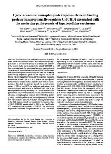
RT-PCR was used to determine whether the CT receptor was present in the hypothalamic cell cultures. Expression of the CT receptor was similar in adult mediobasal hypothalamic tissue and in fetal hypothalamic cell cultures (Fig. 2, bottom panel). Both the C1a and C1b forms of the CT receptor were expressed, as evidenced by the 545-bp and 656-bp PCR products, respectively. No PCR product was detected with the omission of reverse transcriptase. The identity of the PCR products were confirmed by Southern blot analysis using a
FIG. 2. CT receptor expression in fetal hypothalamic cell cultures (lanes 2–5) and in the mediobasal hypothalamus of ovariectomized rats (lanes 7– 8), as determined by RT-PCR and visualized with ethidium bromide staining (bottom panel). A 100-bp ladder was included in lane 1. Two forms of the CT receptor are expressed and are similar in adult hypothalamic tissue and fetal hypothalamic cells. The 545-bp PCR product denotes expression of CT receptor C1a, whereas the 656 bp PCR product denotes expression of CT receptor C1b. Note the absence of PCR product with the omission of reverse transcriptase. The identity of the PCR products was confirmed by Southern blot analysis with a specific probe for the CT receptor (top panel). Similar expression was observed in a total of four mediobasal hypothalamic samples and seven different cell cultures.
Endo • 1999 Vol 140 • No 7
specific 32P-labeled probe for the CT receptor (Fig. 2, top panel). Because the cloned C1a and C1b CT receptors display different sensitivities to human CT (8), the effects of sCT and human CT on TH activity were compared with increasing concentrations of peptide. Human CT was less effective than sCT in stimulating TH activity with the dose-response curve for human CT shifted to the right (Fig. 3). Analysis of the role of cAMP in the sCT-induced increase in TH activity
The role of cAMP in the sCT-induced increase in TH activity was examined by blocking the cAMP-protein kinase A pathway with Rp-cAMPS, a cAMP antagonist (36, 37), or H-8, a cyclic nucleotide kinase inhibitor (38). After 1 h sCT treatment, TH activity was increased to 175% of control values. The stimulatory action of sCT was blocked by Rp-cAMPS in a concentration-dependent fashion (Fig. 4, top panel). TH activity was not significantly different from control values in the presence of 100 mm Rp-cAMPS with or without sCT. The 1 mm concentration of Rp-cAMPS completely abolished the response and also reduced basal TH activity to 69% of control values. In a similar manner, H-8 also suppressed the sCTinduced elevation in TH activity in a concentration dependent manner (Fig. 4, bottom panel). The response was significantly suppressed with 0.5 mm H-8 and completely blocked with 5 mm H-8. At the highest concentration (50 mm), H-8 alone suppressed TH activity to 82% of control values, in addition to inhibiting the sCT-induced rise. These data suggest that cAMP and protein kinase A are involved in the sCT-induced increase in TH activity in hypothalamic cells. The ability of Rp-cAMPS and H-8 to block a known cAMP response in the fetal hypothalamic cells was assessed with forskolin, an activator of adenylate cyclase. Forskolin (1 mm) increased TH activity in the hypothalamic cells to 213 6 17% (n 5 5) of control values. Rp-cAMPS (1 mm) reduced the
FIG. 3. TH activity (DOPA accumulation) in fetal hypothalamic cell cultures after a 1-h treatment with different concentrations of sCT and human CT. Control values were normalized and are indicated by the dashed line. Experimental values are represented as percent of control and are indicated by the solid lines. A significant increase was observed with 1 nM sCT and 10 nM human CT. Each value is a mean 6 SE of determinations from three different cell culture experiments. *, Significantly different from control (P , 0.05).
CT INCREASES TYROSINE HYDROXYLASE ACTIVITY
3277
FIG. 5. The effect of 10 nM sCT and/or 1 mM Rp-cAMPS treatment for 1 h on radiolabeled phosphate incorporation into TH immunoprecipitable protein. sCT increased radiolabeled phosphate incorporation into TH protein and Rp-cAMPS completely prevented the effect. Control values were normalized and are indicated by the dashed line. Experimental values are represented as percent of control. Each value is a mean 6 SE of determination from five different cell culture experiments. *, Significantly different from control. **, Significantly different from sCT.
FIG. 4. Top panel, The effect of Rp-cAMPS, a cAMP antagonist, on the 10 nM sCT-induced increase in TH activity in fetal hypothalamic cell cultures. sCT increased TH activity to 175% of control values, whereas 1 mM Rp-cAMPS completely prevented the effect. Bottom panel, The effect of H-8, a cyclic nucleotide protein kinase inhibitor on the 10 nM sCT-induced increase in TH activity in fetal hypothalamic cells. sCT increased TH activity to 161% of control values and H-8 (0.5 mM and 5 mM) reduced the sCT-induced rise to 125% and 103% of control values, respectively. Control values were normalized and are indicated by the dashed line. Experimental values are represented as percent of control. Each value is a mean 6 SE of determinations from five to seven different cell culture experiments. *, Significantly different from control (P , 0.05). **, Significantly different from sCT (P , 0.05).
marked forskolin-induced increase to 122 6 12% of control values, whereas 50 mm H-8 decreased the rise to 142 6 3% of control values. Effect of sCT on TH phosphorylation
Given the rapid action of sCT on TH activity, the next experiment was to examine whether there was a change in radiolabeled phosphate incorporation into the TH molecule after sCT treatment and if cAMP was involved in the changes in phosphate incorporation. The autoradiographic film showed a band at approximately a molecular weight of 60,000, corresponding to TH. As shown in the inset in Fig. 5, the intensity of this band increased after sCT treatment and was reduced by prior and concomitant treatment with RpcAMPS. The 60,000 molecular weight band was completely
absent when TH antiserum was omitted during the immunoprecipitation procedure (data not shown). sCT treatment increased 32P incorporation into TH to 169% of control levels and 1 mm Rp-cAMPS completely prevented this increase (Fig. 5). Rp-cAMPS alone had no effect on phosphate incorporation. Analysis of the role of the phospholipase C/calcium mobilization/protein kinase C pathway in the sCT effect on TH activity
Calphostin C, a specific inhibitor of protein kinase C, was used to examine the role of protein kinase C activation in the sCT-induced increase in TH activity. Although 1 mm Calphostin C alone decreased TH activity to 77% of control values, neither 0.1 mm or 1 mm Calphostin C had a significant effect on the sCT-induced increase in TH activity (Fig. 6, top panel), indicating that sCT is not coupled to the protein kinase C pathway to exert its effect on TH activity in the hypothalamic cells. The ability of Calphostin C to block a known protein kinase C-dependent response was assessed with PMA, an activator of protein kinase C. PMA caused a concentration dependent decrease in TH activity to 18% of control levels with the 1 mm concentration (Fig. 6, bottom panel). In contrast, 4-a-PMA, a negative control for phorbol ester activation of protein kinase C, did not alter TH activity. Calphostin C (1 mm) completely prevented the decrease in TH activity induced by 1 and 10 nm PMA and partially blocked the inhibitory effect of 0.1 and 1 mm PMA. The involvement of intracellular calcium mobilization was evaluated in a calcium-free EGTA containing medium by depletion of the intracellular calcium stores and by chelating intracellular calcium. The hypothalamic cells were preloaded
3278
CT INCREASES TYROSINE HYDROXYLASE ACTIVITY
FIG. 6. Top panel, The effect of Calphostin C, a protein kinase C inhibitor, on the 10 nM sCT-induced increase in TH activity in fetal hypothalamic cell cultures. sCT increased enzyme activity to 143% of control values. Calphostin C (0.1 and 1.0 mM) did not significantly reduce the sCT-induced increase in DOPA accumulation in the hypothalamic cell cultures. Bottom panel, The effect of phorbol 12-myristate 13-acetate (PMA), an activator of protein kinase C, and 4-aPMA, the negative control for phorbol ester activation of protein kinase C, on TH activity and the effect of calphostin C on the PMAinduced decrease in TH activity. A significant decrease in TH activity was observed with PMA, but not 4-a-PMA. Calphostin C reversed the inhibitory effect of PMA. Control values were normalized and are indicated by the dashed line. Experimental values are represented as percent of control. Each value is a mean 6 SE of determinations from 5– 8 different cell culture experiments. *, Significantly different from control (P , 0.05). **, PMA 1 calphostin C significantly different from PMA (P , 0.05).
with BAPTA/AM, a cell permeant calcium selective chelator (39), for 45 min and then incubated with sCT for 1 h to determine TH activity. Treatment with 10 nm sCT increased TH activity in wells without BAPTA/AM treatment to 152% of control values (Fig. 7, top right panel). Pretreatment of the cells with 1 mm BAPTA/AM did not significantly alter TH activity or the ability of sCT to increase TH activity. However, even though basal TH activity was decreased to 68% of control values after 10 mm BAPTA/AM pretreatment, sCT increased TH activity to 163% of the BAPTA/AM-treated
Endo • 1999 Vol 140 • No 7
FIG. 7. Top panel, The effect of BAPTA/AM, a intracellular calcium chelator, pretreatment and thapsigargin, an inhibitor of the endoplasmic reticulum calcium ATPase, treatment on TH activity with calcium-free, EGTA-containing medium. sCT increased TH activity to 152% of control. Although 10 mM BAPTA/AM suppressed both basal and sCT-stimulated TH activity, sCT increased TH activity to 163% of the respective basal levels. Thapsigargin did not alter the sCTinduced increase in TH activity. Bottom panel, The effect of U-73122, a phospholipase C inhibitor, and U-73343, a structurally similar compound without phospholipase C inhibiting activity, on the sCTinduced increase in TH activity. U-73122 did not alter basal or sCTstimulated TH activity, whereas the inactive U-73343 cause a modest decrease in both parameters. Each value is a mean 6 SE of determination from five to six different cell culture experiments. *, Significantly different from control. **, Significantly different from sCT.
wells. Thapsigargin, an inhibitor of the endoplasmic reticulum calcium ATPase, an enzyme responsible for pumping the calcium into intracellular stores (40) was used to evaluate the depletion of intracellular calcium stores. Thapsigargin caused a modest decrease in TH activity, but did not alter the sCT-induced increase in TH activity (Fig. 7, top left panel). It should also be noted that sCT caused a similar increase in TH activity with calcium-containing medium (Fig. 7, bottom panel) and calcium-free, EGTA-containing medium (Fig. 7, top left panel), suggesting that extracellular calcium does not have a role in the response. U-73122, an inhibitor of phopholipase C (41), was used to determine if phospholipase C activation contributes to the sCT-induced increase in TH activity. The ability of sCT to increase TH activity in the hypothalamic cells was not altered
CT INCREASES TYROSINE HYDROXYLASE ACTIVITY
when U-73122 was included in the preincubation and incubation medium (Fig. 7, bottom panel). The structurally related compound, U73343, which does not inhibit phopholipase C, modestly reduced basal and sCT-induced levels of TH activity. Discussion
We are reporting that sCT has a profound stimulatory effect on TH activity in fetal hypothalamic cells and that enhanced phosphorylation of TH protein contributes to the sCT-induced elevation in enzyme activity. Our data also indicate that the hypothalamic cells express both the C1a and C1b isoforms of the CT receptor and that sCT treatment causes an augmentation of cAMP production, suggesting that at least one of these receptor isoforms is linked to adenylate cyclase activation in the hypothalamic cell cultures. Moreover, cAMP mediates the sCT-induced increase in TH catalytic activity and phosphorylation state, as supported by the ability of specific protein kinase inhibitors, which are targeted to the adenylate cyclase/cAMP/protein kinase A signal transduction pathway, to prevent the effect on TH. Thus, this study identified a specific neuronal subtype in the hypothalamus as a target for CT and the signal transduction mechanism involved. In a broader sense, these data may have implications in the control of anterior pituitary hormone secretion. This study identifies dopaminergic neurons as a site of action for CT in fetal hypothalamic cells. The marked stimulatory effect was observed on the enzyme, TH, which serves as a key control point in dopamine biosynthesis. sCT caused a significant increase in TH activity at concentrations as low as 100 pm and a near maximal effect at 1–10 nm. The concentration dependence for the effect of sCT on TH was very similar to concentrations of sCT that displaced 125I sCT binding to brain or hypothalamic membranes (13, 14) and the cloned rat CT receptor (7, 8). Thus, the sensitivity for the biological response reported in this study corresponds to CT receptor binding characteristics reported by other investigators (13, 14). Both the homologous rat CT and the heterologous sCT were effective in increasing TH activity, even though the amino acid sequences of these peptides are not identical (5). It is not clear what peptide serves as the endogenous ligand for the sCT-sensitive receptor(s) in the hypothalamus. The principal product of the CT gene itself in the brain is calcitonin gene-related peptide (CGRP), which has binding sites with a specificity and distribution distinct from the conventional CT binding sites (1). However, peptides that are biologically and immunologically similar to sCT have been identified in the brain (5, 42). Moreover, the identification of a sCT-like immunoreactive peptide, which is synthesized and release by the anterior pituitary (4, 18), raises the possibility of pituitary control of hypothalamic function. Both the C1a and C1b forms of the CT receptor were expressed similarly in the hypothalamic cell cultures and the mediobasal hypothalamus of adult rats, suggesting that the findings in this study may have implications in the control of hypothalamic function in vivo. This high level of CT receptor expression in this study agrees with previous reports of significant CT receptor messenger RNA levels (8, 22) and
3279
a high density of CT binding sites (1, 6) in the hypothalamus. The primers used in this study were designed by Albrandt et al. (7) and flank the 111 bp nucleotide insert in the C1b receptor isoform found predominantly in the brain (7, 8). Thus, the primers were able to recognize both forms of the receptor and differentiate between the isoforms by the size of the PCR product. The specific receptor subtype associated with activation of the dopaminergic neurons was not identified in this study, but the receptor recognizes both rat CT and sCT. These data argue against the involvement of the C1b isoform, which has a poor interaction with human and rat CT (8). Furthermore, the concentration response characteristics for human CT and sCT are similar to sCT binding and cAMP production profiles of the cloned C1a CT receptor (8). In general, these data support the notion that the C1a CT receptor mediates the effects of sCT on the hypothalamic dopaminergic neurons. This study did not address the issue of whether sCT receptors are located on dopaminergic neurons, but the response occurs rapidly, supporting the notion of a direct action. Our results indicate that a CT receptor coupled to adenylate cyclase activation is present in the hypothalamic cell culture, as evidenced by the sCT-induced concentration dependent increase in cellular cAMP content. These data contrast with previous studies showing that high concentrations of sCT cause a decrease in adenylate cyclase activity in hypothalamic membrane preparations (13, 14). However, when both of the identified isoforms of the CT receptor were transfected into COS cells, sCT caused a concentration-dependent increase in cAMP production (7, 8). Thus, the use of intact cells may be required for functional coupling to adenylate cyclase. The increased TH activity in hypothalamic dopaminergic neurons appears to be exclusively due to activation of protein kinase A. The involvement of the adenylate cyclase/cAMP/ protein kinase A pathway based is supported by a couple of lines of evidence. First, sCT caused a concentration-dependent increase in cellular cAMP content, indicating activation of adenylate cyclase by sCT. Moreover, the range of sCT concentrations necessary to elicit increased cAMP content was similar to that required to increase TH activity in the hypothalamic neurons. Second, two specific inhibitors to this signal transduction pathway blocked the sCT-induced increase in TH activity. H-8 is a potent inhibitor of cyclic nucleotide kinases, acting by competing with the ATP binding site (43). H-8 caused a significant reduction in the sCTinduced increase in TH activity at 0.5 mm and completely blocked the response at 5 mm. Although the present study used intact cells, the effect H-8 concentrations are consistent with the reported Ki of 1.2 mm for cAMP-dependent protein kinase and well below the reported Ki of 15 mm for protein kinase C (43). However, H-8 has an even greater potency (Ki50.5 mm) inhibiting cGMP-dependent protein kinase (43). Strong support for the involvement of cAMP-dependent protein kinase was provided with Rp-cAMPS, a competitive antagonist for cAMP (36, 37), which completely blocked the sCT-induced increase in TH activity. The high concentrations required for complete inhibition of the response likely stem from the relative inability of this compound to penetrate cell membranes.
3280
CT INCREASES TYROSINE HYDROXYLASE ACTIVITY
The involvement of the phospholipase C/protein kinase C signal transduction pathway in the sCT-induced increase in TH activity is not supported by the data in the present study. Specific inhibitors for each step in the signal transduction pathway were not effective in blocking the ability of sCT to increase TH activity. U-73122, a phospholipase C inhibitor, did not alter the stimulatory effect of sCT at a dose equal to or above that required to inhibit other phospholipase Cmediated events (41, 44). Our data also argue against the involvement of calcium mobilization to increase TH activity. The ability of sCT to increase TH activity was retained when available calcium was lowered by depleting the calcium stores with thapsigargin or chelating intracellular calcium with BAPTA/AM. The 1 mm concentration of thapsigargin has been reported to deplete intracellular stores in less than 10 min (45). It would be anticipated that the intracellular calcium stores would remain depleted during the present study because thapsigargin was included during both the 45-min preincubation and the 1-h incubation period. Additionally, calcium was nominally absent from the extracellular medium to prevent restoration of the calcium stores. The 10 mm concentration of BAPTA/AM suppressed basal TH levels, but did not alter the stimulatory effect of sCT. Although the absolute levels of TH activity after sCT treatment were lower and not significantly different from control levels in the 10 mm BAPTA/AM-treated group, sCT’s ability to stimulate TH activity was apparent. This concentration of BAPTA/AM has been reported to completely suppress the increase in dopamine release in striatal slices induced by ionomycin, but not the nitric oxide (46). Finally, protein kinase C involvement is not supported. In contrast to the sCTinduced increase in TH activity, phorbol ester activation of protein kinase C resulted in a marked decrease in TH activity that was specific to the active isomer. Thus, the effects of PMA and sCT on hypothalamic TH activity are diametrically opposed. Furthermore, calphostin C, a potent and specific inhibitor of protein kinase C (38), did not alter the ability of sCT to increase TH activity in the hypothalamic cells. The Ki for Calphostin C with respect to protein kinase C is 0.05 mm and 1 mm Calphostin C completely inhibits protein kinase C, whereas 50 mm has no effect on protein kinase A (38). In the present study, concentrations as high as 1 mm did not alter sCT’s action on TH activity. In contrast, calphostin C reversed the inhibitory effect of PMA on TH activity. While we cannot rule out that sCT affects multiple hypothalamic cell types and activates different signaling pathways in other cells, it appears that sCT regulation of TH is governed entirely by a cAMP-dependent mechanism. A role for augmented phosphorylation of the TH protein in response to sCT stimulation is implicated in the present study. Indeed, sCT caused a marked increase in radiolabeled phosphate incorporation into a 60,000 molecular weight protein immunoprecipitated with a specific TH anti-TH sera. Moreover, the enhanced phosphorylation state is driven by a cAMP-dependent mechanism, as evidence by the ability of Rp-cAMPS, a cAMP antagonist, to prevent the change. The involvement of a phosphorylation/dephosphorylation mechanism is not surprising, given the very rapid action of sCT on TH activity. Indeed, TH is a well established target
Endo • 1999 Vol 140 • No 7
for cAMP-dependent protein kinase, which phosphorylates TH on serine 40 and results in enzyme activation (47–52). The data in this study may have broader implications in the neuroendocrine control of PRL release. The tuberoinfundibular dopaminergic neurons in the hypothalamus have a primary role in the tonic inhibition PRL secretion (20), and an increase in dopaminergic neuronal activity would result in suppressed PRL levels. Indeed, sCT administration has been reported to reduce circulating PRL levels in some cases (15, 16). Although the endogenous ligand is not known, an anti-sCT serum causes a marked increase in circulating PRL levels in vivo but caused a more modest increase in PRL release of anterior pituitary cells in vitro (18). This suggests an extra-pituitary action of a sCT-like peptide, possibly on the hypothalamic dopaminergic neurons. This prospect requires further investigation. In conclusion, our data support the notion that the adenylate cyclase/cAMP/protein kinase A signaling pathway mediates the marked sCT-induced increase in TH activity in hypothalamic dopaminergic neurons. Furthermore, phosphorylation of the TH protein is a major intracellular mechanism for the enhanced enzyme activity and is driven by cAMP-dependent protein kinase. The identification of the dopaminergic neurons as a specific target for sCT supports an indirect hypothalamic action for sCT to influence PRL secretion. Acknowledgments We would like to thank Dr. Elliot Cohen (American Cyanamid Co., Pearl River, NY) for the gift of brocresine. We would also like to thank Dr. Thomas Cox for helpful discussions on calcium signaling and Carrie Campion for technical assistance.
References 1. Sexton PM 1991 Central nervous system binding sites for calcitonin and calcitonin gene-related peptide. Mol Neurobiol 5:251–273 2. Shah GV, Rayford W, Noble MJ, Austenfeld M, Weigel J, Vamos S, Mebust WK 1994 Calcitonin stimulates growth of human prostate cancer cells through receptor-mediated increase in cyclic adenosine 39,59-monophosphates and cytoplasmic Ca21 transients. Endocrinology 134:596 – 602 3. Shah GV, Kennedy D, Dockter ME, Crowley WE 1990 Calcitonin inhibits thyrotropin-releasing hormone-induced increases in cytosolic Ca21 in isolated rat anterior pituitary cells. Endocrinology 127:613– 620 4. Shah GV, Deftos LJ, Crowley WR 1993 Synthesis and release of calcitonin-like immunoreactivity by anterior pituitary cells: evidence for a role in paracrine regulation of prolactin secretion. Endocrinology 132:1367–1372 5. Hilton JM, Mitchelhill KI, Pozvek G, Dowton M, Quiza M, Sexton PM 1998 Purification of calcitonin-like peptides from rat brain and pituitary. Endocrinology 139:982–992 6. Henke H, Tobler PH, Fischer JA 1983 Localization of salmon calcitonin binding sites in rat brain by autoradiography. Brain Res 272:373–377 7. Albrandt K, Mull E, Brady EMG, Herich J, Moore CX, Beaumont I 1993 Molecular cloning of two receptors from rat brain with high affinity for salmon calcitonin. FEBS Lett 325:225–232 8. Sexton PM, Houssami S, Hilton JM, O’Keeffe LM, Center RJ, Gillespie MT, Darcy P, Findlay DM 1993 Identification of brain isoforms of the rat calcitonin receptor. Mol Endocrinol 7:815– 821 9. Lin HY, Harris TL, Flannery MS, Aruffo A, Kaji EH, Gorn A, Kolakowski Jr LF, Lodish HF, Goldring SR 1991 Expression cloning of an adenylate cyclase-coupled calcitonin receptor. Science 254:1022–1024 10. Goldring SR, Gorn AH, Yamin M, Krane SM, Wang J-T 1993 Characterization of the structural and functional properties of cloned calcitonin receptor cDNAS. Horm Metab Res 25:477– 480 11. Chabre O, Conklin BR, Lin HY, Lodish HF, Wilson E, Ives HE, Catanzariti L, Hemmings BA, Bourne HR 1992 A recombinant calcitonin receptor independently stimulates 39,59-cyclic adenosine monophosphate and Ca21/inositol phosphate signaling pathways. Mol Endocrinol 6:551–556 12. Judd AM, Kubota T, Kuan SI, Jarvis WD, Spangelo BL, MacLeod RM 1990 Calcitonin decreases thyrotropin-releasing hormone-stimulated prolactin re-
CT INCREASES TYROSINE HYDROXYLASE ACTIVITY
13. 14. 15. 16. 17. 18. 19.
20. 21. 22. 23. 24. 25. 26. 27. 28. 29. 30.
31.
lease through a mechanism that involves inhibition of inositol phosphates production. Endocrinology 127:191–199 Rizzo AJ, Goltzman D 1981 Calcitonin receptors in the central nervous system of the rat. Endocrinology 108:1672–1677 Twery MJ, Seitz PK, Nickols GA, Cooper CW, Gallagher JP, Orlowski RC 1988 Analogue separates biological effects of salmon calcitonin on brain and renal cortical membranes. Eur J Pharmacol 155:285–292 Olgiati VR, Guidobono F, Luisetto G, Netti C, Bianchi C, Pecile A 1981 Calcitonin inhibition of physiological and stimulated prolactin secretion in rats. Life Sci 29:585–594 Cressent M, Elie C, Milhaud G 1984 The effect of calcitonin on prolactin secretion during gestation. Life Sci 34:1621–1626 Chihara K, Iwasaki J, Iwasaki Y, Minamitani N, Kaji H, Jujita T 1982 Central nervous system effect of calcitonin: stimulation of prolactin release in rats. Brain Res 248:331–339 Shah GV, Pedchenko V, Stanley S, Li Z, Samson WK 1996 Calcitonin is a physiological inhibitor of prolactin secretion in ovariectomized female rats. Endocrinology 137:1814 –1822 Shah GV, Wang W, Grosvenor CE, Crowley WE 1990 Calcitonin inhibits basal and thyrotropin-releasing hormone-induced release of prolactin from anterior pituitary cells: evidence for a selective action exerted proximal to secretagogueinduced increases in cytosolic Ca21. Endocrinology 127:621– 628 Ben-Jonathan N 1994 Dopamine: a prolactin-inhibiting hormone. Endocr Rev 6:564 –589 Bjo¨rklund A, Moore RY, Nobin A, Stenevi U 1973 The organization of tuberohypophyseal and reticulo-infundibular catecholamine neuron systems in the rat brain. Brain Res 51:171–191 Sheward WJ, Lutz EM, Harmar AJ 1994 The expression of the calcitonin receptor gene in the brain and pituitary gland of the rat. Neurosci Lett 181:31–34 Arbogast LA, Voogt JL 1997 Prolactin (PRL) receptors are colocalized in dopaminergic neurons in fetal hypothalamic cell cultures: effect of PRL on tyrosine hydroxylase activity. Endocrinology 138:3016 –3023 Beyer C, Kolbinger W, Reisert I, Pilgrim C 1994 Activation of cultured rat hypothalamic dopaminergic neurons by long-term but not short-term treatment with prolactin. Neurosci Lett 180:231–234 Sarkar DK 1989 Evidence for prolactin feedback actions on hypothalamic oxytocin, vasoactive intestinal peptide and dopamine secretion. Neuroendocrinology 49:520 –524 Beyer C, Eusterschulte B, Pilgrim C, Reisert I 1992 Sex steroids do not alter sex differences in tyrosine hydroxylase activity of dopaminergic in vitro. Cell Tissue Res 270:547–552 Arbogast LA, Soares MJ, Robertson MC, Voogt JL 1993 A factor(s) from a trophoblast cell line increases tyrosine hydroxylase activity in fetal hypothalamic cell cultures. Endocrinology 133:111–120 Levitt M, Spector S, Sjoerdsma A, Udenfriend S 1965 Elucidation of the rate-limiting step in norepinephrine biosynthesis in the perfused guinea-pig heart. J Pharmacol Exp Ther 148:1– 8 Ahmed Z, Walker PS, Fellows RE 1983 Properties of neurons from dissociated fetal rat brain in serum-free culture. J Neurosci 3:2448 –2481 Arbogast LA, Voogt JL 1991 Hyperprolactinemia increases and hypoprolactinemia decreases tyrosine hydroxylase messenger ribonucleic acid levels in the arcuate nuclei, but not the substantia nigra or zona incerta. Endocrinology 128:997–1005 Cahill AL, Perlman RL 1984 Phosphorylation of tyrosine hydroxylase in the superior cervical ganglion. Biochim Biophys Acta 805:217–226
3281
32. Porter JC 1986 In situ activity and phosphorylation of tyrosine hydroxylase in the median eminence. Mol Cell Endocrinol 46:21–27 33. Bradford MM 1976 A rapid and sensitive method for the quantitation of microgram quantities of protein utilizing the principle of protein-dye binding. Anal Biochem 72:248 –254 34. Zar JH 1984 Biostatistical Analysis, ed 2. Prentice-Hall, Englewood Cliffs, NJ 35. Gerald KB 1990 Common multiple comparison procedures. Nurse Anesthesia 1:162–165 36. Parker Botelho LH, Rothermel JD, Coombs RV, Jastorff B 1988 cAMP analog antagonists of cAMP actions. Methods Enzymol 159:159 –172 37. Van Haastert PJM, Van Driel R, Jastorff B, Baraniak J, Stec WJ, De Wit RJW 1984 Competitive cAMP antagonists for cAMP-receptor proteins. J Biol Chem 259:10020 –10024 38. Casnellie JE 1991 Protein kinase inhibitors: probes for the functions of protein phosphorylation. Adv Pharmacol 22:167–203 39. Tsien RY 1980 New calcium indicators and buffers with high selectivity against magnesium and protons: design, synthesis, and properties of prototype structures. Biochemistry 19:2396 –2404 40. Missiaen L, Wuytack F, Raeymaekers L, De Smedt H, Droogmans G, Declerck I, Casteels R 1991 Ca21 extrusion across plasma membrane and Ca21 uptake by intracellular stores. Pharmacol Ther 50:191–232 41. Smith RJ, Sam LM, Justen JM, Bundy GL, Bala GA, Bleasdale JE 1990 Receptor-coupled signal transduction in human polymorphonuclear neutrophils: effects of a novel inhibitor of phospholipase C-dependent processes on cell responsiveness. J Pharmacol Exp Ther 253:688 – 697 42. Sexton PM, Hilton JM 1992 Biologically active salmon calcitonin-like peptide is present in rat brain. Brain Res 596:279 –284 43. Hidaka H, Kobayashi R 1992 Pharmacology of protein kinase inhibitors. Annu Rev Pharmacol Toxicol 32:377–397 44. Smallridge RC, Kiang JG, Gist ID, Fein HG, Galloway RJ 1992 U-73122, an aminosteroid phospholipase C antagonist, noncompetitively inhibits thyrotropin-releasing hormone effects in GH3 rat pituitary cells. Endocrinology 131:1883–1888 45. Kerper LE, Hinkle PM 1997 Cellular uptake of lead is activated by depletion of intracellular calcium stores. J Biol Chem 272:8346 – 8352 46. Stewart TL, Michel AD, Black MD, Humphrey PPA 1996 Evidence that nitric oxide causes calcium-independent release of [3H] dopamine from rat striatum in vitro. J Neurochem 66:131–137 47. Funakoshi H, Okuno S, Fujisawa H 1991 Different effects on activity caused by phosphorylation of tyrosine hydroxylase at serine 40 by three multifunctional protein kinases. J Biol Chem 266:15614 –15620 48. Joh TH, Park DH, Reis DJ 1978 Direct phosphorylation of brain tyrosine hydroxylase by cyclic AMP-dependent protein kinase: mechanism of enzyme activation. Proc Natl Acad Sci USA 75:4744 – 4748 49. Richtand NM, Inagami T, Misono K, Kuczenski R 1985 Purification and characterization of rat striatal tyrosine hydroxylase. J Biol Chem 260:8465– 8473 50. Vulliet PR, Langan TA, Weiner N 1980 Tyrosine hydroxylase: a substrate of cyclic AMP-dependent protein kinase. Proc Natl Acad Sci USA 77:92–96 51. Harris JE, Baldessarini RJ, Morgenroth III VH, Roth RH 1975 Activation by cyclic 39:59-adenosine monophosphate of tyrosine hydroxylase in the rat brain. Proc Natl Acad Sci USA 72:789 –793 52. Haycock JW, Wakade AR 1992 Activation and multiple-site phosphorylation of tyrosine hydroxylase in perfused rat adrenal glands. J Neurochem 58:57– 64