eISSN 1308-4038
International Journal of Anatomical Variations (2013) 6: 176–178
Case Report
3D reconstruction computerized tomography findings of an asymptomatic Eagle’s Syndrome patient: a case report Published online September 27th, 2013 © http://www.ijav.org
Hadi SASANI Ozgur CEYHAN [2] Serra SENCER [1] Mehdi SASANI [3]
Abstract
[1]
Istanbul University, Istanbul Faculty of Medicine, Department of Radiology, Istanbul [1], Florence Nightingale Hospital, Department of Radiology, Istanbul [2], Trakya University, Faculty of Medicine, Department of Anatomy, Edirne [3], TURKEY.
Eagle’s Syndrome is a condition and aggregate of symptoms due to elongation and ossification of the styloid process resulting in related symptoms. Despite being symptomatic in the 4-10.3% of general population, it may be asymptomatic. Diagnosis of the Eagle’s Syndrome depends on clinical findings and radiological imaging modalities.
© Int J Anat Var (IJAV). 2013; 6: 176–178.
Hadi Sasani, MD Istanbul University Istanbul Faculty of Medicine Department of Radiology Capa, 34093 Istanbul, TURKEY. +90 (212) 414 20 00 / 31142
[email protected]
Received November 16th, 2012; accepted April 4th, 2013
Key words [Eagle’s Syndrome] [3D reconstruction] [computerized tomography]
Introduction
Discussion
Eagle’s Syndrome (ES) is a condition caused by the elongation and ossification of the styloid process, and the related symptoms due to the close relationship between neurovascular structures around it [1]. About 4% of the population has long styloid process more than 3 cm; however, only 4-10.3% of them are symptomatic [2]. In this article we report a case of an asymptomatic ES with a general literature review, clinical and radiological findings.
ES which was defined by Eagle, is a condition related to the elongation and ossification of the styloid process that results in orofacial pain [1]. There are two clinical presentations including classic stylohyoid and stylocarotid syndromes. The classic stylohyoid syndrome, due to affection of lower cranial nerves, presents in the form of cervicofacial pain accompanied by dysphagia and foreign body sensation. The stylocarotid syndrome, due to carotid artery compression, may be associated with a neurological focus and even syncope [3, 4]. Embryologically four different segments are present in the stylohyoid apparatus including the tympanohyal, the stylohyal, the ceratohyal and the hypohyal segments. The ligamentous part, has its origin from the ceratohyal cartilage, extends from the stylohyal to the lesser horn of hyoid bone. There is a potential for ossification of the ligament, and when ossified, segmentation and pseudoarticulation may occur [1, 5–7]. The internal carotid artery, the internal jugular vein, and the IX-XIIth cranial nerves (glossopharyngeal, vagus, accessory,
Case Report
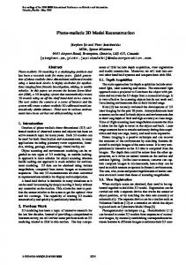
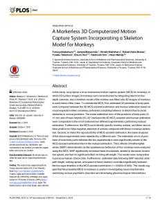
A 45-year-old male patient with the history of accidental trauma presented to the emergency department. On physical examination there was pain on the cervical region, but not any other specific finding or neurological problem detected. The laboratory tests were unremarkable. The cervical computerized tomography (CT) was done revealing the block vertebra appearance on C5 and C6 vertebral level (Figure 1). Beside this, bilateral elongated styloid processes incidentally were detected, which extended until C3 vertebral level on the right side (78 mm) and C2 on the left (50 mm) (Figure 2).
Asymptomatic Eagle’s Syndrome
Figure 1. CT scan revealed block vertebra on C5 and C6 vertebral level (arrowheads).
177 tongue, change in voice or sensation of hypersalivation and particularily pain during the neck rotation [9, 10]. Eagle [1] described a group of patients with symptoms of spastic and nagging pain in the pharynx radiating into the mastoid region. A feeling of a foreign body in the throat, dysphagia, or distortion of taste sensation was also experienced. All of the patients had had a history of tonsillectomy, and there was dense scar tissue in the tonsillar fossa. A hard mass was palpated in the fossa, which on radiography proved to be an ossified stylohyoid ligament. The closed neighborhood of styloid process to ECA may result in compression, even dissection of ECA which has been reported in literature [3]. ES should be suspected by presenting symptoms and physical examination. Elongation and ossification of the styloid process is diagnosed by imaging modalities through craniocervical radiograph, particularly the 3D reconstruction CT that provides high quality imaging demonstrating the anatomical and the contact with adjacent structures [11]. Diagnosis is made both radiographically and by physical examination. Palpation of the styloid process in the tonsillar fossa is mainly indicative of elongated styloid process. Normally it should not be palpated. As the confirmatory test radiographic studies should be done [12]. Also, most frequently, a panoramic radiograph is used to determine whether the styloid process is elongated, if it is more than 3 cm in length [13]. On the other hand, elongated styloid process is not only pathognomonic for ES, because many patients with incidental findings of an elongated styloid process are asymptomatic. Lateral and anterior-posterior radiographs, panoramic radiographs and 3D reconstruction CT imaging are helpful in the diagnosis of an elongated styloid process, and in providing complementary information with clinical status. The length of the styloid process may vary in size. As in this case the right side styloid process was extended until C3 vertebral level.
Conclusion
Figure 2. Bilateral elongated styloid processes are more prominent on the right side (red arrows) than the left side (yellow arrows), and extended to C3 vertebral level.
hypoglossal nerves) are medial and close to the stylohyoid apparatus. The external carotid artery (ECA) is lateral and is also closely related [8]. The normal stylohyoid process is 2.5-3 cm long, but considerable variation occurs (5-7.5 cm). It is important knowing about the symptomatic elongation of the styloid process, the ES, which should be considered in the differential diagnosis of obscure causes of head and neck pain. ES may present unilaterally or bilaterally. The most frequent symptoms are dysphagia, headache, pain on extension of the
Depending on the clinical symptoms (4-10.3% symptomatic, as in this case), ES may present itself asymptomatically and can be detected incidentally. In revealing the elongated styloid process imaging modalities are essential, especially cross-sectional imaging such as 3D reconstruction CT.
References [1]
Eagle WW. Elongated styloid process. Report of two cases. Arch Otolaryngol. 1937; 25: 584–587.
[2]
Murtagh RD, Caracciolo JT, Fernandez G. CT findings associated with Eagle syndrome. AJNR Am J Neuroradiol. 2001; 22: 1401–1402.
[3]
Cano LM, Cardona P, Rubio F. Eagle syndrome and carotid dissection. Neurologia. 2010; 25: 266–267. (Spanish)
Sasani et al.
178 [4]
Eagle WW. Symptomatic elongated styloid process. Report of two cases of styloid processcarotid artery syndrome with operation. Arch Otolaryngol. 1949; 49: 490–503.
[9]
Baugh RF, Stocks RM. Eagle’s syndrome: a reappraisal. Ear Nose Throat J. 1993; 72: 341–344.
[5]
Kohler A, Zimmer EA. Borderlines of the normal and early pathologic in skeletal roentgenology. 3rd Ed., New York, Grune & Stratton. 1968: 322–324.
[10] Strauss M, Zohar Y, Laurian N. Elongated styloid process syndrome: intraoral versus external approach for styloid surgery. Laryngoscope. 1985; 95: 976–979.
[6]
Stafne EC, Hollinshead WH. Roentgenographic observations in the stylohyoid chain. Oral Surg Oral Med Oral Pathol. 1962; 15: 1195–1200.
[11] Savranlar A, Uzun L, Ugur MB, Ozer T. Three-dimensional CT of Eagle’s syndrome. Diagn Interv Radiol. 2005; 11: 206–209.
[7]
Gossman JR Jr, Tarsitano JJ. The styloid-stylohyoid syndrome. J Oral Surg. 1977; 35: 555–560.
[12] Rechtweg JS, Wax MK. Eagle’s syndrome: a review. Am J Otolaryngol. 1998; 19: 316–321.
[8]
Lorman JG, Biggs JR. The Eagle syndrome. AJR Am J Roentgenol. 1983; 140: 881–882.
[13] Keur JJ, Campbell JP, McCarthy JF, Ralph WJ. The clinical significance of the elongated styloid process. Oral Surg Oral Med Oral Pathol. 1986; 61: 399–404.