case, an internal cellular 'compass' interprets a shallow ... this chemical compass is called chemotaxis. More than ... two essential cytoskeletal elements support.
concepts
A chemical compass Henry R. Bourne and Orion Weiner
ukaryotic cells often devote much time and energy to business trips. Leukocytes crawl towards sites of infection, soil amoebae find other amoebae with which to form multicellular organisms, and budding-yeast cells find their mating partners. How do these cells know where to go? In each case, an internal cellular ‘compass’ interprets a shallow gradient of an external chemical attractant to orientate the cell’s polarity in the correct direction; migration using this chemical compass is called chemotaxis. More than a billion years of eukaryotic evolution have conserved most components of the compass, yet we have just begun to scratch the surface of the network of positive and negative feedback loops that control gradient interpretation and cell polarity. Only now are we beginning to unravel this mechanism to reveal how a cell’s ability to point in any direction allows it to navigate effectively. The compass responsible for chemotaxis in eukaryotic cells differs from that of bacteria. Bacteria swim in a jerky trajectory, using a temporal strategy to interpret the gradient — they compare the concentration of attractant at one time with that of a moment before. In contrast, eukaryotic cells take advantage of their larger size to implement a spatial strategy — they compare attractant concentrations across the cell surface, point themselves towards the highest concentration, and move directly up the gradient. To do this, leukocytes such as circulating neutrophils in mammals amplify the shallow external gradient of the attractant by converting it to a steeper gradient of internal signals, resulting in a polarized cell with a protruding front that points up the external gradient. This polarity produces asymmetric attractant sensitivity — greater at the cell’s protruding edge than at the trailing one. Consequently, a neutrophil confronted
EYE OF SCIENCE/SPL
E
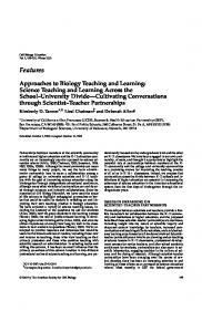
Stretching the point: the single-celled Amoeba proteus demonstrates its locomotor pseudopodia.
with a 1807 reversal in the attractant gradient does not simply transform its trailing edge into a leading one; instead, it follows its highly sensitive ‘nose’ and executes a U-turn. We propose that asymmetric responsiveness to extracellular gradients involves two complementary mechanisms that make the cell’s ‘front’ (pseudopod) distinct from its ‘back’. Each mechanism depends on actin, a cytoskeletal protein, to produce a selforganizing system for creating morphological polarity. The first mechanism uses localized positive feedback to amplify responsiveness to the external signal at the front of the cell. The feedback loop promotes new assembly of actin polymers, which form the pseudopod, push it forwards, and facilitate accumulation of a lipid signal in the plasma membrane. To confine positive feedback to the leading edge, a rapidly diffusing inhibitor acts to inhibit actin polymerization and membrane protrusion outside the pseudopod. This mechanism depends on assemblies of actin and the contractile protein myosin, located in the cell cortex at the rear and sides, which inhibit protrusion. Protrusion and contraction are controlled by different small enzymes that belong to the Rho family of proteins, which hydrolyse the nucleotide GTP (and are therefore called Rho GTPases). These two mechanisms cooperate to generate and stabilize an internal polarity that greatly exceeds the external gradient, allowing cells to detect and crawl persistently up shallow gradients of attractant and, in the case of neutrophils, to polarize in response to uniform attractant (usually in a randomly chosen direction). In the positive feedback loop, the attractant causes an increase in the concentration of phosphatidylinositol-3,4,5-trisphosphate (PIP3), a membrane lipid that promotes Rho GTPase activation. In turn, the GTPases promote actin polymerization in a pseudopod at the leading edge, where the GTPases and new actin polymers together promote further PIP3 accumulation, amplifying the response to attractant at the front of the cell. The feedback loop is best documented in neutrophils, but PIP3 also accumulates at the front of the amoeba Dictyostelium and of fibroblast cells. Inhibition at other parts of the cell confines the positive feedback loop to the leading edge, by promoting cortical actin–myosin assemblies and reducing membrane accumulation of PIP3. Disrupting the actin–myosin assemblies makes the back of a neutrophil as sensitive to attractant as the front — reversing the gradient causes the cell simply to reverse its direction, rather than performing a U-turn. Although necessary for long-term asymmetric sensitivity to chemoattractants, this contractile network is not absolutely required for
NATURE | VOL 419 | 5 SEPTEMBER 2002 | www.nature.com/nature
© 2002 Nature Publishing Group
Cell polarity Only now are we beginning to unravel the mechanisms behind a cell’s ability to point in any direction and navigate effectively. polarization itself — disruption of the network allows neutrophils and Dictyostelium to form PIP3-rich membrane protrusions towards a source of attractant, although the cells do form multiple pseudopodia at their sides, and crawl haltingly in the correct direction. In both cell types, membranes at the rear and sides fail to accumulate PIP3; in Dictyostelium, this resistance results from preferential association of a PIP3-degrading enzyme with these membranes. The actin–myosin network and mechanism of resistance to PIP3 accumulation cooperate, re-enforcing one another to interrupt the positive feedback loop that is necessary for protrusion. During chemotaxis, gradient interpretation is intimately linked to the morphological polarity required for forward movement. The two essential cytoskeletal elements support diametrically opposite effects on PIP3 accumulation and attractant sensitivity, each limiting the proportion of the cell surface that is susceptible to regulation by the other. This conversation between opposite ends of the cell may be a self-organizing property of cytoskeletal systems that create cell polarity. In one example of such a self-organizing system, round fragments of fish keratocytes respond to transient pressure on one side by polarizing and crawling away. Contractile actin–myosin complexes form on the pressured side, and actin polymerizes on the opposite side to form a protruding pseudopod; polarity and motility persist long after the stimulus disappears. In this case, as in chemotaxis, the front and back of the cell probably converse both mechanically (by asymmetric distribution of cortical tension) and biochemically (using Rho GTPases, PIP3 and other signals). A neutrophil decides correctly where to go because its ‘nose’ and its actin–myosin ‘muscles’ reciprocally regulate one another. These clever cells would scorn brains as an unnecessary luxury. ■ Henry Bourne is in the Departments of Medicine and of Cellular and Molecular Pharmacology, University of California, San Francisco, California 94143, USA. Orion Weiner is in the Department of Cell Biology, Harvard Medical School, Cambridge, Massachusetts 02115, USA. FURTHER READING Comer, F. I. & Parent, C. A. Cell 109, 541–544 (2002). Verkhovsky, A. B. et al. Curr. Biol. 9, 11–20 (1999). Weiner, O. D. Curr. Opin. Cell Biol. 14, 196–202 (2002). Zigmond, S. H. J. Cell Biol. 75, 606–616 (1977). 21


![Curious George - UCSF Career - University of California, San Francisco [PDF]](https://m.moam.info/img/260x300/curious-george-ucsf-career-university-of-californi_6479a1db098a9ee37d8b45ab.jpg)


![paul atreides - UCSF Career - University of California, San Francisco [PDF]](https://m.moam.info/img/260x300/paul-atreides-ucsf-career-university-of-california_6479c230098a9efa498b4622.jpg)

