Partial Necrosis Consequence of the Infection Spreading from an Adjacent Apical Periodontitis: A Case Report Saeed Asgary a, Leyla Roghanizadeh b* a Iranian Center for Endodontic Research, Research Institute of Dental Sciences, Shahid Beheshti University of Medical Sciences, Tehran, Iran; b Dental Research Center, Research Institute of Dental Sciences, Dental School, Shahid Beheshti University of Medical Sciences, Tehran, Iran
ARTICLE INFO
ABSTRACT
Article Type: Original Article Received: 08 May 2018 Revised: 05 Jun 2018 Accepted: 13 Jun 2018 doi: 10.22037/iej.v13i3.22089
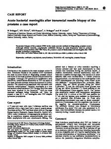
As the dental pulp could not be directly inspected before endodontic treatment, indirect evaluation of the pulp status via (para)/clinical tests should be performed which need careful inspection. This report presents a root-treated right maxillary first molar with recurrent abscess formation and a radiolucent periradicular lesion surrounding the distobuccal root of the right maxillary second molar. The patient underwent surgical retreatment, employing CEM rootend filling, which resulted in no relief from sign/symptoms. In the cone-beam computed tomography (CBCT), the relationship of the lesion with the mesio-buccal root of the second maxillary molar was detected. Despite the latest tooth showed positive responses to pulp sensibility tests, endodontic therapy was planned for it. During treatment, it became clear that the mesiobuccal canal pulp was necrotic, although vital pulp tissues were present in two other root canals. Following treatment, full recovery from all discomforts was obtained and the lesion healed after 18 months. This case showed that a more complicated evaluation such as CBCT should be used for diagnosis of perpetuated lesions. Furthermore, it might be probable that root canals of vital teeth become necrotic due to involvement in the adjacent apical lesion, a phenomenon known as anachoresis.
author: Leyla Dental Research Center, Research Institute of Dental Sciences, Evin, Shahid Beheshti Dental School, Tehran, Iran. Tel: +98-21 22405648 E-mail:
[email protected] *Corresponding
Roghanizadeh,
Keywords: Anachoresis, Apical Periodontitis; Calcium-Enriched Mixture; CEM Cement; Dental Pulp Necrosis; Endodontic; Spread of Infection
Introduction pical periodontitis is the sequel of the dynamic encounter between an endodontic infection and the host’s defense, which results in inflammation and destruction of periradicular tissues [1]. If apical periodontitis remains after root canal treatment, it is considered as a persistent lesion, usually attributed to failure of endodontic treatment [2]. Investigations show such lesions could be the result of persistent primary or secondary intraradicular, or extraradicular infections with species resisting and proliferating in burdensome environments; some inflammatory or immunologic reactions may be responsible, as well [3, 4]. For correct diagnosis and verification the origin of periradicular lesions, assessment of the health status of the pulp in involved teeth is necessary [5]. Pulp sensibility tests (thermal and electrical) have limitations, and false responses can occur. Vitality tests, which are able to directly assess the blood flow within the dental pulp, are better indicators of the
A
pulp health status; however, sensibility tests still have satisfactory validity and accuracy values to indirectly determining the state of pulpal health in common clinical practice [6-8]. Radiographic evaluations play a crucial role in diagnosis of periradicular lesions. The extent of the lesion, number of roots and root canals, detection of the involved root or roots, and whether a lesion around one root has a communication pathway to another root, should be investigated through the radiographic examinations [9]. The limitation of periapical radiographs in rendering information in two dimensions lead clinicians to use computed tomography (CT) scans [10]. This report presents a case of apical periodontitis assumed to be related to a failed conventional root canal therapy of a maxillary first molar, which perpetuated even after surgical retreatment. Finally, it was proved to be attributed to the adjacent partially-vital maxillary second molar, which had given normal positive responses in pulp sensibility tests.
IEJ Iranian Endodontic Journal 2018;13(3): 420-423
421
Asgary & Roghanizadeh
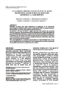
Figure 1. Periapical images: A) Preoperative; B) Postoperative image after distal root-end surgery of right maxillary first molar; C) Postoperative twomonth follow-up; D) Post-RCT image of the maxillary second molar; E) 18-month follow-up: healed periradicular lesion
Figure 2. CBCT views showing obvious relationship between the mesiobuccal root of the maxillary second molar and the lesion
Case Report A 51-year-old male with no history of systemic disease was admitted to the endodontic department of a private dental clinic. He was suffering from recurrent abscess formation in the right maxillary buccal vestibule near the right upper first molar. Radiographic examination (Figure 1A) revealed a circumscribed periapical lesion contiguous to the distobuccal root of the right upper first molar (tooth #16), extended to the mesial of mesiobuccal root of the adjacent second molar (tooth #17). Tooth #16 was root treated and restored with amalgam. The tooth #17, which had an amalgam restoration, had positive responses to an electrical pulp tester (Parkell, Edgewood, NY, USA), and cold test with Endo-Frost (Coltène-Whaledent, Langenau, Germany). In the clinical examination, no sinus tract was found. The patient had some tenderness to palpation of the associated buccal gingiva and expressed pain on percussion on tooth #16. Probing depths of gingival sulcus in both teeth were normal (