... Gugenheim3, Nacer Abbas1, Pierre Bouche3, Yves Agid1,2 and Alexis Brice1,2 ..... Wise, C.A., Garcia, C.A., Davis, S.N., Heju, Z., Pentao, L., Patel, P.I. and.
1993 Oxford University Press
Human Molecular Genetics, 1996, Vol. 5, No. 1
103–106
A de novo case of hereditary neuropathy with liability to pressure palsies (HNPP) of maternal origin: a new mechanism for deletion in 17p11.2? Eric LeGuern1,2,*, Riadh Gouider1,3, Nicole Ravisé1, Judith Lopes1, Sandrine Tardieu1, Michel Gugenheim3, Nacer Abbas1, Pierre Bouche3, Yves Agid1,2 and Alexis Brice1,2 1INSERM
U289, 2Fédération de Neurologie and 3Service d′Explorations Fonctionnelles Neurologiques, Hôpital de la Salpêtrière, 47 Bd de l′hôpital, 75651 Paris cedex 13, France Received August 30, 1995; Revised and Accepted October 13, 1995
Hereditary neuropathy with liability to pressure palsies (HNPP) is an autosomal dominant neuropathy, most often associated with a deletion of the 17p11.2 region, which is duplicated in 70% of patients with Charcot– Marie–Tooth type 1 (CMT1A). Most de novo CMT1A and HNPP cases have been of paternal origin. A rare case of de novo HNPP of maternal origin was analysed to determine the underlying mechanism. Affected individuals in the family carried a deletion corresponding to the CMT1A/HNPP monomer unit associated with a rearrangement of the CMT1A–REP sequences. Segregation analysis of 17p11-p12 markers in the family indicated that the deletion was not generated by unequal crossing over between homologous 17 chromosomes, as in de novo cases from paternal origin, but rather by an intrachromosomal rearrangement. Two distinct mechanisms can therefore lead to the same 17p11.2 deletion. This result suggests that intrachromosomal rearrangement may be specific to maternal transmissions. INTRODUCTION Hereditary neuropathy with liability to pressure palsies (HNPP) is an autosomal dominant peripheral neuropathy (1). HNPP patients present variously located recurrent truncular palsies or sensory loss, precipitated by minor trauma. In most cases, patients recover within days or weeks, but relapses may be frequent, and paresis may last for long periods. Nerve conduction studies reveal a characteristic pattern of decreased motor velocities, prolonged distal latencies and altered sensory nerve action potentials, even in clinically non affected areas or in asymptomatic at risk individuals (2). Peripheral nerve biopsies show a characteristic focal sausage-shaped thickening of the myelin sheath (tomacula) in numerous internodes.
*To whom correspondence should be addressed
The presence of an interstitial deletion of the 17p11.2 region associated with this disorder was demonstrated in most of the HNPP families (3,4). The same region, designated the CMT1A/HNPP monomer unit, is duplicated in a more frequently diagnosed neuropathy, Charcot-Marie-Tooth type 1A disease (CMT1A) (5–8). Thus, it was hypothesized that an unequal crossing-over would generate both a duplication that could lead to CMT1A or a deletion that could result in HNPP. The identification of two homologous sequences flanking the CMT1A/HNPP monomer unit, the CMT1A-REPs, supported this hypothesis (9). Recently, Chance et al. (10) proposed that the CMT1A duplication and the HNPP deletion could both arise from recombination events that occur within a limited region of the CMT1A-REP. De novo deletions have been reported in five HNPP patients, four of paternal origin (3,11,12) and one of possible maternal origin (13). In CMT1A, however, the de novo duplications, where the parental origin could be determined, were paternal (5,14,15). In order to analyse the mechanism underlying maternal de novo deletions in HNPP, we performed a molecular study of a family with a HNPP patient presenting a de novo deletion from maternal origin using 17p11-p12 markers and the CMT1A-REP probe pNEA102. The deletion was found to be associated with a rearrangement of the CMT1A-REPs but, unlike previous reports, haplotype reconstruction strongly supported the hypothesis that an intrachromosomal rearrangement generated the deletion. RESULTS Eight members of the family SAL-902 were genotyped for eight microsatellites and two RFLP markers from 17p11-12. Segregation analysis with the RM11GT microsatellite at D17S122 locus demonstrated the presence of a 17p11.2 deletion in patient 15 who carried allele 5, but received no allele from his mother (2/3) at this locus, showing that, in this family, the phenotype was associated with a deletion within 17p11.2 as described previously (2–4,11–13). The absence of maternal contribution to the patient 15 at the D17S122 locus, demonstrated the maternal origin of this de novo case of HNPP. All tested at risk individuals (7,17,20)
104
Human Molecular Genetics, 1996, Vol. 5, No. 1
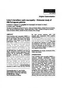
Figure 2. Gene dosage of the CMT1A–REP sequences detected with the pNEA102 probe on EcoRI digested Southern blots. The 7.8 and 6.0 kb EcoRI fragments corresponds to the proximal and distal CMT1A-REP. The 6.0 kb EcoRI fragment was less strongly labelled than the 7.8 kb fragment in patients 15 and 32, but not in controls (C) or unaffected individual 6.
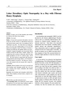
Figure 1. (A) Haplotype reconstruction for 10 17p11-12 markers in family SAL-902. Genotypes are indicated below individuals. Affected subjects are represented in black. *, clinically and electrophysiologically examined subjects from whom blood samples were obtained. The solid line to the left to the loci designation indicates the groups of markers for which the order cannot be resolved with odds >1000:1 (23). Clear boxes correspond to the deleted region deduced by allele segregation of the markers; hatched boxes represent the loci which are not informative of transmission. (B) Schematic map of the 17p11-p12 Généthon markers. The clear box indicates the CMT1A/HNPP monomer unit. The genetic distances are given in cM. D17S122 was physically mapped between D17S953 and D17S839 (15,16) but their relative genetic distances are not known.
shown to be unaffected by clinical and electrophysiological examination were heterozygous for at least one of the markers within the CMT1A/HNPP monomer unit (Fig. 1). Patient 15 did not transmit any alleles of D17S955 and D17S921 to his affected son 32. The deletion encompassed the D17S122 locus, which was mapped within the first 500 kb of the 1.5 Mb CMT1A/HNPP monomer unit at its centromeric extremity (9) and the D17S921 locus which was physically and genetically shown to be the most telomeric marker of the CMT1A monomer unit (16,17). Patients 15 and 32 were heterozygous for D17S953 and D17S922 which flank the CMT1A/HNPP monomer unit. These results indicate that the deletion certainly corresponds to the CMT1A/HNPP monomer unit. In addition, the two affected individuals 15 and 32 were tested with probe pNEA102 which detects, on EcoRI digested Southern blots, two fragments of 6.0 and 7.8 kb corresponding to the distal and proximal CMT1A-REP respectively (10). The hybridization signal for the 7.8 kb fragment was stronger than for the 6.0 kb fragment, suggesting that a part of a distal CMT1A-REP was deleted (Fig. 2). It can be concluded that the deletion is associated with a rearrangement of the CMT1A-REP sequences and confirms that the CMT1A/HNPP monomer unit is involved (10,18,19).
Using the microsatellites D17S842 (afm240xe5), D17S783 (afm026vh7) and D17S953 (afm304xh5) which are centromeric to the CMT1A/HNPP monomer unit and the telomeric marker D17S922 (afm197xh6)(16,17), the most probable haplotypes for the 17p11-p12 region were reconstructed, as shown in Figure 1. The flanking markers on each side of the deletion were inherited intact from the mother and showed no crossing-over. Considering the genetic distances between the tested markers (see Materials and Methods), the probability of a double recombination in patient 15 is very low. Therefore, an unequal crossing-over between the chromosome 17 homologues, as in de novo deletions and duplications of paternal origin associated with HNPP and CMT1A (3,5,11,12,14,15), is very unlikely in this family. DISCUSSION We report a de novo case of HNPP of maternal origin. The deletion detected in affected members is associated with a rearrangement of CMT1A-REP sequences and corresponds to the CMT1A/HNPP monomer unit. However, haplotype reconstruction highly suggests that this deletion was not caused by an unequal crossing-over as described previously (3,11,12). It is highly probable that the de novo deletion observed in family SAL-902 results from an intrachromosomal rearrangement. Our observation provides the first evidence that rearrangements involving identical autosomal sequences can occur either by unequal crossing-over between homologues or by intra-homologue rearrangement. Interestingly, all documented de novo duplications of paternal origin resulting in CMT1A, have been shown by haplotype reconstruction to result from an unequal recombination between homologue chromosomes 17 (5,14,15). In HNPP, both paternal de novo deletions (3,11,12) but also a possible maternal deletion (13) have been reported. The latter received no allele of the pEW401HE 17p11.2 marker (D17S61) from his mother who was shown to be unaffected by clinical and electrophysiological examination. The molecular mechanism underlying this deletion was not analyzed, however. Terminal or interstitial deletions detected by classical cytogenetic techniques are generally caused by the occurrence of one or two breaks respectively, within a chromosome with the loss of the acentric fragment. An intrachromosomal mechanism was also postulated to cause the de novo deletions occurring in males in Duchenne muscular dystrophy or haemophilia A (20). It was demonstrated that some deletions in these genes, which are located on the X chromosome can occur in male gametes. Since male gametes bear only one X chromosome, the origin of these
105 Human Acids Molecular Genetics, Vol.No. 5, No. Nucleic Research, 1994,1996, Vol. 22, 1 1 rearrangements is mitotic and the corresponding process is certainly intrachromosomic. Intrachromosomal rearrangement has also been invoked for deletions of several Mb on autosomes, in particular in the Angelman/Prader–Willi syndrome (21,22). For de novo deletions occurring in males in Duchenne muscular dystrophy or in haemophilia A, the excision of a chromatidal loop during replication was proposed (20). This mechanism was also postulated for the deletions within the beta-globin gene domain resulting in the hereditary persistence of fetal hemoglobin (HPFH) (23). In Angelman/Prader–Willi syndrome, unequal sister chromatid exchange was thought as an alternative to explain the deletion (21,22). Unfortunately, the molecular tools which would distinguish between these two intrachromosomal mechanisms in our family are not yet available. Since deletions in HNPP can result both from crossing-over between homologues and intra-chromosomal rearrangement but duplications only by crossing over, HNPP should be more frequent than CMT1A with duplication. HNPP is apparently less prevalent, however, than CMT1A but this is probably because of underdiagnosis (2). Since unequal crossing-overs resulting in deletion or duplication were observed during male meioses, the intrachromosomal deletion of this region detected in this study may preferentially occur in females, although additional cases must be analyzed before a generalization can be made. MATERIALS AND METHODS
105
visual comparison of the hybridization signals of the two EcoRI fragments hybridized with probe pNEA102. Microsatellite markers The following (CA)n microsatellites, which span 13 cM on chromosome 17p11-p12, were used: D17S122 (RM11GT)(6), D17S842 (afm240xe5), D17S783 (afm026vh7), D17S953 (afm304xh5), D17S839 (afm200yb12), D17S955 (afm317yg1), D17S921 (afm191xh12) and D17S922 (afm197xh6). Genetic distances between the Généthon markers were as follows: (D17S842–D17S783), 6 cM, D17S953, 5 cM, (D17S839– D17S955), 1 cM, D17S921, 1 cM, D17S922 (24). Genotypes were determined with the PCR/blotting technique (25). ACKNOWLEDGEMENTS The authors thank Dr J. R. Lupski who kindly provided the pNEA102 probe. They are grateful to Christiane Penet, Yolaine Pothin and Isabelle Lagroua for technical and medical assistance, and would like to thank Drs Merle Ruberg and Josué Feingold for critical reading of the manuscript. This study was supported by the Association Française contre les Myopathies (AFM), the Assistance Publique des Hôpitaux de Paris (AP–HP) and the Association pour le Développement de la Recherche sur les Maladies Génétiques Neurologiques et Psychiatriques (ADRMGNP).
Clinical data Index case 15 from family SAL-902, examined at age 41, described three episodes of acute sensory-motor truncular deficits without obvious precipitating factors. Several nerves were affected simultaneously during the third episode. Motor nerve conduction (MNCV) and sensory nerve conduction velocities (SNCV) were abnormal bilaterally in the median, ulnar and peroneal nerves. Tomaculous changes involving 36% of internodes were found in superficial peroneal nerve biopsies. Clinical and electrophysiological examinations were performed in all first degree relatives. Individual 32, the son of the index case, presented three episodes of truncular sensory-motor palsies and his brother, 31, described paresthesias with a right ulnary distribution lasting a few hours. Patients 31 and 32 both presented diffuse MNCV and SNCV abnormalities. Parents and siblings never complained of motor or sensory abnormalities. The mother (6) of index case 15 was a 73 year old farmer, who presented with unilateral electrophysiological carpal tunnel syndrome but had no other abnormal electrophysiological features. The father of the index case was not available for examination. All siblings had normal MCV and SCV. Individuals 6 and 15 were genotyped twice with two different blood samples in order to eliminate sampling or typing errors. RFLP markers The genomic probes EW401 (D17S61) detects two MspI alleles with sizes of 5.5 and 4.4 kb, and VAW409R3a (D17S122) three MspI alleles with sizes of 2.8, 2.7 and 1.9 kb. Probe pNEA102, which detects two EcoRI fragments of 6.0 and 7.8 kb corresponding respectively, to the proximal and distal CMT1A-REP which flank the CMT1A/HNPP monomer unit, was also used. The probes were hybridized to Southern blots after preannealing with placental DNA. The CMT1A-REP deletion was assessed by
REFERENCES 1. Windebank, A.J. (1993) Inherited recurrent focal neuropathies. In Dyck, P.J., Thomas, P.K., Griffin, J.W., Low, P.A., Poduslo, J.F. (eds), Peripheral Neuropathy. 3rd edn, Hereditary Motor and Sensory Neuropathies, Saunders, Philadelphia, pp. 1094–1136. 2. Gouider, R., LeGuern, E., Gugenheim, M., Tardieu, S., Maisonobe, T., Léger, J.M., Vallat, J.M., Agid, Y., Bouche, P. and Brice, A. (1995) Clinical, electrophysiological and molecular correlations in 13 families with hereditary neuropathy with liability to pressure palsies and a chromosome 17p11.2 deletion. Neurology, in press. 3. Chance, P.F., Alderson, M.K., Leppig, K.A., Lensch, M.W., Matsunami, N., Smith, B., Swanson, P.D., Odelberg, S.J., Distsche, C.M., Bird, T.D. (1993) DNA deletion associated with hereditary neuropathy with liability to pressure palsies. Cell, 72, 143–151. 4. LeGuern, E., Sturtz, F., Gugenheim, M., Gouider, R., Bonnebouche, C., Ravisé, N., Gonnaud, P.-M., Tardieu, S., Bouche, P., Chazot, G., Agid, Y., Vandenberghe, A. and Brice, A. (1994) Detection of deletion within 17p11.2 in 7 French families with hereditary neuropathy with liability to pressure palsies (HNPP). Cytogenet. Cell Genet., 65, 261–264. 5. Raeymaekers, P., Timmerman, V., Nelis, E., De Jonghe, P., Hoogendijk, J.E., Baas, F., Bakker., D.F., Martin, J.J., De Visser, M., Bolhuis, P.A., Van Broeckhoven, C. and the HMSN Collaborative Research Group (1991) Duplication in chromosome 17p11.2 in Charcot–Marie–Tooth neuropathy type 1a (CMT 1a). Neuromusc. Disord., 1, 93–97. 6. Lupski, J.R., Montes de Oca–Luna, R., Slaugenhaupt, S., Killian, J.M, Garcia, C.A., Chakravarti, A. and Patel, P.I. (1991) DNA duplication associated with Charcot–Marie–Tooth disease type 1A. Cell, 66, 219–232. 7. Raeymaekers, P., Timmerman, V., Nelis, E., Van Hul, W., De Jonghe, P., Martin, J.–J., De Visser, M., Bolhuis, P.A., Van Broeckhoven, C. and the HMSN collaborative research group (1992). Estimation of the size of the chromosome 17p11.2 duplication in Charcot–Marie–Tooth type 1a (CMT1a). J. Med. Genet., 29, 5–11. 8. Brice, A., Ravisé, N., Stevanin, G., Gugenheim, M., Bouche, P., Penet, C., Agid, Y. and the French CMT Research Group (1992) Duplication within chromosome 17p11.2 in 12 families of French ancestry with Charcot–Marie– Tooth disease type 1a. J. Med. Genet., 29, 807–812. 9. Pentao, L., Wise, C.A., Chinault, A.C., Patel, P.L. and Lupski, J.R. (1992) Charcot–Marie–Tooth type 1A duplication appears to arise from recombination at repeat sequences flanking the 1.5 Mb monomer unit. Nature Genet., 2, 292–300.
106
Human Molecular Genetics, 1996, Vol. 5, No. 1
10. Chance, P.F., Abbas, N., Lensch, N.W., Pentao, L., Roa, B.B., Patel, P.L. and Lupski, J.R. (1994) Two autosomal dominant neuropathies result from reciprocal DNA duplication/deletion of a region on chromosome 17. Hum. Mol. Genet., 3, 223–228. 11. Mariman, E.C., Gabreëls-Festen, A.A., van Beersum, S.E., Valentijn, L.J., Baas, F., Bolhuis, P.A., Jongen, P.J., Ropers, H.H. and Gabreëls, F.J. (1994) Prevalence of the 1.5 Mb deletion in families with hereditary neuropathy with liability to pressure palsies. Ann. Neurol., 36, 650–655. 12. Verhalle D., Lofgren, A., Nelis, E., Dehaene, I., Theys, P., Lammens, M., Dom, R., Van Broeckhoven, C. and Robberecht,W. (1994) Deletion in the CMT1A locus on chromosome 17pl1.2 in hereditary neuropathy with liability to pressure palsies. Ann. Neurol., 35, 704–708. 13. Reisecker, R., Leblhuber, F., Lexner, R., Radner, G., Rosenkranz, W. and Wagner, K.A. (1994) Sporadic form of hereditary neuropathy with liability to pressure palsies: clinical, electrodiagnostic, and molecular genetic findings. Neurology, 44, 753–755. 14. Palau, F. , Löfgren, A., De Jonghe, P., Bort, S., Nelis, E., Sevilla, T., Martin, J.–J., Vilchez, J., Prieto, F. and Van Broeckhoven, C. (1993) Origin of the de novo duplication in Charcot–Marie–Tooth disease type 1A: unequal nonsister chromatid exchange during spermatogenesis. Hum. Mol. Genet., 2, 2031–2035. 15. Wise, C.A., Garcia, C.A., Davis, S.N., Heju, Z., Pentao, L., Patel, P.I. and Lupski, J.R. (1993) Molecular analyses of unrelated Charcot–Marie–Tooth (CMT) disease patients suggest a high frequency of the CMT1A duplication. Am. J. Hum. Genet., 53, 853–853. 16. Cudrey, C., Chevillard, C., Le Paslier, D., Vignal, A., Passage, E. and Fontes, M. (1995) Assignment of microsatellite sequences to the region duplicated in CMT1A (17p12): a useful tool for diagnosis. J. Med. Genet., 32, 231–233 . 17. LeGuern, E., Ravisé, N., Gouider, R., Gugenheim, M., Lopes, J., Bouche, P., Agid, Y. and Brice, A. (1995) Microsatellite mapping of the deletion in patients with hereditary neuropathy with liability to pressure palsies (HNPP): new molecular tools for the study of the 17p11–p12 region and for diagnosis. Cytogenet. Cell Genet., in press.
18. Lorenzetti, D., Pareyson, D., Sghirlanzoni, A., Roa, B.R., Abbas, N., Pandolfo, M., Di Donato, S.and Lupski, J. (1995). A 1.5 Mb deletion in 17p11.2-p12 is frequently observed in Italian families with hereditary neuropathy with liability to pressure palsies. Am. J. Hum. Genet., 56, 91–98. 19. LeGuern, E., Gouider, R., Lopes, J., Abbas, N., Gugenheim, M., Tardieu, S., Ravisé, N., Léger, J.-M., Vallat, J.-M., Bouche, P., Agid, Y., Brice, A. and the French CMT collaborative Research Group (1995) Constant rearrangement of the CMT1A–REP sequences in HNPP patients with a deletion in chromosome 17p11.2: a study of 30 unrelated cases. Hum. Mol. Genet., 4, 1673–1674. 20. Darras, T.D. and Francke, U. (1987) A partial deletion of the muscular dystrophy gene transmitted twice by an unaffected male. Nature, 329, 556–558. 21. Goettlieb, W., Rogan, P.K., Driscoll, D.J. and Nicholls, R.D. (1993) Analysis of chromosome breakage mechanisms in Prader–Willi and Angelman syndromes. Am. J. Hum. Genet., 53 (suppl), A553. 22. Nicholls, R.D. (1994) Recombination model for generation of a submicroscopic deletion in familial Angelman syndrome. Hum. Mol. Genet., 3, 9–11. 23. Collins, F.S., Cole, J.L., Lockwood, W.K., et al. (1987) The deletion in both common types of hereditary persistence of fetal hemoglobin is approximately 105 kilobases. Blood, 70, 1797–1803. 24. Gyapay, G., Morissette, J., Vignal, A., Dib, C., Fizames, C., Millaseau, P., Marc, S., Bemardi, G., Lathrop, M. and Weissenbach, J. (1994) The 1993–1994 Généthon human genetic linkage map. Nature Genet., 7, 246–339. 25. Stevanin, G., LeGuern, E., Ravisé, N., Chneiweiss, H., Dürr, A., Cancel, G., Vignal, A., Boch, A.-L., Ruberg, M., Penet, C., Pothin, Y., Haguenau, M., Rancurel, G.,Weissenbach, J., Agid, Y. and Brice, A. (1994) A third locus for Autosomal Dominant Cerebellar Ataxia type 1 maps to 14q24.3-qter. Evidence for the existence of a fourth. Am. J. Hum. Genet., 54, 11–20.