virus in peripheral blood mononuclear cells from persistently infected patients. Journal of Virology 71, 5399â5407. Stuyver, L., De Gendt, S., Van Geyt, C., Zoulim ...
Journal of General Virology (2001), 82, 883–892. Printed in Great Britain ...................................................................................................................................................................................................................................................................................
A novel variant genotype C of hepatitis B virus identified in isolates from Australian Aborigines : complete genome sequence and phylogenetic relatedness Fuminaka Sugauchi,1 Masashi Mizokami,2 Etsuro Orito,1 Tomoyoshi Ohno,1 Hideaki Kato,1 Seiji Suzuki,1 Yoshihide Kimura,1 Ryuzo Ueda,1 L. A. Butterworth3 and W. G. E. Cooksley3 1, 2
Second Department of Internal Medicine1 and Laboratory Medicine2, Nagoya City University Medical School, Kawasumi, Mizuho, 467-8601 Nagoya, Japan 3 Royal Brisbane Hospital Research Foundation, Brisbane, Australia
There have been no reports of DNA sequences of hepatitis B virus (HBV) strains from Australian Aborigines, although the hepatitis B surface antigen (HBsAg) was discovered among them. To investigate the characteristics of DNA sequences of HBV strains from Australian Aborigines, the complete nucleotide sequences of HBV strains were determined and subjected to molecular evolutionary analysis. Serum samples positive for HBsAg were collected from five Australian Aborigines. Phylogenetic analysis of the five complete nucleotide sequences compared with DNA sequences of 54 global HBV isolates from international databases revealed that three of the five were classified into genotype D and were most closely related in terms of evolutionary distance to a strain isolated from a healthy blood donor in Papua New Guinea. Two of the five were classified into a novel variant genotype C, which has not been reported previously, and were closely related to a strain isolated from Polynesians, particularly in the X and Core genes. These two strains of variant genotype C differed from known genotype C strains by 5n9–7n4 % over the complete nucleotide sequence and 4n0–5n6 % in the small-S gene, and had residues Arg122, Thr127 and Lys160, characteristic of serotype ayw3, which have not been reported previously in genotype C. In conclusion, this is the first report of the characteristics of complete nucleotide sequences of HBV from Australian Aborigines. These results contribute to the investigation of the worldwide spread of HBV, the relationship between serotype and genotype and the ancient common origin of Australian Aborigines.
Introduction Hepatitis B virus (HBV) is an aetiological agent of acute and chronic liver disease including fatal fulminant hepatitis, cirrhosis and hepatocellular carcinoma, which is one of the most common human cancers and causes of death worldwide (Lee, 1997). It is estimated that there are currently more than 350 million carriers worldwide. There have been no studies on
Author for correspondence : Masashi Mizokami. Fax j81 52 842 0021. e-mail mizokami!med.nagoya-cu.ac.jp The nucleotide sequence data reported in this paper will appear in the DDBJ/EMBL/GenBank databases under the accession numbers AB048701–AB048705.
0001-7475 # 2001 SGM
DNA sequences of HBV from Australian Aborigines, although the antigen known as the hepatitis B surface antigen (HBsAg) was discovered in Australian Aborigines (Blumberg et al., 1965). It is widely accepted that Australian Aborigines are a high-risk population for HBV infection. In contrast, a low prevalence of HBV marker is found in the non-Aboriginal population (Patterson et al., 1993 ; Campbell et al., 1989). Several studies have shown, by determining the prevalence of serological markers of HBV in different Aboriginal groups around mainland Australia and the Torres Strait islands (Holman et al., 1987 ; Campbell et al., 1989), that the prevalence of HBsAg varies from approximately 3 % to as high as 35 %. Most HBV infection in Aborigines occurs in early life, either by vertical transmission from a mother who is an HBsAg carrier, by horizontal transmission in households that include IID
F. Sugauchi and others
Table 1. Clinicopathological features of Australian Aborigines Abbreviations : ALT, alanine aminotransferase ; AST, aspartate aminotransferase ; CH, chronic hepatitis ; BTH, blood-transfusion history ; , not known ; , not tested. ALT and AST activities are given in units per litre. All locations listed are in Queensland, Australia. Sample
Collection date*
Age
BTH
AustRC AustSJ AustGC AustKW AustDF
7\5\92 8\12\94 25\7\91 14\11\91 2\3\89
54 36 22 53 26
k k k k k
Place of birth
Residence HBeAg Anti-HBe
ALT
AST
Liver biopsy result
Cherbourg Murgon Brisbane Cherbourg Gympie
Ipswich Cherbourg Brisbane Weipa
k j j k j
62 56 108 29 127
317 34 48 19 52
Liver cancer CH with mild fibrosis CH with no fibrosis CH with no fibrosis
j k k j k
* Dates are given in the form day\month\year.
Table 2. HBV strains of genotypes C and D used in this study Accession no. AustSJ AustRC D23684 D23681 M38636 AB014378 D50520 X04615 Y18857 AF068756 X75665 X75656 AustGC AustDF AustKW X65259 X68292 X65258 J02203 X85254 X97849 X80924 M32138 L27106 Y07587 AB033559
Genotype Var-C Var-C C C C C C C C C C C D D D D D D D D D D D D D D
Serotype* ayw3b,c ayw3b,c adrb adrb adra adrb adrb ayra adw2b adra adrq−a adrq−a aywb,c aywb,c aywb,c aywa aywa adw2a ayw3b aywb ayw3a aywa aywb aywb aywa aywa
Country of origin Australia Australia Japan Japan Korea Japan Japan Japan China Thailand New Caledonia Polynesia Australia Australia Australia Italy Italy Italy France Italy UK UK Turkey UK Germany Papua New Guinea
Reference This study This study Horikita et al. (1994) Horikita et al. (1994) Kim et al. (1988) Takahashi et al. (1998) Asahina et al. (1996) Okamoto et al. (1986) Unpublished Monkongdee et al. (1998) Norder et al. (1994) Norder et al. (1994) This study This study This study Unpublished Unpublished Unpublished Galibert et al. (1979) Unpublished Alexopoulou et al. (1996) Unpublished Tong et al. (1990) Hasegawa et al. (1994) Stoll-Becker et al. (1997) Okamoto et al. (1988)
* Serotypes were : a, mentioned in an article or a description of registration ; b, deduced from sequence data ; and\or c, determination by seroreaction.
an HBsAg carrier or as a result of tattooing or intravenous drug use in young adults (Campbell et al., 1989). The genotypic classification of HBV has been extended to seven genotypes, types A–G, on the bass of S gene sequence and complete genome sequences (Norder et al., 1992 ; Okamoto et al., 1988 ; Stuyver et al., 2000). These genotypes reflect the geographical distribution of HBV. Genotype A predominates IIE
in north-western Europe, North America and Africa. Genotypes B and C are found in Asia. Genotype D is the most widespread worldwide and is the predominant genotype of the Mediterranean region. Genotype E is found in East Africa and genotype F is found mainly in the New World (Magnius & Norder, 1995 ; Norder et al., 1993, 1994). Genotype G was found recently in isolates from America and France (Stuyver et
Australian Aboriginal strain of HBV
al., 2000) but it has not yet been characterized. There is, however, no information regarding HBV genotype distribution in Oceania (consisting mainly of Australia). In this study, we analysed the complete genome sequences of HBV strains isolated from Australian Aborigines living mainly in Queensland and compared them with published sequences by molecular evolutionary analysis.
Methods
Samples. Five serum samples positive for HBsAg were collected from Australian Aborigines from different parts of Queensland between 1989 and 1994 and used as the source of HBV DNA for sequencing. The clinicopathological features of these five patients are shown in Table 1. Two were positive for antibody to HBeAg (anti-HBe) but negative for HBeAg. There was no blood-transfusion history for any of the patients. From the results of liver biopsies, one patient was found to have liver cancer and three to have chronic hepatitis. Forty-eight HBV complete nucleotide sequences, representing each of genotypes A–G, and six complete nucleotide sequences of HBV isolated from non-human primates (chimpanzee, gorilla, gibbon and woolly monkey) were obtained from international DNA databases (DDBJ\EMBL\GenBank). The accession numbers, genotypes, serotypes, countries of origin and references for genotype C and D strains are listed in Table 2. The most divergent strains in each basic genotype were included, the accession numbers of which were M57663 for genotype A, D00331 and M54923 for genotype B, X75665 and X75656 for genotype C and X65259 and X68292 for genotype D. Open reading frame analysis
was performed with the same sequences as were used for comparison of the complete nucleotide sequence.
HBV DNA extraction. Serum samples were stored at k80 mC until assay. DNA was extracted from 100 µl of serum by using a DNA extractor kit (Sumitomo, Tokyo).
HBV DNA amplification. The complete genome of HBV was amplified as two overlapping fragments by using the sense primer HB8F and antisense primer HB6R to yield a 3200 bp amplicon (fragment A) and the sense primer HB7F and antisense primer HB7R to yield a 462 bp amplicon (fragment B). For fragment A, nested PCR was performed to amplify 11 overlapping fragments by using the primers listed in Table 3. The primers used in this study were based on the most conserved regions derived from known sequences published in DDBJ\EMBL\GenBank. The amplification reaction was performed in a 96-well cycler (GeneAMP9600, Perkin-Elmer Cetus) and the PCR was initiated by the hot-start technique. The first round of PCR for fragment A was undertaken for 35 cycles (94 mC for 1 min, 55 mC for 1 min and 72 mC for 1n5 min) followed by an extension reaction at 72 mC for 5 min. The first round of PCR for fragment B and the second round of PCR for fragment A were performed for 30 cycles (94 mC for 1 min, 55 mC for 1 min and 72 mC for 1 min) followed by extension at 72 mC for 5 min. The PCR products were analysed by electrophoresis on 2n0 % agarose gels, stained with ethidium bromide and visualized on a UV transilluminator. Standard precautions to avoid contamination during PCR were taken. A negative control serum was also included in each run to ensure specificity.
Sequencing of the full-length sequences of HBV. Twelve overlapping fragments of PCR-amplified HBV DNA were sequenced
Table 3. HBV DNA oligonucleotide primers used for full-length sequencing Primer
Nucleotide sequence (5h
3h)
HB1F HB1R HB2F HB2R HB3F HB3R HB4F HB4R HB5F HB5R HB6F HB6R HB7F HB7R HB8F HB8R HB9F HB9R HB10F HB10R HB11F HB11R HB12F HB12R
AAGCTCTGCTAGATCCCAGAGT GAAACATAGAGGTGCCTTGAGCAG TGCTGCTATGCCTCATCTTC CATACTTTCCAATCAATAGG GCCAAGTCTGTACAACATCTTGAG AGTTGGCGAGAAAGTGAAAGCCTG CCTATTGATTGGAAAGTATGTCA CGGGACGTAGACAAAGGACGT CTCTGCCGATCCATACTGCGGAA TTAACCTAATCTCCTCCCCCA TTGTYTACGTCCCGTCGGCG AACAGACCAATTTATGCCTA GAGACCACCGTGAACGCCCA CCTGAGTGCTGTATGGTGAGG TTCACCTCTGCCTAATCATC ATAGGGGCATTTGGTGGTCT TCAGGCAACTATTGTGGTTTCA GGATAGAACCTAGCAGGCAT CGCAGAAGATCTCAATCTCGG GGGTTGAAGTCCCAATCTGGATT GGGTCACCATATTCTTGGGAA GAACTGGAGCCACCAGCAGG GTGGAGCCCTCAGGCTCAGG CGAGTCTAGACTCTGTGGTA
Position 18–39 557–534 414–433 989–970 760–783 1107–1084 970–992 1434–1414 1256–1278 1761–1741 1421–1440 1803–1784 1611–1630 2072–2048 1824–1843 2314–2278 2190–2211 2654–2635 2417–2437 2987–2965 2814–2834 75–56 3075–3094 256–237
Polarity Sense Antisense Sense Antisense Sense Antisense Sense Antisense Sense Antisense Sense Antisense Sense Antisense Sense Antisense Sense Antisense Sense Antisense Sense Antisense Sense Antisense
IIF
F. Sugauchi and others
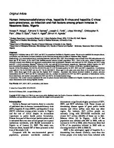
Fig. 1. For legend see facing page.
IIG
Australian Aboriginal strain of HBV directly by the dideoxy method using the Taq Dye Deoxy Terminator cycle sequencing kit with a fluorescent 373A DNA sequencer (Applied Biosystems).
Serotyping for HBsAg. Serotypes of samples from five Aborigines were deduced by using monoclonal antibodies directed against the a, d, y, w and r determinants of the surface antigen with the HBsAg subtype kit from the Institute of Immunology Co. (Tokyo, Japan) and confirmed on the basis of sequence data. The serotypes of the 54 HBV strains used for comparison were obtained from published articles or deduced from the registered sequence.
Phylogenetic analysis. The nucleotide sequences of the five Australian Aboriginal HBV strains were compared with those of the 54 reference strains. Sequences were aligned by using the CLUSTAL W software and confirmed by visual inspection. Genetic distances were estimated by using the six-parameter method and phylogenetic trees were constructed by the neighbour-joining (NJ) method (Saitou & Nei, 1987). To confirm the reliability of the phylogenetic tree analysis, bootstrap resampling and reconstruction were carried out 1000 times. These analyses were carried out using the ODEN program of the
National Institute of Genetics (Mishima, Japan) (Ina, 1994). The HBV genotype was assigned according to classification systems reported previously (Norder et al., 1992 ; Okamoto et al., 1988 ; Stuyver et al., 2000).
Results Genetic distances and phylogenetic relatedness
The five strains AustDF, AustKW, AustGC, AustRC and AustSJ were analysed and found to have nucleotide lengths of 3182, 3182, 3182, 3215 and 3194 bp. Phylogenetic analysis of the complete genome sequences of these five strains compared with those of 54 global HBV strains from DDBJ\ EMBL\GenBank showed that three, AustDF, AustGC and AustKW, were classified into genotype D with bootstrap values of 100 % (Fig. 1 a). Furthermore, the three strains were shown to be most closely related in terms of evolutionary
Fig. 1. Phylogenetic tree analysis of the HBV strains isolated from five Australian Aborigines compared with 54 reference strains. Genetic distances were estimated by the six-parameter method and phylogenetic trees were constructed by the NJ method. The outgroup consisted of the Woolly Monkey strain (WMHBV). Accession numbers are indicated on the tree. Arrows show the Australian Aboriginal strains. Bootstrap values are shown along each main branch. The lengths of the horizontal bars indicate the number of nucleotide substitutions per site. The regions included in the analysis were : the complete genome (a), the ORF of the small-S gene (only the HBsAg region) (b), the ORF of the large-S gene (including preS1, preS2 and HBsAg) (c), the ORF of the P gene (d ), the ORF of the Core gene (including the pre-core region) (e) and the ORF of the X gene ( f ).
IIH
F. Sugauchi and others
Table 4. Mean percentage nucleotide differences for the small-S gene and complete nucleotide sequence between two Australian strains and other HBV genotypes Reference sequences included 48 HBV DNA isolates from DDBJ\EMBL\GenBank. Differences were obtained by pair-wise analysis. Values are mean percentage differences with the range in parentheses. AustSJ
AustRC
Genotype
Small-S gene
Complete genome
Small-S gene
Complete genome
A B C Var-C D E F G
5n91 (5n0–6n5) 5n90 (5n4–6n5) 4n63 (4n0–5n1) 0n60 4n79 (4n3–6n0) 4n50 (4n4–4n6) 7n50 (7n5–7n5) 5n40
10n1 (9n9–10n8) 10n3 (9n8–11n1) 6n9 (6n2–7n4) 1n5 11n9 (10n2–11n7) 10n8 (10n7–10n8) 13n9 (13n7–14n2) 13n0
6n35 (5n4–6n9) 6n36 (5n9–7n0) 5n07 (4n6–5n6) 0n60 5n20 (4n7–6n5) 4n80 (4n6–5n0) 7n80 (7n8–7n8) 5n90
9n9 (9n6–10n5) 9n9 (9n6–10n7) 6n6 (5n9–7n2) 1n5 11n7 (11n1–12n3) 10n8 (10n7–10n8) 13n7 (13n5–13n9) 13n2
distance (0n01441, 0n01334 and 0n02613 substitutions per position, respectively) to a strain isolated from a healthy blood donor in Papua New Guinea (accession no. AB033559 ; Okamoto et al., 1988) that was reported previously. The remaining two strains, AustSJ and AustRC, were classified into a novel variant genotype C (mean nucleotide difference from genotype C 6n7 % ; range 5n9–7n4 %) that has not been reported previously, with bootstrap values of 100 % (Fig. 1 a). The mean percentage nucleotide differences, obtained by pair-wise analysis, in the complete genome sequence between the two Australian HBV strains (AustSJ and AustRC) and all genotypes, including 48 HBV DNA sequences from DDBJ\ EMBL\GenBank, are shown in Table 4. Based on the results of small-S gene analysis, AustSJ and AustRC were classified into a novel genotype that was separate from genotype C, with significant bootstrap values, similar to the relationship between genotypes A and D or A and G (Fig. 1 b). The mean percentage nucleotide differences, obtained by pair-wise analysis, between the small-S genes of the two Australian HBV strains (AustSJ and AustRC) and all genotypes, including 48 HBV DNA sequences from DDBJ\ EMBL\GenBank, are shown in Table 4. The mean percentage nucleotide differences between the Australian strains (AustSJ and AustRC) and strains belonging to the each of the seven genotypes were greater than 4 %. To confirm the relationship, we also analysed the nucleotide sequences of the small-S genes of 91 HBV strains retrieved from DDBJ\EMBL\GenBank, including sequences of 81 strains of genotype C (references not shown), and another phylogenetic tree was constructed (Fig. 2). AustSJ and AustRC were again classified into a novel genotype distinctly separate from the genotype C group with significant bootstrap values. In the genotype D group, strains AustDF, AustGC and AustKW were again clustered with a strain isolated from Papua New Guinea (AB033559 ; Okamoto et al., 1988) based on III
small-S gene analysis (Fig. 1 b). These analyses were also performed for each open reading frame, large-S (pre-S1 to S gene), P, Core and X (Fig. 1 c–f). The results were consistent with those obtained for complete genome sequence analysis. Based on analysis of the Core and X gene sequences (Fig. 1 e, f), two Australian strains (AustSJ and AustRC), which were classified into a variant genotype C, were clustered with isolates from New Caledonia (X75665) and Polynesia (X75656) that had been reported previously (Norder et al., 1994). Serotypic relatedness and characterization of the deduced amino acid sequence
The serotype of all five samples was ayw, deduced by using a serum reaction kit for HBsAg. Two of the five strains, however, AustSJ and AustRC, were classified further into the ayw3 serotype, based on the presence of Arg"##, Lys"'! and Thr"#( residues determined by sequence data analysis. The three remaining strains, AustDF, AustGC and AustKW, were classified into the ayw1 serotype based on the presence of Arg"##, Lys"'!, Pro"#( and Tyr"$% residues (Fig. 3). There was no specific mutation in the ‘ a ’ determinant, encompassing residues 124–147 of HBsAg of the five Australian strains (Fig. 3). Within the pre-S1 region, an 11 amino acid deletion at codons 3–13, which is a specific deletion for genotype D, was found in AustDF, AustKW and AustGC, and a 7 amino acid deletion at codons 1–7 was found in AustRC, which was the pathogenic agent of a liver cancer (data not shown). Within the pre-S2 region, one substitution, of residue Thr%', was found in the variant genotype C strain sequences and this was not present in the other genotype sequences (data not shown). Within the X region, three substitutions were found in the variant genotype C strain sequences that were not present in the other genotype sequences (data not shown). No specific substitutions were found in the pre-core and core regions (data
Australian Aboriginal strain of HBV
Fig. 2. Phylogenetic analysis of the HBV strains isolated from two Australian Aborigines compared with 91 strains on the basis of small-S gene sequence, using the same methods as outlined in the legend to Fig. 1.
not shown). Within the P region, the YMDD motif was conserved in all of the Australian strains, but it was shown that a Val&&& Leu mutation occurred downstream of the YMDD motif, as in genotype F strains (data not shown).
Discussion On the basis of phylogenetic analysis of the complete genome sequences of five HBV strains from Australian Aborigines and comparison with the complete genome sequences of global strains from international databases, a novel genotype C variant strain was identified that belonged
to the ayw serotype and has not been reported previously in the genotype C group. Phylogenetically, based on nucleotide divergence, HBV strains are considered to be genotypically different if individual virus strains differ by more than 8 % from other HBV strains over the complete genome sequence (Okamoto et al., 1988). We conclude that these strains belong to a novel variant genotype C (Variant C), based on analysis of the complete genome sequence, according to standard HBV genotyping. Interestingly, based on analysis of the small-S gene region in two Australian strains (AustSJ and AustRC), they were classified into a novel genotype that was distinctly separate from the genotype C group on the basis of sequence IIJ
F. Sugauchi and others
Fig. 3. The amino acid sequences deduced from the small-S genes of the different HBV genotypes using 18 human HBV strains and one chimpanzee HBV strain. Dots indicate nucleotides identical to those of the genotype A sequence.
comparison, genetic distances and phylogenetic analysis performed using bootstrap values. Based on analysis of complete genome sequences, subtypes have been reported of the following genotypes : A (accession no. M57663), B (M54923 and D00331), C (X75665 and X75656) and D (X65259 and X68292). Based on small-S gene sequences, however, the nucleotide divergence of these individual virus strains between each basic genomic group was less than 4 %. The fact that, in this study, the genetic distance was more than 4 % from the basic group based on small-S gene analysis is important when the evolutionary process, virus pathogenicity and clinical outcome are considered. It should be noted that the patient who was infected with this variant genotype C virus strain had liver cancer. Further investigation is necessary to determine the relationship between variant genotype C and clinical characteristics. The genotypic classification of HBV is likely to correlate with the geographical origin of strains. Genotype D, although IJA
it has been found to be the most widespread strain, predominates in the Mediterranean region. This genotype was also found in parts of Asia and in aboriginal populations in Papua New Guinea. In our study, three (AustGC, AustDF and AustKW) of five strains isolated from Australian Aborigines were classified into genotype D. Furthermore, they were clustered with a strain isolated from Papua New Guinea (AB033559) with closer genetic distances, based on the complete genome sequence and ORF analysis. Australia was first settled from South-east Asia more than 40 000 years ago (Roberts et al., 1990) in a migration to the ancient continent of Sahul, which comprised Australia and Papua New Guinea (White & O’Connell, 1982). Seven thousand years ago, Papua New Guinea and Australia became islands separated by the Torres Strait. Analysis of α-globin haplotypes indicated a close association between Australian Aborigines and Papua New Guinea highlanders (Roberts-Thomson et al., 1996). In this study, a common ancestral source population of these
Australian Aboriginal strain of HBV
aboriginal populations was indicated on the basis of molecular evolutionary analysis of HBV. Further investigations of the route of infection of HBV using phylogenetic analysis should lead to very interesting results regarding the affinity between Australian Aborigines and Papua New Guinea populations. On the other hand, AustSJ and AustRC, which were classified into a genotype C variant, clustered with isolates from New Caledonia (X75665) and Polynesia (X75656), exhibiting close genetic distances based on analysis of the X and Core genes. Further investigation is required to determine the relationship between these Polynesian peoples and Australian Aborigines. The amino acid residues specifying d\y and w\r have been shown to be present at positions 122 and 160 of HBsAg (Okamoto et al., 1988). Nine serological groups have been designated : adw2, adw4, adr, adrq, ayw1, ayw2, ayw3, ayw4 and ayr (Courouce! et al., 1976 ; Courouce! -Pauty et al., 1978). It has been shown that these nine different HBsAg serotypes may belong to either one or several HBV genotypes (Magnius & Norder, 1995 ; Norder et al., 1993). Most ayw serotypes are grouped in genotype D. In the present study, however, we identified two ayw strains isolated from Australian Aborigines that were grouped in a variant genotype C. There have been no previous reports on the sequence variability of the S gene for strains of genotype C with the ayw serotype. The serotype of the strains isolated from Australian Aborigines was ayw ; however, studies regarding the serotypic distribution of HBV using a large number of subjects in Australian Aborigines may need to consider that patients with variant genotype C or genotype D of ayw serotype may present different clinical and virological features. The serotypic subtype of HBsAg determined by the serological reaction of the product of the HBV S gene does not always agree with molecular evolutionary classification at the gene level. Further investigation is required, since these strains occur not only in Australian Aborigines but in other parts of the world. In conclusion, we report for the first time interesting characteristics of the complete genome sequences of HBV strains isolated from the HBsAg-positive serum of Australian Aborigines. These results will contribute to the investigation of the worldwide spread of HBV, the relationship between serotype and genotype and the ancient common origin of aboriginal Australians. It will be interesting to try to understand the migration of Australian Aborigines by analysing the spread of HBV. This study was supported by grants from the Ministry of Education, Science and Culture of Japan (11691222).
References Alexopoulou, A., Karayiannis, P., Hadziyannis, S. J., Hou, J., Pickering, J., Luo, K. & Thomas, H. C. (1996). Whole genome analysis of hepatitis
B virus from four cases with fulminant hepatitis : genetic variability and its potential role in disease pathogenicity. Journal of Viral Hepatitis 3, 173–181.
Asahina, Y., Enomoto, N., Ogura, Y., Sakuma, I., Kurosaki, M., Izumi, N., Marumo, F. & Sato, C. (1996). Complete nucleotide sequences of
hepatitis B virus genomes associated with epidemic fulminant hepatitis. Journal of Medical Virology 48, 171–178. Blumberg, B. S., Alter, H. J. & Visnich, S. (1965). A ‘ new ’ new antigen in leukemia sera. Journal of the American Medical Association 191, 541–546. Campbell, D. H., Sargent, J. W. & Plant, A. J. (1989). The prevalence of markers of infection with hepatitis B virus in a mixed-race Australian community. Medical Journal of Australia 150, 489–492. Courouce! , A. M., Holland, P. V., Muller, J. Y. & Soulier, J. P. (1976). HBs antigen subtypes. Bibliotheca Haematologica 42, 1. Courouce! -Pauty, A.-M., Lemaire, J. M. & Roux, J. F. (1978). New hepatitis B surface antigen subtypes inside the ad category. Vox Sanguinis 35, 304–308. Galibert, F., Mandart, E., Fitoussi, F., Tiollais, P. & Charnay, P. (1979).
Nucleotide sequence of the hepatitis B virus genome (subtype ayw) cloned in E. coli. Nature 281, 646–650. Hasegawa, K., Huang, J., Rogers, S. A., Blum, H. E. & Liang, T. J. (1994). Enhanced replication of a hepatitis B virus mutant associated
with an epidemic of fulminant hepatitis. Journal of Virology 68, 1651–1659. Holman, C. D. J., Bucen, M. R., Quadros, C. F. & Reid, P. M. (1987).
Occurrence and distribution of hepatitis B infection in the aboriginal population of Western Australia. Australia and New Zealand Journal of Medicine 17, 518–525. Horikita, M., Itoh, S., Yamamoto, K., Shibayama, T., Tsuda, F. & Okamoto, H. (1994). Differences in the entire nucleotide sequence
between hepatitis B virus genomes from carriers positive for antibody to hepatitis B e antigen with and without active disease. Journal of Medical Virology 44, 96–103. Ina, Y. (1994). ODEN : a program package for molecular evolutionary analysis and database search of DNA and amino acid sequences. Computer Applications in the Biosciences 10, 11–12. Kim, K. T., Hyun, S. W., Kim, Y. S. & Rho, H. M. (1988). Complete nucleotide sequence of hepatitis B virus (subtype adr). Korean Journal of Biochemistry 21, 319–331. Lee, W. M. (1997). Hepatitis B virus infection. New England Journal of Medicine 337, 1733–1745. Magnius, L. O. & Norder, H. (1995). Subtypes, genotypes and molecular epidemiology of the hepatitis B virus as reflected by sequence variability of the S-gene. Intervirology 38, 24–34. Monkongdee, P., Boonchird, C., Balachandra, K., Thawaranantha, D., Watanaseree, J. & Pantuwatana, S. (1998). Cloning and sequence
analysis of hepatitis B virus genome of adr subtype isolated in Thailand. Journal of the Science Society of Thailand 24, 155–167. Norder, H., Hammas, B., Lo$ fdahl, S., Courouce! , A.-M. & Magnius, L. O. (1992). Comparison of the amino acid sequences of nine different serotypes of hepatitis B surface antigen and genomic classification of the corresponding hepatitis B virus strains. Journal of General Virology 73, 1201–1208. Norder, H., Hammas, B., Lee, S.-D., Bile, K., Courouce! , A.-M., Mushahwar, I. K. & Magnius, L. O. (1993). Genetic relatedness of hepatitis B viral strains of diverse geographical origin and natural variations in the primary structure of the surface antigen. Journal of General Virology 74, 1341–1348. Norder, H., Courouce! , A.-M. & Magnius, L. O. (1994). Complete genomes, phylogenetic relatedness, and structural proteins of six strains of the hepatitis B virus, four of which represent two new genotypes. Virology 198, 489–503. IJB
F. Sugauchi and others Okamoto, H., Imai, M., Shimozaki, M., Hoshi, Y., Iizuka, H., Gotanda, T., Tsuda, F., Miyakawa, Y. & Mayumi, M. (1986). Nucleotide sequence
method for reconstructing phylogenetic trees. Molecular Biology and Evolution 4, 406–425.
of a cloned hepatitis B virus genome, subtype ayr : comparison with genomes of the other three subtypes. Journal of General Virology 67, 2305–2314.
Stoll-Becker, S., Repp, R., Glebe, D., Schaefer, S., Kreuder, J., Kann, M., Lampert, F. & Gerlich, W. H. (1997). Transcription of hepatitis B
Okamoto, H., Tsuda, F., Sakugawa, H., Sastrosoewignjo, R. I., Imai, M., Miyakawa, Y. & Mayumi, M. (1988). Typing hepatitis B virus by
homology in nucleotide sequence : comparison of surface antigen subtypes. Journal of General Virology 69, 2575–2583. Patterson, F., Bumak, J. & Batey, R. (1993). Changing prevalence of hepatitis B virus in urbanized Australian aborigines. Journal of Gastroenterology and Hepatology 8, 410–413. Roberts, R. G., Jones, R. & Smith, M. A. (1990). Thermoluminescence dating of a 50,000-year-old human occupation site in northern Australia. Nature 345, 153–156. Roberts-Thomson, J. M., Martinson, J. J., Norwich, J. T., Harding, R. M., Clegg, J. B. & Boettcher, B. (1996). An ancient common origin
of aboriginal Australians and New Guinea highlanders is supported by αglobin haplotype analysis. American Journal of Human Genetics 58, 1017–1024. Saitou, N. & Nei, M. (1987). The neighbor-joining method : a new
IJC
virus in peripheral blood mononuclear cells from persistently infected patients. Journal of Virology 71, 5399–5407. Stuyver, L., De Gendt, S., Van Geyt, C., Zoulim, F., Fried, M., Schinazi, R. F. & Rossau, R. (2000). A new genotype of hepatitis B virus :
complete genome and phylogenetic relatedness. Journal of General Virology 81, 67–74. Takahashi, K., Akahane, Y., Hino, K., Ohta, Y. & Mishiro, S. (1998).
Hepatitis B virus genomic sequence in the circulation of hepatocellular carcinoma patients : comparative analysis of 40 full-length isolates. Archives of Virology 143, 2313–2326. Tong, S. P., Li, J. S., Vitvitski, L. & Trepo, C. (1990). Active hepatitis B virus replication in the presence of anti-HBe is associated with viral variants containing an inactive pre-C region. Virology 176, 596–603. White, P. J. & O’Connell, J. F. (1982). A Prehistory of Australia, New Guinea and Sahul. Sydney : Academic Press. Received 13 October 2000 ; Accepted 11 December 2000