CPC published: 05 June 2015 doi: 10.3389/fneur.2015.00129
A pediatric patient with refractory seizures and a mesial temporal lobe lesion Marisa McGinley 1 , Haiyan Chen 2 , Douglas Anderson 3 , Jorge Asconape 1 and José Biller 1 * 1
Department of Neurology, Loyola University Medical Center, Maywood, IL, USA, 2 Department of Pathology, Loyola University Medical Center, Maywood, IL, USA, 3 Department of Neurosurgery, Loyola University Medical Center, Maywood, IL, USA
A 12-year-old adolescent presented with refractory seizures and was found to have a mesial temporal lobe lesion. The patient underwent biopsy and was diagnosed with an arteriovenous malformation. Supratentorial lesions in the pediatric population can have a large variety of underlying etiologies, which can be challenging to differentiate on neuroimaging. In this report, we discuss the key features on MRI of several neoplastic, vascular, and infectious processes that can aide in the diagnosis. Keywords: arteriovenous malformation, seizures, mesial temporal lobe lesion, magnetic resonance imaging
Edited by: Augusto Miravalle, University of Colorado, USA Reviewed by: Valentina Garibotto, Université de Genève, Switzerland Lauren Frey, University of Colorado, USA *Correspondence: José Biller, Department of Neurology, Loyola University Medical Center, 2160 South First Avenue, Maywood, IL 60153, USA
[email protected] Specialty section: This article was submitted to Neurology Education, a section of the journal Frontiers in Neurology Received: 13 November 2014 Accepted: 19 May 2015 Published: 05 June 2015 Citation: McGinley M, Chen H, Anderson D, Asconape J and Biller J (2015) A pediatric patient with refractory seizures and a mesial temporal lobe lesion. Front. Neurol. 6:129. doi: 10.3389/fneur.2015.00129
Frontiers in Neurology | www.frontiersin.org
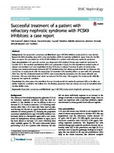
Clinical Case A 12-year-old adolescent presented to his primary care physician with new onset staring episodes. His family stated the patient would be unresponsive during these episodes and would make a “wringing” motion with his hands. The episodes lasted about 1 min and were not preceded by an aura. After the episodes, the patient felt tired. At the time of presentation, the episodes were occurring daily. He was initially started on carbamazepine by his primary care physician who thought the episodes were consistent with seizures and he was referred to neurology. His past medical history was unremarkable. He was a full-term birth and had normal development. He was performing well in school and involved in several athletic activities. On presentation to neurology, it was thought his episodes were consistent with complex partial seizures. His neurologic examination was unremarkable. His seizures were well controlled on the carbamazepine, which was continued and an EEG and MRI brain with and without contrast were ordered. The EEG was unremarkable, but the MRI brain showed a left mesial temporal lobe lesion (Figure 1). At this time, he was referred to neurosurgery for further evaluation and management. There was concern that this lesion was consistent with an underlying neoplasm, specifically a glial tumor, ependymoma, or dysembryoplastic neuroepithelial tumor (DNET). He underwent a left pterional craniotomy with partial resection of the left temporal lobe lesion. The pathology from this lesion was not diagnostic for any specific pathology. It demonstrated a firm collagenized reactive process that could represent an infectious, neoplastic, or reaction to a vascular malformation. He did well post-operative. He continued to have occasional seizures every couple of months and levetiracetam was added. After the addition of levetiracetam, he had better control, but over the next several years both his carbamezepine and levetiracetam were increased after an increase in seizure activity. The patient continued to have an annual MRI brain with and without contrast that demonstrated no change in the lesion. Five years after the initial surgery, the patient continued to have seizures (approximately 2–3/month) and repeat surgery was discussed for possible better seizure control. He underwent a second left temporal craniotomy for resection of the remaining mesial temporal lobe lesion. His immediate post-operative course was complicated by right-sided
1
June 2015 | Volume 6 | Article 129
McGinley et al.
Mesial temporal lobe lesion
FIGURE 1 | MRI brain with and without contrast obtained at onset of seizures demonstrating left mesial temporal lobe lesion. (A) Axial DWI (B) Axial T2 (C) Axial T1 post-contrast (D) Coronal T2 (E) Coronal T1 pre-contrast (F) Coronal T1 post-contrast.
hemiplegia with left-sided intraventricular hemorrhage and an ischemic stroke affecting the left thalamus and left middle cerebral peduncle. Pathology from this biopsy was consistent with a congenital arteriovenous malformation (AVM) with secondary leptomeningeal fibrosis. He was discharged to an inpatient rehabilitation facility and he had partial recovery of strength on his right side to the point and he was able to ambulate with a cane. He was seen in follow-up and at 28 months post-surgery his levetiracetam was discontinued over 6 weeks and at 34 months post-surgery the carbamazepine was discontinued over 12 weeks. He has remained seizure free over the 3 years since surgery.
Discussion This case demonstrates the difficulty in distinguishing neoplastic, vascular, and infectious lesions on imaging. Additionally the patient presented in this case had an initial biopsy that was inconclusive, which further supports the importance of gleaning as much information as possible from imaging studies. In this report, we provide a brief summary of the classic imaging characteristics of a variety of neoplastic, vascular, and infectious etiologies and discuss how this may have helped with the initial work-up and diagnosis of this patient. In the pediatric population, supratentorial cortical based tumors represent 25–40% of all brain tumors (1). Most of these tumors are low grade resulting in a slow progression with a mean latency to diagnosis of 8 months (1). Table 1 provides a summary of the typical imaging characteristics of the low- and high-grade tumors often found in pediatric patients. In general, low-grade tumors appear more homogeneous, whereas high-grade tumors often have a more heterogeneous appearance because of cystic, hemorrhagic, and necrotic portions. Additionally, most tumors are hyperintense on T2/FLAIR. The patient presented in this case had a lesion that was primarily hypointense on T2, which is in contrast to most neoplastic lesions, but could be consistent with a low-grade pilocytic astrocytoma, pleomorphic xanthoastrocytoma, or meningioma. Of these three tumor types, the only one that demonstrates strong contrast enhancement, as in the patient reported, is a meningioma. The patient’s lesion did not appear dural based, but this is not always readily apparent on imaging.
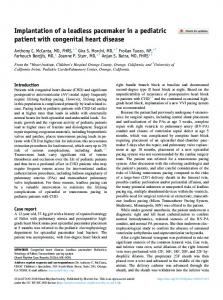
Pathology A biopsy of the left mesial temporal lobe lesion was analyzed (Figure 2). On microscopic examination, the cortex was slightly hypercellular. There was evidence of gliosis with focal brightly eosinophilic Rosenthal fiber formation. Additionally, there was evidence of abnormal vessels with thickened walls within the brain parenchyma and one with extensive calcification. The GFAP and CD34 immunostain demonstrated cortex with reactive gliosis and intraparenchymal thin-walled as well as thickened blood vessels with intervening brain tissue. The leptomeninges showed extensive fibrosis and mineralization. A synaptophysin stain showed positive process in the neuropil, but is negative in the neuronal cell bodies. A Ki-67 proliferation index was