Dental Materials Journal 2008; 27(3): 440-447 Original Paper
A Prospective Study on the Clinical Performance of Polysiloxane Soft Liners: One-year Results
Mustafa Murat MUTLUAY1,2, Serra OGUZ3, Finn FLØYSTRAND1, Erik SAXEGAARD1, Arife DOGAN3, Bulent BEK4 and I. Eystein RUYTER2 1
Department of Prosthodontics, Faculty of Dentistry, University of Oslo NIOM, Nordic Institute of Dental Materials, Haslum, Norway 3 Department of Prosthodontics, Faculty of Dentistry, Gazi University, Ankara, Turkey 4 Department of Prosthodontics, Faculty of Dentistry, Cumhuriyet University, Sivas, Turkey Corresponding author, M. Murat MUTLUAY; E-mail:
[email protected] 2
Objective: The aim of the present study was to evaluate the clinical performance of four denture soft liners up to 12 months. Materials and methods: Thirty-three edentulous patients who experienced difficulties when using hard denture bases because of changes in denture-supporting tissues were accepted for the study and randomly received Molloplast B, GC Reline Soft, Silagum Comfort, or Mollosil Plus relines. Performance of the materials was evaluated using nine criteria at 3, 6, and 12 months: physical integrity, surface detail, adhesion, color, odor, plaque accumulation, resilience, hygiene, and mucosal condition. A four-point categorized scale (1=poor, 2=fair, 3=good, 4=excellent) was used. Unscheduled maintenance events and the presence of fungal colonization were also recorded. Results: The percentage of patients available at 3, 6, and 12 months were 91%, 91%, and 66%. Main reasons for dropouts and discontinuation were fractured dentures and patient dissatisfaction. At 6 months, 96% of the performance scores were good or excellent and the largest changes were observed for physical integrity, surface detail, color, and fungal colonization. Fungal colonization was the most commonly observed problem and was the only reason of failure at 12 months. Conclusions: The clinical performance of all soft liners was slightly impaired over the 12-month observation. Except for cases showing extensive fungal colonization, the observed changes in clinical performance did not necessitate remaking of the dentures. Mollosil Plus showed a performance comparable to that of Molloplast B, and the other materials had slightly lower performance especially in terms of fungal colonization. Key words: Polysiloxane, Removable denture, Fungal colonization
INTRODUCTION For patients who cannot tolerate a hard denture base, soft liners are an important adjunct treatment to help the patients adapt to their new dentures1). A recent report indicated that some 75% of patients needed relining of the implant-supported overdentures after an average of 7.8 months, and soft liners were often favored as an alternative to conventional hard relining materials2). Use of soft lining materials was previously considered as a temporary measure3). However, several authors have reported on a long service life of soft liners, sometimes remaining on the denture for its full service life4-8). A soft liner’s assessment criteria as listed by many authors were, namely, resilience, tear resistance, biocompatibility, lack of odor and taste, adhesive bond strength, color stability, and resistance to abrasion9-11). Although some of the material types were reported to maintain these qualities for a longer time than the others, loss of bonding to denture base polymers and fungal colonization on and within the
Received Sep 18, 2007: Accepted Dec 19, 2007
soft liners continue to be the main problems limiting their use9-11). In the last decade, soft liners have been improved and new materials have become available12). However, there are few published studies which effectively test the clinical performance of these liners and provide documentation about their serviceability. The specific aim of this study was to assess the clinical performance of three recently introduced materials with relatively new formulations and application methods, and to compare their clinical performance with that of a well-studied, conventional material serving as a control. MATERIALS AND METHODS Soft liner materials The materials used in this study were, Molloplast B, GC Reline Soft, Silagum Comfort, and Mollosil Plus (Table 1). All the four materials are based on polysiloxanes. They differ mainly in terms of application technique, polymerization, and/or curing
DMJ 2008; 27(3): 440-447
441
cycle (Tables 1 and 2). Molloplast B is a heatcured, compression-molded material, and the other soft liners are chairside relining materials which polymerize at room temperature.
informed consent was obtained. The study protocol was reviewed and approved by the Human Ethics Committee at Gazi University’s Faculty of Medicine, Ankara, Turkey.
Study population Subjects accepted for this study were edentulous male (15) and female (18) patients (median age for all patients was 65 years) who came to the prosthodontics clinic at Faculty of Dentistry, Gazi University in Ankara, Turkey, with complaints relevant to clinical indications for a soft liner therapy. These problems arose from changes in the denture-bearing hard and soft tissues, i.e., alveolar resorption or poor quality of the denture-bearing mucosa1). Patients who needed local relief of pressure for wound healing following a surgical intervention or patients with other acute problems were not included in this study, since such indications usually require only short-term use of the material limited to a few months. The distribution of the patients is given in Table 3. Subjects were accepted for this study after their
Treatment protocol During the first appointment, a detailed examination form was filled out and the oral condition of the patient was recorded. Subjects were distributed according to a randomly generated treatment allocation to four soft liner groups and received the liners accordingly. For the Molloplast B group (n=9), the tissue surface of the patient’s denture was ground to obtain the necessary space required. A thickness of 2-3 mm was aimed at, but because of anatomical variations and variations among the different patients’ dentures, this was not always achieved. Consequently, the dentures were border-molded and an impression was taken with a eugenol-free impression paste (Cavex Outline, Cavex, Holland). At the laboratory, the recommended primer was applied as per the
Table 1
Materials used for the study
Brand names (Code)
Manufacturer
Processing method
Molloplast B (MLP)
Detax GmbH, Ettlingen, Germany
Heat-cure
Primo adhesive
Detax GmbH, Ettlingen, Germany
Recommended for Molloplast B. Apply and let dry for 60-90 min.
GC Reline Soft (GCS)
GC Co, Tokyo, Japan
Autopolymerizing
GC Reline Primer R
GC Co, Tokyo, Japan
Recommended for GC Reline soft. Apply to the surface and gently dry with air.
Silagum Comfort (SLC)
DMG, Hamburg, Germany
Autopolymerizing
Silagum Comfort Primer
DMG, Hamburg, Germany
Recommended for Silagum AM Comfort. Apply and let solvent dry for 1 min.
Mollosil plus (MLS)
Detax GmbH, Ettlingen, Germany
Autopolymerizing
Mollosil plus Primer
Detax GmbH, Ettlingen, Germany
Recommended for Mollosil plus. Apply and let solvent dry for one minute
Table 2
Chemical ingredients of the materials according to manufacturers’ information
Material
Components
Primer
MLP
Condensation polysiloxane material, PMMA, γ-methacryloyloxypropyltrimethoxysilane
Mixture of methoxy and ethoxy silane derivatives
GCS
Silicon dioxide, Vinyl dimethyl polysiloxane, Hydrogen polysiloxane
SLC
Vinyl polysiloxane, Hydrogen polysiloxane, aerosil, additives
MLS
Polydimethylsiloxane with functional groups, filler, pigments, platinum catalyst PMMA=Poly (methyl methacrylate)
Ethyl acetate >90% Ethyl acetate, modified polyacrylate, additives Ethyl acetate 60-100%
442
DMJ 2008; 27(3): 440-447
manufacturers’ recommendations and a standardized curing cycle was used for processing the Molloplast relines. The denture was then delivered to the patient and necessary adjustments were made at chair-side. For chairside relining materials (n=8 for each group), the dentures were ground on the tissue contacting surface in order to obtain adequate space for the soft liner. This was done to a depth of 2 mm as long as the denture thickness allowed. They were coated with primer, relined with the soft liner, and delivered to the patient at the same appointment according to the manufacturers’ instructions. Blinding in this study was not possible because the different relining materials had very distinct color differences. The clinicians had no previous experience with the proprietary materials used; this would reduce any bias introduced by familiarity with one of the products. Patients were instructed to clean the tissue contacting surfaces of their dentures after every meal using liquid soap and a regular toothbrush under Table 3
running water. They were thus instructed because this was the most commonly used method which did not require the patients to purchase any additional material. The instructions were repeated during every recall for a continuous level of motivation. Data collection Patient demographics included gender, age, and occupation (Table 3). Furthermore, patients’ health and medication usage, smoking history and their oral hygiene practices were recorded. An intraoral examination was carried out, and the results were entered in an examination form. A second form was filled out during the visits where the condition of the liner was subjectively evaluated according to the nine primary criteria (Table 4): physical integrity, surface detail, adhesion, color, odor, plaque accumulation, resilience, hygiene, mucosal condition of the patient occasionally, and signs of fungal colonization5). Fungal colonization was recorded as P=present or NP=not present; the other parameters were assigned scores on a four-point categorized scale (Table 4).
Data collected during the study
Demographics
Gender distribution Age distribution (mean=65.5)
History
Smoking habits Medication usage
Denture hygiene practices before the study
Denture Table 4
Upper Lower
Male Female 40-49 50-59 60-69 >70
15 18 2 9 9 13
Smoking Non-smoking Using at least one medication regularly Not on medication Toothbrush and toothpaste Toothbrush and soap Soap only Immersing into NaOCI Cleaning tablets
3 30 14 19 2 7 19 2 3 4 29
Criteria used for the evaluation of the liners and the scoring system used to record the condition of the liner
Mucosal condition evaluation Liner conditon evaluation
1-4 rating 1) Physical integrity: Tearing loss of material 2) Surface detail: Loss of surface texture, roughening 3) Adhesion: Failure of the bond 4) Color: Comparison with fresh unused material 5) Odor: Presence of any distinctive odor
1-4 rating (1=poor, 2=fair, 3=good, 4=excellent
6) Plaque accumulation: Surface coverage with plaque 7) Hygiene: Surface coverage with food particles 8) Resilience: Comparison with fresh unused material 9) Fungal colonization: Presence of colonization
P-NP rating (P=present, NP=not present)
443
DMJ 2008; 27(3): 440-447 The worst site where the most obvious changes were observed was also recorded where applicable. Patients were recalled at 3, 6, and 12 months for an evaluation of the liner, and unscheduled maintenance events were also recorded. Dropouts from the study population were also categorized and recorded at the time of the event either as patient not satisfied, patient lost to follow-up, adverse reaction, or fracture of denture.
RESULTS Overall performance The recall rate at 3, 6, and 12 months were 91%, 91%, and 66%. Table 5 lists the dropouts and discontinuers. The reason for discontinuing was severe fractures which required remaking or rebasing of the dentures. Five lower dentures (15%) had fractured between baseline and 12 months. The main reason for dropout was patient dissatisfaction with the denture. Only one adverse reaction was recorded during the study: a female patient using the MLS reported a burning sensation on the mucosa and generalized redness was observed. Among the followed relines, four were on upper dentures. Two of these patients were lost to follow-up at 6 and 12 months. Therefore, a proper statistical analysis was not possible. Nonetheless, no recognizable differences were observed between upper and lower dentures in terms of material performance.
Statistical analysis Collected data were analyzed using a SPSS statistical package (version 14.0). The outcomes were changes in primary end-points and fungal colonization. A comparison of the material performance over time and among the material types was done by chisquare and Wilcoxon signed rank tests. The 12month controls had too much lost data. Therefore, the main statistical analysis was carried out on data obtained from the 3 and 6-months controls.
Table 5
Dropouts and discontinued patients and reasons 3 months
6 months
12 months
MLP 0 0 2d,d a GCS 1 0 4b,b,d,d SLC 0 0 1a MLS 2a,c 0 1d Superscript letters show the reasons for dropouts and discontinuers: a patient not satisfied, b lost to follow up, c adverse reaction, and discontinued patients d severe fracture of denture requiring new prosthesis. Table 6
Some of the parameters followed during the study. The values show the distribution of scores for every material type at each recall
Good
Excellent
n
Poor
Fair
Good
Excellent
n
Poor
Fair
Good
Excellent
12 months
Fair
6 months
Poor
3 months
n
Physical integrity
MLP GCS SLC MLS
0 0 0 1
0 0 0 0
2 5 3 0
7 2 5 5
9 7 8 6
0 0 0 1
0 0 0 1
2 5 3 1
7 2 5 3
9 7 8 6
0 1 0 0
0 0 1 1
2 1 2 1
5 1 4 3
7 3 7 5
Surface detail
MLP GCS SLC MLS
0 0 1 0
0 1 0 0
0 1 1 1
9 5 6 5
9 7 8 6
0 0 1 0
0 2 0 1
2 0 4 2
7 5 3 3
9 7 8 6
0 1 1 0
0 0 0 1
2 1 3 1
5 1 3 3
7 3 7 5
Adhesion to denture base
MLP GCS SLC MLS
0 0 1 0
0 1 0 0
0 1 1 1
9 5 6 5
9 7 8 6
0 0 1 0
0 2 0 1
2 0 4 2
7 5 3 3
9 7 8 6
0 0 1 0
0 1 0 1
2 1 3 1
5 1 3 3
7 3 7 5
Color stability
MLP GCS SLC MLS
0 0 0 0
0 0 0 0
4 2 3 1
5 5 5 5
9 7 8 6
0 0 0 0
1 0 1 1
5 3 3 2
3 4 4 3
9 7 8 6
0 0 0 0
1 0 1 1
3 2 3 1
2 1 3 3
7 3 7 5
444
DMJ 2008; 27(3): 440-447
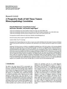
Performance outcomes Physical integrity: Average physical integrity score of the materials changed after 3 months (Table 6). The greatest decrease was with the GCS material where five of the seven dentures (71%) had problems related to physical integrity. This change was significantly different from MLP which had changes on only two dentures (22%) after 3 and 6 months (p=0.049). No other significant differences were observed among the materials. Of the 33 relined dentures, an average point of 3.5 was rated with the lowest score of 2 which was given to one denture only, and for 13 dentures (39%) the score was still 4 after one year. The MLP material had more scores of 4 than the other materials. SLC also showed an early decrease in physical integrity where three of the samples had a decreased score after 3 months. Failures were usually seen where the material was thin and not bonded to the denture base. These
areas were limited in size and did not require any maintenance (Fig. 1). Surface detail: Scores given to visually assessed changes in surface detail or deterioration in surface texture, changed significantly after 3 and 6 months (p=0.008 for both). No significant differences were found among the materials. The changes were often observed as roughening of the surface and loss of shiny appearance ― which was first observed when the liner surface was cured against mucosa. Changes in surface detail were mostly observed in the posterior area. Adhesion: Adhesion to denture base polymer was not significantly changed after 6 months compared to baseline, and no significant differences were found among the materials. 56% of the dentures still had the excellent score after 12 months, and only three (9%) of the relines showed small failures on the borders of the denture which did not require
Fig. 1
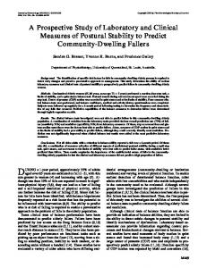
Fig. 2
Table 7
Clinical example of a MLP soft liner at 12 months’ recall. Note the good condition of the liner in terms of physical integrity and surface detail as well as fungal colonization.
Clinical example of a GCS material at 12 months’ recall. Note the fungal colonization and problems concerning physical integrity.
Percentages of soft liners with fungal colonization and failures 3 months
Fungal colonization
Failure because of fungal c.
6 months
12 months
(+)
n
(+)
n
(+)
n
MLP
0
9
22
9
14
7
GCS
13
7
30
7
66
3
SLC
13
8
75
8
83
7
MLS
0
6
0
6
20
5
MLP
0
9
0
9
14
7
GCS
0
7
0
7
33
3
SLC
0
8
0
8
56
7
MLS
0
6
0
6
20
5
DMJ 2008; 27(3): 440-447 repeating the relining procedure (Fig. 1). Color change: Sixteen of the dentures (48%) were rated one or two points lower at 3 months. Scores at 3 and 6 months were significantly different from baseline (p=0.014). No significant differences were found among the materials. Fading of the material was uncommon, and the main reason for color change was staining. The typical staining color was yellowbrown, and this was patient-dependent rather than a general finding. For three of the patients, an intense color change was observed and the predominant color was brown. The average scores after 6 and 12 months were 3.5 and 3.3 respectively. Plaque accumulation and hygiene: The amount of plaque found on the dentures, as well as hygiene, were assessed visually. The surfaces of all the liners were partly covered with plaque, with very little variation among the patients. No significant differences were found among the different materials (p>0.05). Resilience: Resilience of the materials was assessed by comparing the liner to an unused sample, and any detectable change was recorded. After 12 months, none of the soft liners showed any change that could be detected by the clinician. Condition of the mucosa: No indications of change were observed after 3, 6, and 12 months, except for one adverse reaction observed after 24 hours. Colonization of the soft liner surface: Fungal colonization of the soft liner surface was observed at 3 months in two patients (6%). At 12 months, 36% of the dentures were affected with fungal colonization at different levels (Table 7, Fig. 2). For all the time periods, SLC and GCS had a higher degree of colonization than the others. At 12 months, seven patients (21%) were recorded as failure because of extensive colonization (Table 6). The largest differences among the materials were observed for the colonization scores. At 12 months, only one of the dentures relined with MLS showed visually detectable colonization and MLP had colonization on only two of the dentures. At 6 months, MLS was significantly different from GCS (p=0.03) and SLC (p=0.02). DISCUSSION The survival of an oral restoration is affected by operator-dependant factors, design factors, restoration material factors, intraoral location factors, and patient-related factors13). Operator factors are generally believed to be the most important of these14). Our study was carried out in a dental faculty environment by clinicians who are competent in the field of prosthodontics. Applicability of the clinical results to routine general dental
445
practice may thus be limited. The percentages of patients available for followup were 91%, 91%, and 66% for 3, 6, and 12 months respectively. At 12 months, three of the patients did not want to continue and three of the dropouts were patients that could not be reached. The dropout rate appeared to be moderate, considering the relatively high age of the patients. Fatigue fracture of the denture base has frequently been reported as the main reason for discontinuing and this is ascribed to the decreased thickness of the denture5,14,15). In the present study, 15 percent of the dentures fractured within one year. Polysiloxanes are used in many biomaterial applications because of their stability and lack of toxicity. Although rarely reported and usually not properly diagnosed, some adverse reactions to polysiloxanes in different regions of the body have been reported15-17). One of the patients participating in our study showed a reaction to the polysiloxane soft liner MLS after 24 hours of contact, resembling a type 1 allergy to this material or to some other materials used during the chairside relining procedure. This patient wanted to withdraw from the study, and her denture was relined with a conventional denture base polymer. A history of allergy was also recorded when this patient was first examined. Although no additional information was available for this patient, an allergic reaction was possible and this should be kept in mind during treatments. The term ‘physical integrity’ is related to the general soundness and durability of the soft liner materials5). It may also reflect the durability of the materials during cleaning procedures where brushing or other means of cleaning subject the reline to various stresses. Failure is generally observed as tearing of the material and loss of parts of the reline during clinical use. MLP showed slightly better performance at 12 months than the other materials and significantly better than GCS. The clinical significance of this finding was questionable because the physical integrity problems were small. Failures were observed where the thickness of the soft liner was less than adequate or not properly bonded to the denture base. A recent study by Oguz et al.18) published a ranking of tensile strength and tear resistance of MLP, GCS, and SLC. The results with and without thermocycling indicated that the tensile strength and tear resistance results of MLP were not higher than the others. The reason for performing better under clinical conditions might be the result of differences in the application procedure of the materials. The MLP material was cured in the laboratory using a conventional compression mold technique. As a result, it was possible to maintain an ideal thickness and optimum physical conditions during curing.
446
DMJ 2008; 27(3): 440-447
The other materials (MLS, GCS, and SLC) might have been negatively affected by less controllable curing conditions during chairside application and curing. The variable factors in terms of humidity, temperature, and pressure present during the curing of these materials could have caused poor crosslinking and varying material thicknesses. An intimate contact between a denture and the supporting tissues is considered as one of the key features of a successful removable denture. Dentures must follow the contour and texture of the mucosa for both retention purpose and hygiene maintenance. The ability of a material to maintain this is important for long-term clinical success. The materials used in our study showed changes in the surface texture starting from the third month. This type of degradation problem was frequently reported in previous publications4,5,8). MLP maintained the surface texture better than the other materials, which might be an indication of long-term success; however, this difference was not statistically significant. This was comparable to a study where the dentures relined with MLP needed replacement after 4-9 years of clinical service5). Degradation of the materials resulting in changes in surface detail and roughening was mostly observed in the posterior region, which was probably a result of forces of mastication affecting this region. Soft liners rely on good adhesion to the denture base to function properly. Adhesion of polysiloxane soft liners to denture base polymer has occasionally been pronounced as the weak link19). In particular with the chairside polysiloxane soft liners, this has been restated by many authors1,9,10,20,21). However, some studies also reported on long-term clinical serviceability of the MLP material4,5,7,8). After one year of clinical service, the average score for the adhesion of soft liners to denture base polymer was still 3.9 for the current study. This result strongly suggested that the adhesion obtained by these systems was durable enough to withstand clinical use for one year. Failure was observed only marginally, and no relines required replacement after 12 months of use. This was also supported by a previous study which reported only slight edge failure of adhesion for MLP4). No indications of adhesion failure were reported for dropouts. The type of color change observed throughout the study was staining of the materials rather than fading of the color. It did not affect the appearance of the subject when using the denture. Three of the dentures showed heavy staining and these were related to smoking as expected. All the other dentures showed an acceptable resistance to staining after one year. Other evaluation criteria such as plaque retention, oral hygiene, condition of the mucosa, and
resilience remained optimal during the 12-month period. These properties did not show any statistical changes compared to baseline and did not indicate any failure related to material performance. Fungal colonization of the soft liner surface was frequently observed in this study. Colonization started at 3 months for GCS and SLC and at 6 months for MLP. Although the colonization of the materials by oral fungi presents a problem regarding useful service life, no effects of such colonization on the mucosal condition of the patients were observed. The lack of such a relation has been similarly reported in two published studies4,7). This lack of mucosal response to fungal colonization could be explained by the fact that the patients chosen for our study had no acute problems or no indications of denture stomatitis; they were generally healthy at the beginning of the study22). Moreover, most of the dentures followed in our study were lower dentures, and the inflammatory changes related to denture stomatitis were reported to occur rarely under the lower dentures23). In the present study, fungal colonization was assessed by visual inspection of the dentures. For colonized micro-organisms to be visible, they would have formed a plaque of significant size. Colonization of the soft liner surface begins with the initial adhesion of the micro-organisms24). For forming plaques or biofilms by cell division and agglutination, surface roughness may be an important contributing factor25-27). This was partly confirmed in our study, where visible colonization increased with changes in surface detail (Tables 6 and 7). Changes in surface detail resulted in a very rough surface with many small cracks in the material, which thus provided a larger surface area and a more protected environment for colonization. The number of failures recorded in this study suggested that fungal colonization of the materials, probably a result of roughened soft liner surface, was the main reason for replacement (Fig, 2). However, this was not a general finding and was probably related to the patient’s oral conditions, which in this case included diet, saliva secretion, saliva quality, and efficiency of the cleaning procedures. CONCLUSIONS Within the confines of this clinical study, the following conclusions were drawn: 1. Among the chairside materials tested, MLS showed comparable performance to the compression-molded and heat-cured MLP, followed by SLC and GCS. The easier/simpler application procedure may thus render this material as an alternative. 2. After one year of clinical service, adhesion
DMJ 2008; 27(3): 440-447
3. 4.
5. 6.
failure was observed only marginally and no relines required replacement because of adhesion problem. After one year, SLC and GCS showed fungal colonization sooner and more extensively than the other materials. Reducing fungal colonization on the surface and improving mechanical strength may further improve the clinical performance of soft liners. For all the investigated liners, the results were good or excellent for majority of the patients after six months of clinical use. For early detection of fungal colonisation, every patient with a relined denture should be called back after three months of use. ACKNOWLEDGEMENTS
The authors wish to express their gratitude to Professor Leiv Sandvik for statistical advice. We also thank Professor Dag Ørstavik and Professor Timo Närhi for helpful discussions and valuable suggestions during the preparation of the manuscript. The materials were in part supplied by the manufacturers, and this was also much appreciated. REFERENCES 1) Mack PJ. Denture soft lining materials: clinical indications. Aust Dent J 1989; 34: 454-458. 2) Attard NJ, David LA, Zarb GA. Immediate loading of implants with mandibular overdentures: Oneyear clinical results of a prospective study. Int J Prosthodont 2005;18: 463-470. 3) Wright PS. Soft lining materials: their status and prospects. J Dent 1976; 4: 247-256. 4) Wright PS. The success and failure of denture soft-lining materials in clinical use. J Dent 1984; 12: 319-327. 5) Wright PS. Observations on long-term use of a soft-lining material for mandibular complete dentures. J Prosthet Dent 1994; 72: 385-392. 6) Schmidt WF, Jr., Smith DE. A six-year retrospective study of Molloplast-B-lined dentures. Part I: Patient response. J Prosthet Dent 1983; 50: 308-313. 7) Schmidt WF, Jr., Smith DE. A six-year retrospective study of Molloplast-B-lined dentures. Part II: Liner serviceability. J Prosthet Dent 1983;50: 459-465. 8) Jepson NJ, McCabe JF, Storer R. The clinical serviceability of two permanent denture soft linings. Br Dent J 1994; 177: 11-16. 9) Braden M, Wright PS, Parker S. Soft lining materials ― a review. Eur J Prosthodont Restor Dent 1995; 3: 163-174. 10) Braden M. Polymeric Dental Materials, SpringerVerlag, Berlin, 1997, pp.125-137. 11) Garcia LT, Jones JD. Soft liners. Dent Clin North
447
Am 2004; 48: 709-720. 12) McCabe JF. A polyvinylsiloxane denture soft lining material. J Dent 1998; 26: 521-526. 13) Nikawa H, Jin C, Hamada T, Makihira S, Kumagai H, Murata H. Interactions between thermal cycled resilient denture lining materials, salivary and serum pellicles and Candida albicans in vitro. Part II. Effects on fungal colonization. J Oral Rehabil 2000; 27: 124-130. 14) Jokstad A, Bayne S, Blunck U, Tyas M, Wilson N. Quality of dental restorations. FDI Commission Project 2-955. Int Dent J 2001;51: 117-158. 15) Shim JS, Watts DC. An examination of the stress distribution in a soft-lined acrylic resin mandibular complete denture by finite element analysis. Int J Prosthodont 2000; 13: 19-24. 16) Jordan DR, Nerad JA. An acute inflammatory reaction to silicone stents. Ophthal Plast Reconstr Surg 1987; 3: 147-150. 17) Davenport JC. An adverse reaction to a silicone rubber soft lining material. Report of a case. Br Dent J 1970; 128: 545-546. 18) Oguz S, Mutluay MM, Dogan OM, Bek B. Effect of thermocycling on tensile strength and tear resistance of four soft denture liners. Dent Mater J 2007; 26: 296-302. 19) Mutluay MM, Ruyter IE. Evaluation of bond strength of soft relining materials to denture base polymers. Dent Mater 2007; 23: 1373-1381. 20) Kutay O, Bilgin T, Sakar O, Beyli M. Tensile bond strength of a soft lining with acrylic denture base resins. Eur J Prosthodont Restor Dent 1994; 2: 123126. 21) McCabe JF, Carrick TE, Kamohara H. Adhesive bond strength and compliance for denture soft lining materials. Biomaterials 2002; 23: 1347-1352. 22) Okita N, Orstavik D, Orstavik J, Ostby K. In vivo and in vitro studies on soft denture materials: microbial adhesion and tests for antibacterial activity. Dent Mater 1991; 7: 155-160. 23) Wright PS, Clark P, Hardie JM. The prevalence and significance of yeasts in persons wearing complete dentures with soft-lining materials. J Dent Res 1985; 64: 122-125. 24) Radford DR, Challacombe SJ, Walter JD. Denture plaque and adherence of Candida albicans to denture-base materials in vivo and in vitro. Crit Rev Oral Biol Med 1999; 10: 99-116. 25) Quirynen M. The clinical meaning of the surface roughness and the surface free energy of intra-oral hard substrata on the microbiology of the supra- and subgingival plaque: results of in vitro and in vivo experiments. J Dent 1994; 22: 13-16. 26) Radford DR, Sweet SP, Challacombe SJ, Walter JD. Adherence of Candida albicans to denture-base materials with different surface finishes. J Dent 1998; 26: 577-583. 27) Verran J, Maryan CJ. Retention of Candida albicans on acrylic resin and silicone of different surface topography. J Prosthet Dent 1997; 77: 535539.