for Electron Microscopy of Paraffjm-Embedded. Tissue. Sibylle Wid&n, HTL, and Lars-Gunnar Kindblom, MD. Department of Pathologg, Sahlgren Hospital,.
New Bchniques A Rapid and Simple Method for Electron Microscopy of Paraffjm-Embedded Tissue
Ultrastruct Pathol Downloaded from informahealthcare.com by Mr. OR. Dr. Wolfgang MUSS on 06/20/12 For personal use only.
Sibylle Wid&n, HTL, and Lars-Gunnar Kindblom, MD Department of Pathologg, Sahlgren Hospital, University of G&&org, SO413 45 G&&org, Sweden
A new rapid and simple method for electron microscopy of paraffinembedded material is described. The paraffinembedded tissue is deparaffinized in xylene, stained in a 0.01% toluidine bluelabsolute ethanol solution, infiltrated in a propylene oxidelresin mixture, and embedded in epoxy resin. This quick method, which excludes rehydration, postosmification, and dehydration, takes about 3 h and produces results equal to previously described more laborious re-embedding techniques.
KEY WORDS: electron microscopy, paraffin-tissue, re-embedding technique.
INTRODUCTION
dehydration in ethanol, and embedding in In routine histopathologic, diagnostic work epoxy resin. Although these methods have it is often desirable or necessary t o extend been useful, we have found them somethe morphologic investigation t o the ultra- what laborious and timeconsuming, usustructural level. As the primary fixation of ally at least 36 h are needed. We have surgical material in glutaraldehyde has not therefore tried t o find a simpler and more become routine, the electron microscopic rapid method of re-embedding. The examination often has t o be done on present study was undertaken t o answer formaldehyde-fixed tissue and sometimes the question of whether it is really necesonly paraffin-embedded blocks may be sary t o postfix the deparaffinized speciavailable, especially in retrospective stud- mens in osmium tetroxide and, if not, t o ies of older material. The usefulness of find the most simple and rapid method such paraffinembedded tissue for ultra- possible. structural studies has been emphasized in numerous reports by other a u t h o r ~ " ~ ' " ~ and our own The previously used MATERIAL procedure of deparaffinizing and re- Eighteen paraffin blocks from 17 different embedding formaldehyde-fixed paraffin- human soft tissue tumors of various types embedded tissue has mainly been per- were used. All were primarily fixed in 4% formed according t o the method described formaldehyde. by Hultquist and Karlsson' or with modifications according t o Johannessen' and our~elves.~ These ,~ procedures are based METHOD on the rehydration of the deparaffinized Representative areas from routinely specimens in diluted ethanol and buffer, stained paraffin sections were chosen unthe postfixation in osmium tetroxide, the der the light microscope and marked with Ultrastructural Pathology, 12: 131- 136, 1988 Copyright
0 1988 by Hemisphere Publishing Corporation
131
Ultrastruct Pathol Downloaded from informahealthcare.com by Mr. OR. Dr. Wolfgang MUSS on 06/20/12 For personal use only.
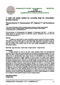
'suoii3as uiqiwiln 6uiun3 JOJ Apes pue uisaJ Axoda ui pappaqwa-aJ uawpads paziuit-eJedap aqi JO a m d uiqi y (p) sa3ad Jet -n6ueiaa~llews oiui in3 do1 uo eaJe paiDalas aqi q i ! ~uawpads aqi JO aield ui#eJed uiql ( 3 ) ;bcO in3 51 q3!qM $0 do1 aqi *p!ueJAd unnop-appdn flews e JO adeqs ui $noin3 eaJe pai3qas aqi JO iUi3Wi36JeIU3 (q) Apnis leJn$mJis -eJiIn aqi JOJ pai3alas eaJe atp 6 u i ~ o q s uawpads pappaqwa-uigeJed (e) 1
... .. ....
*sm
133
Ultrastruct Pathol Downloaded from informahealthcare.com by Mr. OR. Dr. Wolfgang MUSS on 06/20/12 For personal use only.
New Techniques: Paraffii-Embedded Tissue for Electron Microscopy
specimens were stained in “blue ethanol” (0.01 % toluidine blue in absolute ethanol) for 10-30 min directly after deparaff inization in xylene and soaked in propylene oxide after the staining. f o r resin infiltration (Agar Resin 100, Agar Aids) the specimens were transferred into a 1:l mixture of propylene oxide and resin for 30 min, which was followed by a 30-min period in pure resin at 4OOC. The tissue was embedded and polymerized at 100°C for 60 min. The time needed for this procedure was about 3 h including staining (Fig. Id). The polymerized specimens were sectioned and routinely stained: the 1 pmsections, stained with 0.1 % toluidine blue, were observed under the light microscope to check whether or not the correct region of the previous paraffin block had been prepared. Ultrathin sections were cut on a LKB ultrotome, contrast-stained with uranylacetate and lead-citrate and studied under a Philips 400 electron microscope.
The cutting and section quality as well as the contrast and ultrastructural quality of the modified Hultquist and Karlsson method and the developed quick method was compared.
REsms
Cutting and Sectioning There were no problems cutting semithin and ultrathin sections in any of the cases. The quality of 1-pm toluidine blue-stained sections as well as that of the ultrathin sections prepared according t o both methods was identical.
Contrast There were no differences in the contrast of the ultrathin sections, regardless of whether a postosmification was carried out or not.
TABLE 1 Summary of the Previously Used and the New Quick Method for Reprocessing Paraffin-embedded Material for Electron Microscopy Previous method Procedure
Medium
Time
Deparaffinization
Xylene Xylenelpropylene oxide (“blue ethanol”) Abs. ethanol
-
Rehydration
95%-50% ethanol Buffer
Postfixation
1% oso4 Buffer
Dehydration
Infiltration
Polymerization
70%-95% ethanol Abs. ethanol Propylene oxide
Propylene oxidel Epoxy resin Pure resin Pure resin
90min
Quick method
Temperature
Time
Temperature
rt
4 0 min 1 0 min (10-30 min
rt
90min 90min 12h
rt rt
120 min 1 0 min
rt
60min 60min 10min
rt
-
rt
-
rt
1 0 min
120 min
rt
30 min
120 min
rt
30 min
4OoC
6OoC
60 min
1oooc
12h’
~
rt)
rt
36 h rt, Room temperature.
rt
rt
rt
3-3.5 h ~
~
_
_
Ultrastruct Pathol Downloaded from informahealthcare.com by Mr. OR. Dr. Wolfgang MUSS on 06/20/12 For personal use only.
134
S. Widkhn and L . 4 . Kiudblom
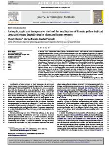
FIG. 2 Electron micrographs showing equal ultrastructural preservation and contrast of re-embedded material from the same paraffin block of a leiomyosarcoma processed according to the previously used method (a) and the quick method (b). Ultrastructural details such as myofilaments, longitudinal densities, attachment sites, and pinocitotic vesicles are well preserved.
Ultrastructure Specimens from the same paraffin block prepared according t o both methods showed no ultrastructural differences (Fig. 2). There were, however, strong variations in the degree of ultrastructural preservation from case to case (Fig. 3).
Time The time saved by the quick method was 2 working days (Table 1).
DISCUSSION The present study clearly shows that postfixation in osmium tetroxide can be omitted when paraffinembedded tissue is re-
processed for electron microscopy. As a result, the prior rehydration performed for the treatment with buffered osmium tetroxide and the following dehydration can also be omitted. The reason that postosmification is unnecessary is probably because most of the phospholipids and some of the proteins that are given contrast by osmium tetroxide have already been dissolved during the primary formaldehyde fixation and dehydration in organic solvents such as ethanol and xylene used before the paraffin embedding. The method described here is much less laborious and saves time, as much as 2 working days. When the infiltration time in pure resin was gradually shortened to 30 min and the polymerization time to 60 min (at 1OOOC), holes sometimes appeared in the resin. This problem could, however, be over-
New Techniques: Paraffii-Embedded Tissue for Electron Microscopy
135
Ultrastruct Pathol Downloaded from informahealthcare.com by Mr. OR. Dr. Wolfgang MUSS on 06/20/12 For personal use only.
come by raising the temperature during the (fast green or hematoxyline) to the absoinfiltration with pure resin from room tem- lute ethanol used after deparaffinization in perature to 4OOC. xylene. Although the staining of the tissue A disadvantage with this quick method disappeared somewhat in propylene oxide was the problem of handling non-osmium- the specimens remained blue enough to be treated specimens that were often difficult visible in the resin. to see during the preparation and cutting In a recently described time-saving procedures. This was, however, compen- method for the reprocessing of paraffinsated for by adding 0.01% toluidine blue embedded tissue for electron microscopy,
FIG. 3 Deparaffinized re-embedded specimens from paraffin blocks of two rhabdomyosarcomas both processed according to the quick method. (a) Detail of a tumor cell with very well-preserved ultrastructural details, including external lamina, highly organized myofilaments, and glycogen. The examined tumor piece is from the superficial parts of a urinary bladder rhabdomyosarcoma of botryoid type, which proves the neoplastic nature of the cell and excludes the possibility of an intermingled normal muscle cell. (b) Detail of a poorly preserved tumor cell with almost complete loss of organelle structures but with easily identifiable remnants of myofilament bundles with a preserved organization into bands proving the rhabdomyoblastic differentiation of the tumor.
Ultrastruct Pathol Downloaded from informahealthcare.com by Mr. OR. Dr. Wolfgang MUSS on 06/20/12 For personal use only.
136
S. Wid6hn and L.-6. Kindblom
osmium tetroxide was dissolved in xylene whereby deparaffinization and osmification were carried out ~imultaneously.~ A disadvantage with this method is the instability of the xylene-osmium tetroxidesolution and the unnecessary risk of using the toxic osmium tetroxide. The various techniques described for reembedding of paraffin sections&' have in our hands proved t o be more complicated and difficult, although they may be of value in cases in which only a very precise histologic area is of interest. Often the ultrastructural preservation has been found to be less satisfactory when sections have been used. This problem has, however, been overcome by a recently described "free-floating" technique.' The great dflerences seen in terms of ultrastructural preservation from case to case were apparently due to the quality of the primary formaldehyde fixation. Small, immediately fixed tissue samples showed generally very well preserved ultrastructural details, whereas tissue samples obtained from central areas of large tumors showed very poor preservation. From a practical point of view, however, even such poorly preserved material may at times be useful and occasionally necessary for a reliable diagnosis. This has reviously been illustrated by Johannessen who has encouraged the use of paraffinembedded material for diagnostic electron microscopy since "neither the patient nor the clinician care about the disintegration of mitochondria1 matrix as long as they receive a reliable diagnosis as a basis for treatment and the assessment of prognosis.'' This has also been emphasized by our own group.4 In the present study this is
P
exemplified by the poorly preserved rhabdomyosarcoma (Fig. 36)where the microfilament organization was still preserved well enough to indicate a rhabdomyoblastic differentiation.
1. Hultquist GT, Karlsson U: Use of formalin-fixed, paraffin-embedded biopsy or autopsy material for electron microscopy. Path Eur, Vol. 7no 2~97-101, 1972. 2. Johannessen JV: Use of paraffin material for electron microscopy. Pathol Annu 12:189-224, 1977. 3. Kindblom L-G, Save-Soderbergh J: The ultrastructure of liposarcoma; a study of 10 cases. Acta Pathol Microbiol lmmunol Scand (A) 87: 109-1 21, 1979. 4. Seidal T, Kindblom L-G: The ultrastructure of alveolar and embryonal rhabdomyosarcoma. A correlating light and electron microscopic study of 17 cases. Acta Pathol Microbiol lmmunol Scand (A) 92:231-248, 1984. 5. van den Bergh Weerman MA, Dingemans K P Rapid deparaffinization for electron microscopy. Ultrastruct Pathol 7:55-57, 1984. 6. Rossi GL, Luginbiihl H, Probst D: A method for ultrastructural study of lesions found in conventional histological sections. Virchows Arch (A) 350216-224, 1970. 7. Bretschneider A. Burns W, Morrison A: "Popo f f technic. The ultrastructure of paraffinembedded sections. Am J Clin Pathol 76(4):450-453, 1981. 8. Ngai HK, Chan KW, Or SB,Yau WL: A rapid method for reprocessing paraffin sections for diagnostic electron microscopy. J Pathol 145~59-62, 1985. 9. Yau WL, Or SB, Ngai HK: A "free-floating" technique for reprocessing paraffin sections for electron microscopy. Med Lab Sci 42:26-29, 1985. Accepted in revised form 23 April 1987.
Request reprints from Sibylle Widbhn.