Perspectives
History of medicine A rare case of osteoblastoma from medieval Tuscany Division of Paleopathology (G Riccomi, G Fornaciari, S Minozzi, V Giuffra) and Diagnostic and Interventional Radiology (G Aringhieri), University of Pisa, Pisa, Italy
[email protected] We declare no competing interests For more on cancer estimates in antiquity see Int J Paleop 2017; published online March 18. DOI:10.1016/j.ijpp.2017.03.005 For more on medieval and early renaissance medicine see Siraisi NG. Medieval and early renaissance medicine: an introduction to knowledge and practice. Chicago, IL: University of Chicago Press, 1990 For more on benign osteoblastoma see Cancer 1956; 9: 1044–52 and Orthop Traumatol Surg Res 2012; 98 (suppl 6): 98–104 For more on osteoblastoma pathology see Hum Pathol 1994; 25: 117–34 For examples of osteoblastoma of the sinuses see J Clin Neurosci 2015; 22: 445–49 and Eur Rev Med Pharmacol Sci 2012; 16: 1891–94 For more on the study of ancient skeletal tumour remains see Clin Anat 2016; 29: 831–43
In antiquity, tumours were less common conditions than nowadays, probably due to the shorter life expectancy of our ancestors, their different lifestyle and dietary habits, and their reduced exposure to risk factors such as environmental pollution. Additionally, it is not possible to obtain reliable cancer estimates through the analysis of ancient human remains. However, the field of paleopathology is inevitably biased because only bone lesions can be detected, limiting the evidence available to bone tumours, or metastasis to the bones originating from soft-tissue tumours. Consequently, cancer estimates in ancient times might be considerably underestimated. In this context, discoveries of paleopathological cases of cancer represent an opportunity to corroborate their presence in ancient populations and shed light on their evolution. Here, we present a case of a benign bone tumour dated between the 10th and 12th centuries, found in the skeletal remains of a young man aged 25–35 years, buried in the cemetery of Pava, Siena, Italy. In Italy, the period between the 11th and 13th centuries concurs with the heyday of the Medical School of Salerno, considered a precursor of the late medieval universities. However, despite the advances in medical knowledge achieved at the time, diagnosis of bone tumours was not possible in the Middle Ages. In 2004, archaeological excavations were carried out in the parish church of Saint Pietro of Pava (Tuscany, Italy). This medieval church is characterised by an annexed large cemetery that has been intensively used over past centuries. The documentation found, including the architectural plans of the religious building and evidence of different burial typologies, along with the radiocarbon dating of some bone specimens, allowed dating of the first use of the cemetery between the 10th and 12th centuries.
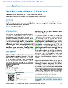
A
B
1 mm
500 μm
Figure: Histology of osteoblastoma from the Pava cemetery (A) Conebeam CT of the osteoblastoma in the right frontal sinus. (B) Toluidine blue staining of the specimen.
26
Among the human skeletal remains buried on the site, a skeleton with pathological evidence of the frontal sinus was brought to light. The skull showed post-mortem damage of the frontal bone, through which an unforeseen, macroscopic, oval bone formation was observed. Cone beam CT confirmed the presence of a tumour in the right frontal sinus, measuring 11 × 4·8 mm, and displaying a central radiolucent area and peripheral radiopaque margins (figure A). Histology of this frontal sinus bone tumour showed an external calcified shell and underlying tissue presenting wide vascular spaces in the absence of mature lamellar bone (figure B). Osteoblastoma is a rare, isolated, well vascularised, distinct type of benign tumour with poorly mineralised woven-bone trabeculae. Osteoblastoma represents approximately 3·5% of benign bone tumours and 1% of all bone neoplasms; it predominantly affects adolescents and young patients during the first three decades of life, with a male-to-female ratio of 3:1. Osteoblastomas typically originate in the vertebral column, the sacrum, or the metaphyseal region of long bones. In the skull, they are rare entities, occurring in 13–21% of all cases, and mainly affect the mandible (cementoblastoma) or other maxillofacial bones. Very few examples of osteoblastoma of the paranasal sinuses are documented in the medical literature, therefore, it is most surprising to find evidence of this type of tumour in ancient human remains. In paleopathology, the detection of paranasal sinuses lesions is mainly incidental due to the presence of postmortem breakages or radiological investigation carried out for other purposes. This case of osteoblastoma in medieval Italy raises questions about which types of tumours were common at the time, and whether those tumours considered rare nowadays were also sporadic in the past. Furthermore, this case highlights the importance of exploiting a variety of diagnostic techniques to increase the chance of tumour detection based on macroscopic studies of ancient skeletal remains. To our knowledge, the case discussed here represents the first paleopathological evidence of osteoblastoma of the frontal sinus in antiquity, enriching our knowledge of neoplastic diseases in this period, and confirms the existence of this type of tumour almost 1000 years ago. Further evaluation of diachronic trends in tumours, and the search for patterns explaining their variability over different periods of time, are crucial goals of paleo-oncology.
*Giulia Riccomi, Gino Fornaciari, Simona Minozzi, Giacomo Aringhieri, Valentina Giuffra
www.thelancet.com/oncology Vol 19 January 2018