intrinsic random events that account for biological aging. Aging and disease are
not synonymous. There are processes of aging and etiologies of disease. The re
...
Copyright 1999 by The Gerontological Society ofAmerica
JOUI7UlI ofGerontology; BIOWGICAL SCIENCES 1999,Vol.54A, No.6, 8255-8259
ESSAY MINI-REVIEW
A View of the Aging-Disease Relationship From Age 85 Herman T. Blumenthal Departmentof Community and FamilyMedicine,St. Louis University Schoolof Medicine, and the Agingand Development Program,Departmentof Psychology, Washington University, St. Louis,Missouri.
T
HIS essay posits a continuum between biological and pathological aging based on the following tenets: (i) Biological aging is not genetically programmed. (ii) Both biological aging and the diseases of the senescent period are caused by intrinsically generated random phenomena. (iii) The same phenotypic disease can occur in the juvenile, mature, and senescent periods of the life span, but with inherited genetic mutations having a progressively diminishing role with advancing age. (iv) The diseases of the juvenile period are of congenital or inherited origin. Those of the period of maturity derive from late-acting inherited genes or from so-called risk factors. During the senescent period, they derive from the same types of intrinsic random events that account for biological aging. Aging and disease are not synonymous. There are processes of aging and etiologies of disease. The relationship between the two are important, but not inevitable. N.W. Shock, 1961 (1) . . . to draw a distinction between disease and normal aging is to attempt to separate the undefined from the undefinable. J.G. Evans, 1988 (2) When I joined the Gerontological Society (now the Gerontological Society of America [GSA]) about 50 years ago, two expressions dominated the discourse relevant to an agingdisease relationship: "the (singular) aging process" and "aging is not a disease." The first of these expressed the objective of finding a unifying mechanism that would account for the diverse manifestations of biological aging. "Aging is not a disease" was based on the concept that the direct causes of the aging-related diseases are independent of "normal' aging phenomena. It would appear that the central objective of both expressions was to establish gerontology as a discipline distinctively different from the disease-oriented ones. I joined the biology and medical science sections because, as a pathologist, although my primary interest was in disease pathogenesis, I was also interested in ascertaining what the links might be between biological aging and the chronic diseases with peak morbidities and mortalities in older personscardio- and cerebrovascular disease, cancer, diabetes, hypertension, and osteoporosis. Alzheimer's disease, now a preoccupation of gerontologists of all stripes, was hardly on the horizon back then. Shock's foregoing statement reflects the orientation of most members of GSA as well as of the medical community at large. However, for some time now it has been recognized that the
many manifestations of aging cannot be accounted for on the basis of a single unifying mechanism. Indeed it is even acknowledged that an encompassing definition of biological aging is difficult, if not impossible, to formulate (3,4). The separation of normal aging from the diseases most prevalent in older persons, however, remains a subject of controversy. Although it appears that most gerontologists continue to hold to a separation of the aging-related diseases from biological aging as posited by Shock, there are some who subscribe to Evans's view of a continuum between biological and pathological aging. Consonant with Shock's statement, Strehler (5) in 1977 offered the criteria of intrinsicality, universality, progressivity, irreversibility, and genetically programmed, intended to separate biological aging from disease. However, not all of these criteria fulfill that objective. Until definitive cures are discovered for the chronic aging-related diseases rather than therapies that slow their progress, the aging-related diseases will remain progressive and irreversible. And with respect to genetic programming, Martin (6) maintains: ''There is no aging program, nor is there an aging gene." The criteria of intrinsicality and universality are addressed later in this essay. From a medical perspective, the following tenets have been offered to separate biological aging from aging-related diseases: Certain diseases have a high prevalence in older persons because the offending extrinsic agent(s) produce disease after a long latent period, or because repeated insults are required. Any disease during the lifetime of an individual is capable of adding "injury" to the "insult" of biological aging. Diseases with a high prevalence in older persons are the consequences of the aging-linked weakening of critical biological defense mechanisms which then permit the effects of extrinsic agents to take hold.
THE ThADmONAL MEDICAL MODEL The foregoing concept of the aging-disease relationship is in the tradition of the medical model that has its origin the era of Pasteur and Koch, when it was discovered that a specific microorganism caused a particular disease. Proof of causation was based on fulfilling Koch's postulates. And even back then it was recognized that the offending organism was a necessary but not a sufficient cause, because not all exposed individuals developed the malady. Because concepts of causation of the aging-related diseases
Downloaded from https://academic.oup.com/biomedgerontology/article-abstract/54/6/B255/691608/A-View-of-the-Aging-Disease-Relationship-From-Age by guest on 24 August 2017
B255
B256
BLUMENTHAL
are not readily applicable to the infectious disease model, risk factors such as smoking, a sedentary lifestyle, dietary indiscretions, obesity, and so forth, have served as proxies for etiology, based largely on risk assessment by statistical analyses. However, there are severalconfoundingfeatures of the risk-factor model of disease causation. One disease may serve as a risk factor for one or more other diseases, such as diabetes and hypertension for cardio- and cerebrovascular disease. Some risk factors are associated with more than one disease, as in the case of cigaretteswith heart diseaseand cancer,diseasesthat have little in common in their pathogenesis. And some risk factors linked with coronary heart disease appear not to apply to cerebrovasculardisease, althoughthe two are similar in their pathogenesis. Moreover, Forbes and Gentleman (7) have identified putative similar pathways between biological aging and smoking-inducedlife shortening, suggestingthat smokingmay, in effect, be an accelerator of aging. Furthermore, these risk factors appearnot to have a significant role in diseaseswith age-at-onset after about age 75, althoughthe prevalence of the aging-related diseases continues to rise in the oldest-old (8). And these risk factors are largelyinapplicable to the genesis of Alzheimer'sdisease at any age-at-onset. Finally with regard to the risk factor concept,age providesthe best statistical correlate. BIOLOOICAL AGING-CAUSES AND DEFENSES
Contrary to Shock's distinction between processes of aging and etiologies of disease, Martin's (6) contention that there is no aging program nor aging genes rests on a body of evidence showing that intrinsically generated random phenomena over the life span playa causal role in the genesis of many of the manifestations of biological aging and of the aging-relateddiseases. As Cutler (9) has recorded: ''All mammalian species express the same temporal patterns of physiological aging and onset frequency of disease." Among the spontaneous random events now regarded as causes of biologicalaging are DNA and RNA coding errors, errors in transcription, microsatellite instabilities, failure of appropriate epigenetic methylation, oxygen free radicals and advancedglycosylationend (AGE)products (10). It is also of note that oxygen free radicals and AGE products can induce errors in transcription and translation, and that chromosomal aberrations associated with biological aging and some aging-related diseases may be the result of errors in the proteins of the cytoskeletal fibrils to which chromosomes attach, separate, and migrate during mitosis. To these can be added the post-translational misfolding of proteins,as discussed below with regard to the genesis of amyloidosis. Holliday (10) also provides a list of defense mechanisms. They include apoptosis, suppressor genes, redundancy of genetic information, DNA and RNA editing mechanisms, DNA repair enzymes, free radical scavengers, mechanismsfor the removal of defective proteins, and the immune system. But as Holliday also notes, these defense mechanisms deterioratewith advancing age by virtue of the same causal mechanisms as those noted above. Moreover, he posits that these causal mechanisms, and the failure of the defenses aqainst them, apply to both biological aging and the intrinsic genesis of aging-related diseases.
A DIFFERENT AGING-DISEASE PARADIGM
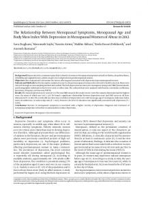
A demographic perspective of disease causation and attendant mortalities over the life span is provided by Figure 1. Several investigators (11-14) have offered interpretations of this bi-modal plot. They are all in agreement that the trough, roughly covering the span between ages 15-30, represents the period during which evolution has endowed the human species with its maximum reproductive capacity, and with the maximum capacityof its defense mechanismsagainst aging and disease. Although Burnet (11) does not take into account the diseases of early age-at-onset, Childs and Scriver (12) interpret these diseasesto be of inherited or congenitalorigin. Interpretations differ, however, as to the meaning of the ascending curve. Burnet (11) posits that as the defenses progressivelyweakenin subsequentyears,intrinsically generatedmutations accumulate and give rise to diseases with a mortality rate essentially in compliance with theGompertz plot, whereasChilds and Scriver(12) regard the diseases of this segment to be of extrinsicorigin, withinheritedgenesplayinga progressively diminishingrole. Brody and Schneider (13) posit that if the prevalence of a disease does not decline at an advanced age, then that disease derives from biological aging. They show that coronary heart disease complies with this interpretation. Furthermore, Brody (14) provides epidemiological evidence that the age-atonsetof the dementias also complies withthisinterpretation. Takingthese various views into account, the following interpretation (15) of this bi-modal plot has been proposed:
/
/
99' /
$1 ,,
o
10
20
30
~
LOG.
2b
i
Ati
50
60
10
80
ib i milo
Figure 1. The bi-modal plot of mortality versus age with a linear age scale and when the age scale is changed to a logarithmic one to show the approach to the straight line of a Gompertz plot.
Downloaded from https://academic.oup.com/biomedgerontology/article-abstract/54/6/B255/691608/A-View-of-the-Aging-Disease-Relationship-From-Age by guest on 24 August 2017
AGING-DISEASERELATIONSHIPFROMAGE 85
Segment I represents mortalities from diseases of early age-at-onset and of inherited or congenital origin, includingjuvenile neoplasms and the syndromes of precocious aging, some of which exhibit diseases comparable to the aging-relateddiseases oflater life. Segment 2 represents the period of maximum reproductive capacity, of maximum capacity of defense mechanisms, and a nadir in mortalities from disease. Segment 3, the lower half of the ascending curve, represents mortalities from diseases of maturity-ageat-onset deriving from late-acting inherited genes or from those attributableto risk factors. Segment 4, the upper portion of the ascending curve, represents mortalities from diseases of senescenceage-at-onset, and posited to derive from biological aging mechanisms.
THE AMYLoIDOSIS MODEL
As Fogle (16) has written with regard to the genotype-phenotype relationship: "DNA dictates the information necessary to delineate the phenotypic structure, and the cellular machinery is the mode through which it is expressed. In this model, all the information in the phenotype is encoded in the genotype." This concept is in accord with the definition of phenotype as an observable expression of a trait affecting structure, physiology, or behavior that results from activity of RNA or protein molecules transcribed from DNA. But Fogle also observes that small changes in the cellular machinery may alter its ability to make sense of the genetic program. The amyloidosis model provides an example that the same phenotypic lesion can derive from a varietyof pathogeneticpathways.
TheAmyloid Phenotype Amyloid is a proteinaceoussubstancedepositedbetween cells in varioustissuesand organs in a wide varietyof clinical settings, ultimately producing pressure atrophy of the parenchymal cells (17).In a landmark review,Glenner (18) detailed the phenotypic characteristics as displayinga pink-orangecolorationwith congo red stain with conventionallight microscopy, a pink fluorescence with ultraviolet light,and apple green crystalswith polarizedlight On electron micrographs amyloid exhibits branched and unbranchedfibrils of varyinglength,40-120 A in diameter, with the fibrils composedof pairedhelicalfilaments. On x-raycrystallography it displays a betapleatedconfiguration from whichcharacteristicsGlennerhas designated all the amyloids as the beta fibrilloses. TheAmyloid Genotypes The amyloid gene designates and their derivative proteins have been classified as follows: AL-Gamma-type light chain immunoglobulin AA-Nonimmunoglobulin apolipoprotein AF-Familial hereditary amyloidoses (gamma trace, beta protein,and transthyretin variants) AE-Endocrine amyloids (amylin of the islets of Langerhans, precalcitonin of the C cells of the thyroid,prolactinof the pituitary, and atrialnatriuretic peptideof the heart)
B257
Beta-2-Microglobulin in patientson hemodialysis ASc-Senescence-related transthyretin variants and beta proteins To emphasize the diversity of pathogenetic pathways, Saraiva (19) has identified 40 variants of transthyretin linked with the familial and senile amyloidoses. In accord with the prevalence of two or more concomitant diseases in aged subjects, there are also patients with combinations of two or more amyloid types. Nongenetic aging-relatedenhancers such as free radicals and AGE proteins have also been linked with amyloidogenesis (20,21), including the observation that senescence-accelerated mice exhibit an enhanced oxidative stress due to an impaired transport of Cu-Zn superoxide dismutase (22,23). These mice develop a universalaccelerated senile amyloidosis.
The Final Commonality The post-translation folding of proteinsthat providethem with their highly specific three-dimensional configuration has been designated the second half of the genetic code (24). Misfoldings of proteins appear to constitute a commonality of all the amyloids, although they are not identical in their molecular composition. As Tauben (25) notes, when normal protein folding goes awry, incompletely folded protein molecules coalesce and form insoluble fibril aggregates.Disorders attributableto protein misfolding have been designatedconformational diseases(26). Amyloidosis and the Criterionof Universality The criterion of universality has been applied in two ways as an aging-life-terminating mechanism applicable across all species, or as a mechanism limited to all individuals within a species. As to the first, aging itself does not comply with the criterion of universality because not all species exhibit senescence (27), and the application of universality across species does not take into account that as evolutionhas progressed there has been an increasing complexity of biological mechanisms with more opportunities for the latter to go awry, thereby providing a diversityof life-terminatingevents. Death certificates are notoriously unreliable as to causes of death in humans, and although autopsies provide more reliable data, they too have limitations, including a current decline in the autopsy rate, particularly in aged subjects, and even more so in nursing horne patients. Even when autopsies are performed, analyses for the presence of amyloid deposits are generally carried out only when there are clinical indications of an amyloidrelated disease. Nevertheless, a composite of several reports representing several hundred aged subjects (28-30) showed senescence-related (ASc) amyloid atrial deposits in 80-100% of cases. A single study (31) of 100 aged subjects revealed that 91 had amyloid deposits in the heart. Amyloid deposits in the heart, whether of immunoglobulin light chain, transthyretin variants, atrial natriuretic polypeptide (ANP), or of ASc derivation are particularly relevant to cause of death in old age. ANP level is an independent predictor of mortalities in the oldest-old (32). There are two consequences of amyloid deposits in the heart. They can be the cause of a clinically undetected lethal arrhythmia or of heart failure. Deposits of atrial amyloid can also produce a restrictive cardiomyopathy that results in heart failure (33). Deaths from amyloid deposits such as these are often erroneously attributedclinicallyto heart failure from coronary artery
Downloaded from https://academic.oup.com/biomedgerontology/article-abstract/54/6/B255/691608/A-View-of-the-Aging-Disease-Relationship-From-Age by guest on 24 August 2017
B258
BLUMENTHAL
disease or as death from so-called natural causes. Although such observations do not preclude death in the absence of disease at the end of the human life span, they support the conclusion that few of advanced age may be vouchsafed such an end. Amyloidosis and the Bi-Modal Plot Because the life-span distribution of the amyloid genotypes in accord with the bi-modal plot has been previously detailed (15), it is not repeated here. Suffice it to note that the presence of some diseases and their derivative amyloids in both the maturity and senescence onset categories reflects the fact that there is no sharp age demarcation between these two periods of the life span.
THE AMYLOIDOGENIC DEMENTIAS Alzheimer's disease (AD) provides an example of the application of the tenets noted in the abstract to a single clinical and pathological entity. Moreover, AD appears to represent the most prevalent disease of those of advanced age. There are no identifiable risk factors except for head trauma. The life-span distribution of this category of the dementias conforms to the bimodal plot and to the principle of diverse pathogenetic pathways leading to the same endpoint lesion. There is also a strong connection with biological aging, and the amyloidogenic dementias provide an example of a progressively diminishing role of genetic markers with advancing age. As to the life span distribution, the amyloid plaques of AD are present at an early age in the Down syndrome (OS). As discussed below, genetic markers for this disease identified thus far apply mostly to AD of maturity age-at-onset. AD, with its highest prevalence in the oldest-old, appears to have few, if any, genetic markers. As to the diversity of pathogenetic pathways, whereas the amyloid plaques of DS derive from the overproduction of the amyloid precursor protein (APP), which derives from the trisomy 21, the plaques in AD of maturity age-at-onset derive from several mutations of APP or other late-acting genes. As also discussed below, the plaques of AD of senescence age-atonset may derive directly from biological aging phenomena. The lesions of the amyloidogenic dementias consist of the deposits of beta protein plaques in the hippocampus and a cerebral amyloid angiopathy (CAA), the two often takinq place together. But CAA with dementia also occurs in the absence of plaques. The beta protein of CAA has a different amino acid composition from the plaques of AD (34). The principal genetic markers thus far identified in AD are overexpression and mutations of APP, two presenilin gene mutations, and mutations of APOE4. APOE is a 299-amino acid lipid transport protein that plays an important role in plasma cholesterol homeostasis. In humans there are three common alleles designated APOE 2,3, and 4. The three alleles differ by only a single amino acid. The APOEs are highly expressed in the liver and brain. In the brain they are synthesized by astrocytes and some microglia. Mutations of APP and of the two presenilin genes are directly involved in the amyloidogenic process, whereas APOE4 is a risk factor for AD and the most common genetic marker. But the mutations of these genes account for less than 35% of AD cases, and with the exception of DS, they fall mostly in the maturity-onset category, although a few cases are in the senescence-onset group (35). Thus they do not account for the vast
majority of AD cases, nor for the dementias of CAA, both of which occur mostly after age 80. With regard to the life-span distribution of APOE4 in AD cases, a study by Asada and colleagues (36) on 224 demented Japanese subjects over age 90, including 47 centenarians, revealed that the APOE4 allele was present in 38% of the demented subjects under age 60, in 22% of those aged 80-89, and in none of the of the centenarians with dementia in whom the prevalence rate of AD was 70%. Thus, in accord with the progressive disappearance with age of genetic markers, the frequency of the APOE4 allele appears to decline with age and to have no impact on AD in centenarians. As yet, no genetic markers have been identified in AD in centenarians. The observation that random-frameshift or assembly-line mutations in the genetic code (37) result in a build up of beta-protein plaques provides an example of a disease deriving directly from a biological aging phenomenon. As has been noted (38), ''typically Alzheimer's disease has a gradual onset, arising by minute degrees over a period of time. Prevalence and incidence estimates will vary qreatly according to where along a continuum from normality to severe disease one places a diagnostic cut-point between nondisease and disease." But the same conundrum confronts the pathologist because the presence of amyloid plaques in clinically nondemented aged subjects (39) leads to the necessity of subjectively correlating the number and distribution of plaques with the clinical manifestations of AD. CONCLUSION
In accordance with the tenor of the time, when the National Institute of Aging (NIA) was created, its assigned mission was to support the study of biological and other aspects of aging, thereby distinguishing it from the disease-oriented institutes. Its support of AD has been viewed by some as a departure from this mission. It may be that initially the support of AD research was adopted by the NIA as a tactical measure to increase its funding, but as subsequent research has evolved, and as has been demonstrated here, there are valid biological reasons for extending this policy to the support of studies on other agingrelated diseases. The concept of causation of the aging-related diseases proposed here rests on the four tenets noted in the abstract and amplified in the text. They represent a significant departure from the traditional medical model of disease causation on which the aging-disease relationship has heretofore been based. The amyloidotic diseases, including the amyloidotic dementias, have been presented as models of causation of the aging-related diseases. They are particularly appropriate because, for the most part, conventional risk factors do not apply to their pathogenesis, and many of the amyloidogenic diseases are of intrinsic origin. Moreover, in humans, amyloid lesions of the heart come close to complying with the criterion of universality. It remains to be demonstrated that other major aging-related diseases such as vascular disease, cancer, diabetes, hypertension, and osteoporosis also conform to this paradigm-a process in which I am now engaged. The paradigm presented here also has implications for the future planning of our health care system. The demographic projections of the future growth of our elderly population are based largely on current concepts of causation of the aging-re-
Downloaded from https://academic.oup.com/biomedgerontology/article-abstract/54/6/B255/691608/A-View-of-the-Aging-Disease-Relationship-From-Age by guest on 24 August 2017
AGING-DISEASE RELATIONSHIP FROMAGE 85
lated diseases and on preventive and therapeutic strategies based on these concepts. The manipulation of genes associated with these diseases is widely viewed as the most promising approach to new preventive and therapeutic measures. However, if the paradigm presented here is validated for other agingrelated diseases, then a strategy based on genetic manipulation, like the strategy of risk-factor avoidance, although eminently desirable, if successful, would not assure that the same or another aging-related diseases would not emerge in the period of senescence. In this scenario, the future growth of our aged population, and the required changes in our health care system, would be greater than current projections. The importance of conducting research that leads to an understanding of the agingdisease relationship poses a challenge that gerontologists of all stripes should address. ACKNOWLEDGMENTS
Address correspondence to Herman T. Blumenthal, PhD, MD, Department of Community and Family Medicine, St. Louis University School of Medicine, 1402South Grand Blvd., St. Louis, MO 63104.
REFERENCES 1. Shock NW. Physiological aspects of aging. Ann Rev Physiol. 1961;23:97-122. 2. Evans JG. Ageing and disease. In: Everid D, Whalen J, eds. Researchand the Aging Population. Ciba Foundation Symposium No. 134. Chichester, UK: John Wiley and Sons; 1988:38-57. 3. Adelman RC. Definition of biological aging. In Haynes SC, Feinleib M, eds. SecorulConferenceon the EpidemiologyofAging. Nlli Publ. No. 80969. Bethescta,MD: NationalInstitutesof Health; 1980:9-13. 4. Gerhard GS, Cristofalo VJ. The limits of biogerontology. In Sprott RL, Warner HB, Williams TF, eds. The Biology ofAging. New York:Springer; 1993:107-118. 5. Strehler BL. Time, Cells and Aging. 2nd ed. New York:Academic Press: 1977. 6. Martin GM. The genetics of aging. Hosp Practice. 1987;32:47-55. 7. ForbesWF, Gentleman JB. A possible similar pathway between smokinginduced life-shorteningand naturalaging. I Gerontol. 1973;28:302-311. 8. Peds TT. The oldest-old.Sci Am. 1995;272:70-75. 9. Cutler RG. Nature of aging and life maintenanceprocesses.In: Cutler RG ed. CellularAgeing: Conceptsand Mechanisms. Interdisciplinary Topics in Gerontology. von Hahn HF, seriesed. Basel,NY: S. Karger; 1976:83-133. 10. Holliday R. Understanding Ageing. Cambridge, UK: Cambridge UniversityPress; 1995. 11. Burnet FM. Intrinsic Mutagenesis: A Genetic Approach to Aging. New York: John Wiley and Sons; 1974. 12. Childs B, Scriver CR. Age at onset and causes of disease. Perspect Biol Med.1986;29:437-460. 13. Brody JS, Schneider EL. Diseases and disordersof aging: an hypothesis.I ChronDis. 1986;39:871-876. 14. Brody JS. An epidemiologistviews senile dementia-facts and fragments. AmI Epidemiol. 1992;115:155-162. 15. BlumenthalHT, PremachandraBN. Bridgingthe aging-disease dichotomy. I. The amyloidosismodel.PerspectBioIMed. 1990;33:402-420.
B259
16. Fogle, T. Information metaphors and the human genome project. Perspect BioI Med. 1995;38:535-547. 17. Cotran RS, Kumar V, Robbins SL. Pathologic Basis of Disease. 5th ed. Philadelphia:w.B. Saunders, 1994:231-240. 18. Glenner GG. Amyloid deposits and amyloidosis: The beta fibrilloses. N Engl I Med. 1980;302:1285-1291, 1333-1343. 19. Saraiva MJ. Transthyretin mutations in health and disease. Hum Mut. 1995;5:191-196. 20. Harman D. Free radical theory of aging: Effect of free radical inheritor on the mortalityrate in male Laf-1 mice. I Gerontol. 1968;23:476-482. 21. Smith MA, Tanaka S, Rickey PL, et al. Advanced Maillard reaction end products are associated with Alzheimer's disease pathology. Proc Natl Acad Sci USA. 1994;91:5710-5714. 22. Park J-W, Choi C-H, Kim M-S, Chung MH. Oxidative status in senescence-accelerated mice. I GerontolBiol Sci. 1996;51A:B337-B345. 23. Higuchi K, Naiki H, Kitigawa K, et al. AS (SAM) amyloidosis presents universally as systemic age-associated amyloidosis. Virch Arch B Cell Pathol. 1991;60:231-239. 24. Kolata G. Trying to crack the second half of the genetic code. Science. 1986;233:1037-1039. 25. TaubenG. Misfoldingthe way to disease.Science. 1996;271:1493-1495. 26. CarrellRW, LomasDA.Conformational disease. Lancet.1997;350:134-138. 27. Finch CEo Variations in senescence and longevity include the possibility of negligiblesenescence.I GerontolBiol Sci. 1998;53A:B235-B239 28. Pitkanen P,Westermark P, Cornwell GG m. Senile systemic amyloidosis. AmI Pathol. 1984;117:391-399. 29. Cornwell GG Ill, Westermark P, Murdoch W, Pitkanen P. A third distinctive type of age-related cardiovascular amyloid. Am I Pathol. 1982;108: 135-139. 30. Cooper lM. Selective staining of amyloid as a function of amyloid composition and structure.Lab Invest. 1974;31:232-238. 31. Kawamamura S, TakahashiM, Ishuhara T, Uchino E. Incidence and distribution of isolated atrial amyloid: histologic and immunochemical studies of 100 aging hearts.PatholInt. 1995;45:335-342. 32. Knight EL, Kiely DK, Fish LC, et al. Atrial natriureticlevel contributes to a model of future mortality in the oldest old. I Am Geriatr Soc. 1998;46:453-457. 33. Kushwaha SS, Fallon IT, Fuster V. Restrictivecardiomyopathy. N Engl 1. Med. 1997;336:267-276. 34. Prelli F, Castano E, Glenner GG, Frangioni B. Differencesbetween vascular and plaque core myloid. I Neurochem. 1988;51:648-651. 35. Marshall E. The Alzheimer's gene puzzle.Science. 1998;280:1002-1004. 36. Asada T, Yamagatam Z, Kinoshita T, et al. Prevalence of dementia and distributionof APOE alleles in Japanese centenarians:an almost complete survey in YamanushiPrefecture.I Am GeriatrSoc. 19%;44:151-155. 37. van Leewen FW, de Kieljn DPY, van den Jark HH, et al. Frameshift mutants of beta amyloid precursor protein and ubiquitin-Bin Alzheimer and Down patients.Science. 1998;279:242-247. 38. Evans DA. The epidemiology of dementia and Alzheimer's disease: an evolvingfield.I Am GeriatrSoc. 1996;44:1482-1483. 39. von Dras D, BlumenthalHT. Dementia of the aged: disease or atypical accelerated aging? Biopathological and psychological perspectives. I Am Geriatr Soc. 1992;40:285-294.
Received Iuly 7, 1998 Accepted January20, 1999
Downloaded from https://academic.oup.com/biomedgerontology/article-abstract/54/6/B255/691608/A-View-of-the-Aging-Disease-Relationship-From-Age by guest on 24 August 2017