A Wearable Physiological Sensor Suite for Unobtrusive Monitoring of Physiological and Cognitive State Robert Matthews, Neil J. McDonald, Paul Hervieux, Peter J. Turner, and Martin A. Steindorf
Abstract—This paper describes an integrated Physiological Sensor Suite (PSS) based upon QUASAR’s innovative noninvasive bioelectric sensor technologies that will provide, for the first time, a fully integrated, noninvasive methodology for physiological sensing. The PSS currently under development at QUASAR is a state-of-the-art multimodal array of sensors that, along with an ultra-low power personal area wireless network, form a comprehensive body-worn system for real-time monitoring of subject physiology and cognitive status. Applications of the PSS extend from monitoring of military personnel to long-term monitoring of patients diagnosed with cardiac or neurological conditions. Results for side-by-side comparisons between QUASAR’s biosensor technology and conventional wet electrodes are presented. The signal fidelity for bioelectric measurements using QUASAR’s biosensors is comparable to that for wet electrodes.
Q
I. INTRODUCTION
UASAR is currently developing an integrated Physiological Sensor Suite (PSS) for monitoring the physiological and cognitive states in operational (i.e. non-clinical) settings. The PSS described in this paper has been designed to be wearable and unobtrusive, with an emphasis on the capability of long-term monitoring of physiological signals. These factors are of considerable importance in operational settings where high end-user compliance is required. The opportunities for measuring physiological variables have been recognized by two recent major programs: the Defense Advanced Research Projects Agency’s (DARPA) Augmented Cognition (AugCog) program [1] and the U.S. Army’s Warfighter Physiological Status Monitor WPSM program [2]. However, those programs were limited in the number of physiological variables they could simultaneously measure and by inadequate development of fully deployable noninvasive sensors and a full complement of integrated sensors. In order to be truly unobtrusive, the PSS (including the sensors) should be donned or doffed quickly by the wearer,
Manuscript received April 16, 2007. This work was supported in part by the U.S. Department of Defense. R. Matthews is President of QUASAR, San Diego, CA 92121 USA (e-mail:
[email protected]). N. J. McDonald is with QUASAR, San Diego, CA 92121 USA. (e-mail:
[email protected]). P. Hervieux is with QUASAR, San Diego, CA 92121 USA. (e-mail:
[email protected]). P. J. Turner is with QUASAR, San Diego, CA 92121 USA. (e-mail:
[email protected]). M. A. Steindorf is with QUASAR, San Diego, CA 92121 USA. (e-mail:
[email protected]).
be easy to use, and require no skin preparation for the sensors to operate with sufficient fidelity. Furthermore, in order to be deployed in operational settings, the electrodes must operate for extended periods with no maintenance and no discomfort for the user. The electronics used to digitize and record the signals need to be small and lightweight, and operate for a minimum of 24 hours using small batteries that are easy to obtain. Finally, the motion of the user during normal activities must not be impeded by the presence of the PSS. The PSS is based on two revolutionary noninvasive bioelectric sensor technologies pioneered by QUASAR: a capacitive bioelectrode capable of through-clothing measurements of electrocardiograms (ECG) [3], [4], and measurements of electromyograms (EMG) and electrooculograms (EOG); and a hybrid (capacitive/resistive) bioelectrode capable of measurements of through-hair electroencephalograms (EEG) [4]. No modification of the skin’s outer layer is required for the operation of either sensor technology. This represents a considerable advantage with respect to conventional electrode technology, which requires the use of conductive pastes or gels, often with abrasive skin preparation of the electrode site. The biosensor technologies are comfortable, easy to use and can be incorporated into items worn by the subject [3], [5]. These sensors are combined with miniature, ultra-low power microprocessor-controlled multichannel data acquisition (DAQ) units and miniaturized wireless transceiver (WT) units that will allow data acquisition to be done close to the locations of the electrodes [5], [6]. Each physiological measurement (e.g. ECG, EEG) is performed by a single module (comprising sensors, DAQ and WT units) that communicates wirelessly to a data logger worn by the subject. The small size and light weight of the DAQ and WT units minimize the impact upon the user. Furthermore, this approach reduces the amount of wiring in the system, thereby improving the mobility of the subject without a consequent increase in signal artifact. The modular nature of the PSS modular approach additionally provides a convenient method for the integration of commercial-off-the-shelf (COTS) sensor technologies for the measurement of other physiological parameters, such as accelerometers for body position/acceleration, thermometers for body temperature, or pulse oximeters for blood oxygenation. There are applications for the PSS in both the military and
civilian spheres. Military applications include monitoring of physiological states for Dismounted Infantrymen, or cognitive state monitoring for Command & Control personnel. Civilian applications include outpatient monitoring of patients diagnosed with cardiac or neurological conditions, or computer interfaces for gaming. This paper includes a description of both sensor technologies and the ancillary electronics required for implementation as part of the PSS. The results described herein include physiological measurements using each of QUASAR’s sensor technologies. The physiological measurements (ECG, EEG, EOG, EMG) have been included because of their value in monitoring physiological and cognitive states.
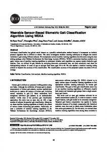
enabling through-hair measurements of EEG without any skin preparation. The hybrid biosensor contacts the skin with a set of ‘fingers,’ each of which is small enough to reach through hair without trapping hair beneath the finger (thereby preventing electrical contact to the scalp). This sensor is also suitable for measurements of EMG through facial hair or stubble.
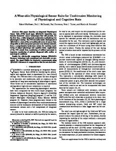
Fig. 1 QUASAR capacitive biosensor with 10c coins for scale.
II. HARDWARE The individual system components for the PSS are described in this section. The capacitive and hybrid biosensor technologies are discussed separately. QUASAR’s proprietary common mode follower (CMF) technology and innovative signal processing techniques are used to reduce pickup and susceptibility to common-mode signals on the body [3]. The DAQ unit is discussed with particular reference to input noise, common-mode rejection ratio (CMRR) and power supply requirements. Preliminary performance data are presented for the WT. Specific details for the data logging unit are also presented. A. QUASAR Capacitive Biosensors QUASAR has recently developed novel capacitive biosensors, which possess sufficient signal fidelity for operational recordings of ECG, EOG, and EMG signals. The QUASAR sensors couple to the electric potential of the body capacitively, requiring no skin preparation or direct electrical contact with the skin. QUASAR’s capacitive sensors are in fact capable of measuring bioelectric signals through several layers of fabric [7]. Detection of human bioelectric signals using purely capacitive coupling was first developed in 1967 and patented in 1970 [8], [9]. Unlike prior methods, QUASAR’s capacitive biosensors (Fig. 1) can tolerate very small capacitances to the source, which enables the new electrodes to be operated at a standoff from the skin (up to several millimeters in practice). In contrast, the susceptibility of prior capacitive electrodes to variations in the electrode-subject spacing was such that it was necessary that the electrodes were held tightly against the skin. Consequently, they are little different in terms of operational use from conventional resistive contact electrodes. B. QUASAR Hybrid Biosensors Measuring EEG using capacitive bioelectrodes is problematic when operating through hair because the sensors’ high impedance renders them susceptible to triboelectric charge generation due to rubbing between the sensor and hair. The hybrid biosensor (Fig. 2) uses a combination of high impedance resistive and capacitive contact to the scalp, thereby
Fig. 2 QUASAR hybrid EEG biosensor.
The contact impedance between the scalp and each finger can be as high 107 Ω. Therefore the amplifier electronics are shielded and placed as close as possible to the electrode in order to limit interference caused by the pickup of external signals. C. Common-Mode Follower (CMF) Technology QUASAR’s high impedance sensors are used in combination with QUASAR’s proprietary CMF technology. The CMF is a separate biosensor that is used to reduce the sensitivity of the biosensors to common mode signals on the body. It operates by measuring the potential of the body relative to the ground of the amplifier system. The ultra-high input impedance of the CMF (~1012 Ω) ensures that the output of the CMF tracks the body-ground potential with a high degree of accuracy. The output of the CMF is then used as a reference for bioelectric measurements by QUASAR biosensors. In this way, the common-mode signal appearing on the body is dynamically removed from the measurement. This typically achieves a common-mode rejection ratio (CMRR) of 50 to 70 dB. D. Miniature Low Power Data Acquisition (DAQ) The DAQ units have been designed to address the general requirements for multichannel ECG, EEG, EOG, and EMG data acquisition. The prototype board in Fig. 3 is a multichannel data acquisition system that can simultaneously acquire up to 10 channels of EEG data with 16-bit resolution. However, PSS modules requiring fewer channels of physiological data will be smaller than the prototype in Fig. 3.
logger is further configurable to communicate with external systems via wireless (specifically 802.11a,b or g; Bluetooth), or via Ethernet, USB 2.0 or RS-232. In this manner, physiological or cognitive status can be updated in real time, or data can be stored for download at a later date. Fig. 3 Prototype Data Acquisition board with credit card for scale.
Fig. 4 Prototype miniature wireless transmitter
In environments with high levels of electromagnetic interference, the resulting common-mode signals may not be completely removed by the CMF. Therefore, the DAQ has been designed to have a high CMRR between channels. Each analog channel has been matched to better than -72 dB below 50 Hz. Data acquisition is performed by 16-bit sigma-delta digital-to-analog converters (ADC). The input noise of the DAQ channels has been measured to be 400 nV/√Hz for a sampling frequency of 4 kHz. The timing error between ADC channels is less than 1 μs (i.e. a phase error less than -80 dB below 100 Hz). Aliasing of out-of-bandwidth signals is less than -80 dB below 50 Hz. The total harmonic distortion is less than -75 dB at 35 Hz. In order to conserve power, the microprocessor operates in a low-power “sleep” mode when not acquiring data. Power consumption for a PSS module is dominated in approximately equal parts by the DAQ and WT (see below). Current estimates show that the run time for a 10-channel EEG module is in excess of 72 hours from 2 AAA batteries. E. Miniature Low Power Wireless Transceiver (WT) The prototype wireless transceiver shown in Fig. 4 uses a Gaussian Frequency Shift Keying (GFSK) protocol and has the ability to use 125 channels of the RF range starting from 2.400 GHz up to 2.525 GHz of the Worldwide ISM band. In order to conserve power, the wireless transceiver transmits information in a data “burst” mode. Wireless data rates up to 2.5 ksamples per second have been achieved using the current prototype operating in the low power mode. F. Data Logger The prototype data logger has been designed using a similar approach to the DAQ and WT units. It is a small, low power microprocessor-based unit that communicates wirelessly with each module and is worn on the subject’s hip or carried in a backpack. The data logger can be configured to do either of the following with physiological data received from the sensor modules: process data for identification of physiological/cognitive status, or write data to FLASH RAM. The data
III. PHYSIOLOGICAL MEASUREMENTS Each of the bioelectric signals measured by the PSS, namely ECG, EEG, EOG and EMG, possesses a utility across a variety of applications. For example, the morphology of the ECG signal during long-term monitoring can be used to identify patients with increased risk of cardiac disease, or in evaluating the success of antiarrhythmic drug therapy. Heart rate derived from the ECG is an indicator as to whether a subject experiences life- or health-threatening levels of physiological strain. The heart rate variability (HRV) is currently used to provide information about a subject’s cognitive state. The EEG is most frequently used in clinical settings for monitoring and diagnosis of epilepsy and subjects with sleep disorders. The PSS provides an opportunity to extend measurements of the EEG to non-clinical settings, where monitoring for epilepsy diagnosis or sleep disorders can be performed on an outpatient basis. Additionally the EEG can be used in operational settings, in which knowledge of an individual’s cognitive state can be used to predict impending cognitive failure and trigger appropriate countermeasures [10], [11]. The main applications for the EOG are in opthalmological diagnosis and recording eye movements. Eye movements and an estimation of blink rate are related to the level of fatigue or sleepiness of the individual [12], [13], and can be used in the determination of cognitive state. The EOG is also used for the removal of artifact signals in EEG signals. The EMG can be used for monitoring the physiological state of the subject such as muscle fatigue and shivering [14], and is also used in clinical sleep studies. Additionally, chin EMG signals are used as indicators of stress. A. ECG Results for QUASAR’s capacitive biosensors measuring ECG have been presented elsewhere [3]. These results demonstrated the biosensors’ ability to measure through clothing ECG. Furthermore, a comparison between QUASAR biosensors and standard pre-gelled ECG electrodes were, in the opinion of a cardiologist who analyzed the data, “at least equivalent, and often superior to the standard wet electrodes. The morphologies of the individual components (P, QRS and T waves) were easily recognized and very similar with the two sets of electrodes” [15]. A clinical trial was subsequently conducted to identify skin compatibility issues for the capacitive sensors and also to quantify ECG signal quality for sensors worn for extended periods. A total of six subjects each wore QUASAR capacitive biosensors mounted in a belt around the torso, as
in Fig. 5, for a period of 30 days. Lead-I ECG data was collected each day with the subject seated (300 seconds per day for each subject) and walking in place (60 seconds per day per subject). The data acquisition system for these measurements was a National Instruments NI-4472 (24-bit, 8channel delta-sigma) card installed in a desktop computer running a LabView-based data acquisition application. The sample frequency of the data was 1200 Hz.
Fig. 5. (left) ECG belt used during clinical trial of QUASAR capacitive biosensors. (right) CMF used during clinical trial. 3
ECG Signal (mV)
2 1 0 -1 4
5
6
7
8
9
45
46
47
Time (s) 3
ECG Signal (mV)
2 1 0 -1 42
43
44 Time (s)
Fig. 6. ECG data from a single subject collected during clinical trial of QUASAR capacitive biosensors. ECG measurements were made while the subject was walking in place. (top) First day of clinical trial. (bottom) 30th day of clinical trial.
ECG signal quality was evaluated using the following criteria: (i) unambiguous representation of rate from QRS complex, (ii) unambiguous identification of T-wave, (iii) identification of QRS configuration and QRS duration, (iv) detection of P-waves and their unambiguous association with QRS complex, (v) determination of rhythm from QRS configuration and P-wave association with QRS complex, and (vi) identification of QT interval. A cardiologist ranked ten second segments of ECG data as: Excellent (i to vi), High (i to v), Medium (i to iii), Low (i to ii), Poor (i only) and Unusable (unable to identify rate). An example of ECG data for a subject walking in place is presented in Fig. 6. No degradation in signal quality is evident in the ECG signal between the 1st day of the trial and the end of the trial. This is reflected in the scores assigned
by the cardiologist to the data: 98.96% of the seated data were classified as Excellent, and none were classified as worse than Medium; 81.17% of the walking data were classified as Excellent, and the remainder were classified as Medium. However, the walking results are adversely affected by a single subject that possessed a barely discernible P-wave in the absence of any artifact signal, and therefore was particularly susceptible to lower clinical scores while the subject was moving. At least 90% of the ECG segments for each of the other subjects were classified as Excellent. No skin compatibility issues with the QUASAR biosensors were observed during the period of the clinical trial. B. EEG The measurements described in this section were performed in order to achieve the following: (a) characterize the performance of QUASAR’s new hybrid bioelectrode technology with respect to conventional ‘wet’ EEG electrode technology, and (b) characterize the noise of the hybrid bioelectrodes on unprepared skin. The results for (a) were obtained using a procedure similar to that used to obtain results for QUASAR’s hybrid biosensors that have been reported elsewhere [4], [5]. Six channels of EEG were recorded simultaneously: three hybrid biosensors and three ‘wet’ electrodes. The hybrid electrodes were positioned at the nominal Cz, Fz and F3 positions, and the wet electrodes were positioned 20 mm anterior to the hybrid electrodes (Fig. 7). Preparation of the scalp for the wet electrodes included abrasion with Nu-Prep, followed by cleaning with alcohol and then application of Grass EC2 electrode paste. No preparation of the scalp was performed at the QUASAR electrode sites. The CMF was placed on the right earlobe of the subject and the ground for the ‘wet’ electrodes was a standard pre-gelled disposable Ag-AgCl electrode placed upon a prepared site on the mid-posterior aspect of the subject’s right pinna (Fig. 7). The results for (b) were obtained by placing two hybrid bioelectrodes on unprepared sites on the subject’s scalp. The electrodes were positioned side-by-side (spacing approximately 20 mm) in order to minimize the differential EEG signal detected by each electrode. The data acquisition system for both measurements was a National Instruments NI-4472 (24-bit, 8-channel deltasigma) card installed in a desktop computer running a Lab-
Fig. 7. (left) Cap for simple deployment of hybrid biosensors for EEG measurements. Conventional wet electrodes are positioned anterior to the hybrid sensors (for side-by-side comparisons with hybrid sensors). (right) CMF used during EEG measurements.
sample frequency of the data was 1200 Hz.
100 Hybrid Cz
Wet Cz
EEG Signal (uV)
50
0
-50
-100 197
198
199
200
201
Time (s)
Fig. 8. Comparison of alpha wave EEG measured using QUASAR’s hybrid biosensors and conventional wet electrodes. Measurements were made at the Cz location.
Fig. 10. Glasses with integrated QUASAR biosensors for measurement of EOG. 500 QUASAR electrodes Wet electrodes
400 300 EOG Signal (uV)
30 20
Signal (µV)
10 0
200 100 0 -100 -200 -300
-10
-400 -20
-500 19
-30
20
21
22
23
Time (s)
-40 50
51 Still 10.6 µVp-p
52
53
Walking 10.0 µVp-p
54
55
Running 34.6 µVp-p
56
57
58
59
60s Time (s)
Fig. 9. Measured sensor-sensor noise levels for hybrid bioelectrodes under subject motion.
View-based data acquisition application. The sample frequency of the data was 1200 Hz. Results for alpha activity at the Cz location are presented in Fig. 8. The data have been filtered between 5 and 15 Hz using a 12th order Bessel bandpass filter. The signals from both sensor technologies are highly similar, with an average correlation coefficient of 88.5% for the segment shown in Fig. 8. The traces in Fig. 9 are the differential output of the two hybrid sensors. The data have been filtered between 1 and 30 Hz using a 12th order Bessel bandpass filter. The quoted noise figures correspond to peak-to-peak voltage fluctuations that lie within 1.65 standard deviations (90%) of the mean voltage. The results for subject still and subject walking are more than an order of magnitude greater than sensor noise in this bandwidth, and therefore represent skin noise on unprepared skin. The noise for subject running is considerably higher than for less vigorous motion, and is dominated by signals appearing at the frequency of the foot strikes (3 Hz). C. EOG The EOG measurements described in this section were taken using QUASAR capacitive biosensors integrated into the glasses shown in Fig. 10. No preparation of the electrode sites was performed was performed. Data were simultaneously collected using standard pre-gelled disposable AgAgCl electrodes positioned as close as possible to the QUASAR biosensors. The data acquisition system for these measurements was a National Instruments NI-4472 (24-bit, 8 channel delta-sigma) card installed in a desktop computer running a LabView-based data acquisition application. The
Fig. 11. EOG blinking data collected using the glasses in Fig. 10. Data simultaneously acquired using Ag-AgCl electrodes are included for comparison.
Results for the subject blinking are presented in Fig. 11. The data are presented as the difference between the outputs of the two electrodes. The data have been filtered between 1 and 10 Hz using a 12th order Bessel bandpass filter. The structure for the EOG signals in both electrode technologies are similar. The signal-to-noise ratio is more than sufficient for determination of blink rate, or for artifact correction in EEG data. D. EMG The EMG measurements described in this section were taken using QUASAR capacitive biosensors positioned on the subject’s bicep, as in Fig. 12. The spacing between sensors was 50 mm. Data were simultaneously collected using standard pre-gelled disposable Ag-AgCl electrodes positioned as close as possible to the QUASAR biosensors. No preparation of the electrode sites was performed. The data acquisition system for these measurements was a National Instruments NI-4472 (24-bit, 8-channel delta-sigma) card installed in a desktop computer running a LabView-based data acquisition application. The sample frequency of the
Fig. 12. QUASAR capacitive biosensors positioned for measurement
spheres. Measurements of ECG, EOG and EMG using QUASAR’s capacitive biosensors, and measurements of EEG using QUASAR’s hybrid biosensors, have been presented. For each physiological variable, a comparison with conventional wet electrode technology has demonstrated that QUASAR’s biosensors provide data of similar quality.
4
EMG Signal (mV)
3 2 1 QUASAR electrode Wet electrode
0 -1 -2 -3 -4
5
10
15
20
ACKNOWLEDGMENT
25
Time (s)
Fig. 13. EMG data measured using QUASAR’s capacitive biosensors for different levels of muscle activity.
acquisition system for these measurements was a National Instruments NI-4472 (24-bit, 8-channel delta-sigma) card installed in a desktop computer running a LabView-based data acquisition application. The sample frequency of the data was 1200 Hz. A comparison of the EMG signals for a QUASAR electrode and the corresponding Ag-AgCl electrode is presented in Fig. 13 for three levels of muscle effort (relaxed, moderate muscle contraction, strong muscle contraction). The data have been filtered between 20 and 200 Hz using a 12th order Bessel bandpass filter. The low level of EMG activity before 10 seconds and after 20 seconds corresponds to the subject’s bicep in a relaxed state, whereas the intervals between 10 seconds and 15 seconds and 15 seconds and 20 seconds correspond to the subject’s bicep undergoing moderate and strong muscle contraction, respectively. The data for both sensors indicate a clear increase in EMG activity as the muscle effort is increased. The relative changes in muscle activity are similar in both electrode technologies.
The authors would like to thank P. H Chou at the University of California, Irvine, for his assistance with the development if the wireless transceiver technology. REFERENCES [1]
[2] [3]
[4]
[5]
[6]
IV. CONCLUSION This paper has described system components for a Physiological Sensor Suite (PSS) that is currently under development at QUASAR. The PSS is a state-of-the-art multimodal array of sensors using QUASAR’s innovative noninvasive sensor technologies for bioelectric measurements, and is readily expandable to include additional sensor technologies. Physiological measurements are made by individual system modules which communicate wirelessly with a personal data logger, thereby forming a comprehensive body-worn system for real-time monitoring of subject physiology and cognitive status. The range of applications for the PSS is increased by the modular nature of its design. In applications such as the military’s Dismounted Infantryman, the full sensor suite may be employed, whereas the suite may be limited to a single module for outpatient monitoring of patients diagnosed with cardiac (ECG) or neurological (EEG) conditions. Furthermore, the noninvasive nature of the system (i.e. zero skin preparation, minimal wiring) permits greater subject motion and will improve user compliance for the system. The physiological variables described in this paper (ECG, EEG, EOG and EMG) were selected for their utility in a variety of applications in both the military and civilian
[7]
[8] [9] [10] [11]
[12] [13] [14] [15]
M. St. John, D. A. Kobus, J.G. Morrison, and D. Schmorrow, “Overview of the DARPA Augmented Cognition Technical Integration Experiment,” International Journal of Human-Computer Interaction, vol. 17, pp. 131-149, 2004. R. W. Hoyt, J. Reifman, T. S. Coster, and M. J. Buller, “Combat medical informatics: present and future,” Proc. AMIA Symp., pp. 335339, 1993. R. Matthews, N. J. McDonald, I. Fridman, P. Hervieux, and T. Nielsen, “The invisible electrode – zero prep time, ultra low capacitive sensing,” presented at the 11th International Conference on Human Computer Interaction (HCII), Las Vegas, NV, July 22-27, 2005. R. Matthews, N. J. McDonald, H. Anumula, L. J. Trejo, “Novel Hybrid Sensors for Unobtrusive Recording of Human Biopotentials,” in Foundations of Augmented Cognition, 2nd ed., San Ramon, 2006, pp. 91-101. R. Matthews, N. J. McDonald, H. Anumula, J. Woodward, P.J. Turner, M. A. Steindorf, K. Chang, and J. M. Pendleton, “Novel hybrid bioelectrodes for ambulatory zero-prep EEG measurements using multichannel wireless EEG system,” to be presented at the 12th International Conference on Human Computer Interaction (HCII), Beijing, July 2227, 2007. C. Park, P. H. Chou, Y. Bai, R. Matthews, and A. D. Hibbs, “An Ultra-Wearable, Wireless, Low Power ECG Monitoring System,” in Proc. IEEE BioCAS, Nov. 29 – Dec. 1, 2006. The British Library, London. R. Matthews, M. A. Krupka, C. Say, and A. D. Hibbs, “Capacitively coupled non-contact sensors for measurement of human bioelectric signals,” presented at Advanced Technology Applications for Combat Casualty Care (ATACCC), Ft. Walton Beach, FL, September 9-14, 2001. P. C. Richardson, “The insulated electrode: A pasteless electrocardiographic technique,” Proc. Annu. Conf. Eng. Med. Biol., vol. 7, pp. 915, 1968. P. C. Richardson, and A. Lopez, Jr., “Electrocardiographic and bioelectric capacitive electrode,” U.S.Patent 3,500,823. M. St. John, D. A. Kobus, and J. G. Morrison, “DARPA Augmented Cognition Technical Integration Experiment (TIE),” SPAWAR Systems Center, San Diego, CA, Tech. Rep. 1905, December 2003. R. Matthews, N.J. McDonald, L.J. Trejo, “Psycho-physiological sensor techniques: an overview,” presented at the 11th International Conference on Human Computer Interaction (HCII), Las Vegas, NV, July 22-27, 2005. K. Kaneko and K. Sakamoto, “Spontaneous blinks as a criterion of visual fatigue during prolonged work on visual display terminals,” Percept. Mot. Skills, vol. 92, pp. 234-50, 2001. S. K. L. Lal and C. Ashley, “Reproducibility of the spectral components of the electroencephalogram during driver fatigue,” Int. J. Psychophysiol., vol. 55, pp. 137-143, 2005. D. G. Bell, P. Tikuisis, & I. Jacobs, “Relative intensity of muscular contraction during muscle shivering,” J. Appl. Physiol., v 72, 2336-42, 1992. W.A. Scott, Professor of Pediatrics/Cardiology, University of Texas, TX, private communication, 2004.