Activation of rat brain ornithine decarboxylase by GTP. Pekka T. KILPELAINEN and Oili A. HIETALA*. Department of Biochemistry, University of Oulu, FIN-90570 ...
577
Biochem. J. (1994) 300, 577-582 (Printed in Great Britain)
Activation of rat brain ornithine decarboxylase by GTP Pekka T. KILPELAINEN and Oili A. HIETALA* Department of Biochemistry, University of Oulu, FIN-90570 Oulu, Finland
The activity of ornithine decarboxylase (ODC) measured in different regions of rat brain was highest in the hippocampus and lowest in the cerebellum. The ODC activity of a crude extract of the cerebellum was increased by the addition of GTP to the enzyme assay. Following dissociation of the ODC-antizyme complex by Sephadex G-75 chromatography in buffer containing 0.25 M NaCl, the GTP-activatable ODC was found in every brain region analysed. This GTP-activatable brain ODC has greater affinity for antizyme than the non-GTP-activatable brain ODC or the kidney ODC. The irreversible inhibitor of ODC, adifluoromethylornithine (DFMO), inhibited approx. 60 % of the ODC activity of all brain regions, whereas kidney ODC was inhibited totally by DFMO. When extracts of brain and kidney
were incubated at 55 °C, kidney ODC was rapidly inactivated, but brain ODC was more heat-stable. Brain ODC, but not kidney ODC, was activated by GTP and ATP, and also by their deoxy forms. The KIf2 for activation of the enzyme was 2 ,uM for GTP and 40 ,uM for ATP. Using partially purified brain ODC, the activation by GTP was irreversible. These results demonstrate for the first time that the GTP-activatable ODC exists in the brain and is associated with the antizyme. The possible mechanisms of activation by GTP, the significance of this finding for the regulation of brain ODC, and the similarities to and differences from the GTP-activatable ODC found in certain rodent and human tumours are all discussed.
INTRODUCTION
EXPERIMENTAL Preparation of extracts Sprague-Dawley male rats were killed by decapitation. The brains were carefully removed and frozen at -70 °C for 1 h. Dissections were performed on an ice-cooled glass plate by the method of Glawinski and Iversen [29]. Five regions (cerebellum, cortex, hippocampus, hypothalamus and 'midbrain') were stored at -70 °C until used. The brain regions were homogenized in 3 vol. of cold 25 mM Tris/HCl buffer, pH 7.4, containing 0.1 mM EDTA, 5 mM dithiothreitol (DTT) and 0.1 mM pyridoxal 5'phosphate (PLP). The homogenates were centrifuged at 105000 g for 1 h at 4 'C. After double (NH4)2SO4 precipitation (20 %60%) the pellets were dissolved in 0.1 M Tris/HCl buffer, pH 7.1, containing 4 mM EDTA, 4 mM DTT and 0.4 mM PLP.
Ornithine decarboxylase (ODC; EC 4.1.1.17) is a rate-limiting enzyme in the biosynthesis of polyamines in mammalian cells [1-4]. Its activity in the adult mouse brain is very low compared with that in other tissues [5-8]. However, brain contains a high amount of immunoreactive enzyme protein [9]. This apparent contradiction can be explained by the fact that the brain cytosol fraction is rich in ODC-antizyme complex [7], which also displays antigenicity [9,10]. The antizyme inhibits the enzyme by noncovalent binding [11] and mediates polyamine-induced acceleration of ODC degradation [12-17]. The ODC-antizyme complex can be dissociated by treatment with high salt concentrations, and at least part of the enzyme is released as catalytically active
[7,18-22]. DL-a-Difluoromethylornithine (DFMO) is a potent irreversible inhibitor of mammalian ODC [23,24]. However, it has been reported that mouse brain ODC could not be inhibited by doses of DFMO that did not result in side effects [8], and that the basal activity of ODC in the neocortex and the cerebellum of the rat was resistant to DFMO in vivo [25]. Also, the ODC of cutaneous tumours of both mouse and human origin has been reported to be resistant to irreversible inactivation by DFMO [26,27]. In addition, the activity of the ODC isoform found in tumours was increased by the addition of GTP to the enzyme assay. Therefore we decided to determine whether the rat brain also contains the GTP-activatable isoform of ODC. Such an activation has not been described for ODC of normal rodent tissues, but nucleotides are known to activate some bacterial ODCs [28]. In the present study, we demonstrate that a GTP-activatable ODC activity is found in rat brain. This ODC form is associated to a large extent with antizyme and has properties which are comparable with those of an ODC isoform found in tumours, such as GTP activation, sensitivity to heat inactivation and relative insensitivity to inhibition by DFMO. It differs from the tumour isoform by its ability to be activated by ATP.
Chromatographic procedures The dissolved pellets were treated with 250 mM NaCl and applied to a Sephadex G-75 Superfine column (1.6 cm x 21 cm) to separate ODC and ODC antizyme. The column was equilibrated with 0.1 M Tris/HCl buffer, pH 7.1, containing 4 mM EDTA, 4 mM DTT and 0.4 mM PLP, essentially according to McCann et al. [20], and eluted with the same buffer containing 250 mM NaCl at a flow rate of 10 ml/h; 1 ml fractions were collected. NaCl, which decreases ODC activity in assay mixture [30], was removed from fractions using a Sephadex G-25 column (PD-10) before ODC and antizyme assays. Fraction number 0 indicates the end of the void volume of the G-75 column.
ODC and antizyme assays ODC activity was determined by measurement of evolved 14CO2 from [1-14C]ornithine essentially as described by Janne and Williams-Ashman [31]. The reaction mixture contained 12.5 mM Tris/HCl buffer (pH 7.3), 1 mM EDTA, 2.5 mM DTT, 0.2 mM PLP and 0.05-1 mM L-ornithine (1.25 nmol of [14C]ornithine) in
Abbreviations used: ODC, ornithine decarboxylase; DFMO, DL-a-difluoromethylornithine; DTT, dithiothreitol; PLP, pyridoxal 5'-phosphate; PMSF, phenylmethanesulphonyl fluoride. * To whom correspondence should be addressed.
P. T. Kilpelainen and 0. A. Hietala
578
Table 1 ODC activty of rat brain regions ODC activity (pmol of C02/60 min per mg of protein) was assayed in the presence and absence of 0.1 mM GTP, and the fold stimulation of enzyme activity by GTP was calculated (+ GTP/-GTP). ODC activities are the means+ S.D. of 2-4 determinations.
Cerebellum
Cortex
-GTP Cytosol Dialysed (NH4)2SO4 Sephadex G-75 Superfine column eluate Fraction having the greatest stimulation by GTP
+ GTP/-GTP
+ GTP
1.49 + 0.07 2.96 + 0.11 1.99 2.57 + 0.76 4.52 + 0.28 1.76 8.27 + 0.71 21.80 + 2.32 2.64 4.84 + 0.46 20.44 + 1.41
4.22
Hypothalamus
80
-GTP
+ GTP
+ GTP/-GTP
-GTP
+ GTP/-GTP
+ GTP
5.48 + 0.37 6.37 + 0.47 1.16 9.94 + 2.89 12.96 + 3.24 1.30 15.96 + 1.98 33.0 + 3.03 2.07
6.79 + 0.64 9.61 + 0.89 1.42 5.32 + 1.32 4.73 + 2.45 0.89 72.46 + 8.58 94.84 + 4.11 1.31
5.22 + 1.13 13.07 + 0.95 2.50
24.00 + 2.83 58.32 + 6.87 2.43
Midbrain
-GTP
Cytosol Dialysed (NH4)2SO4 Sephadex G-75 Superfine column eluate Fraction having the greatest stimulation by GTP
Hippocampus
+ GTP/-GTP
+ GTP
+ GTP
-GTP
+ GTP/-GTP
11.20+1.32 12.98+1.53 1.16 23.88 + 1.55 26.14 + 0.38 1.09 18.47 + 2.13 36.50 + 0.68 1.98
6.64+0.78 7.50+0.77 1.13 21.24 + 1.80 21.73 + 5.50 1.02 21.12 + 3.04 39.85 + 6.43 1.89
22.66 + 2.67 45.97 + 5.07 2.03
15.55 + 1.76 41.60 + 4.10 2.68
80 - Hypothalamus
80
-
60
60
-
60
-
40
40-
40
-
LO0)
20-
20
20 -
0 C
O
Cerebellum
Cortex
* -GTP o +GTP
._n 0,
0-
Co
0
0
2
4
.
.
6
8
0
1.
10
0
2
4
6
8
10
4
6
8
10
0
2
4
6
8
10
0
o 240
120- Midbrain
-
E Q
j
180
-
90-
as
2
-
60
._
0 a
30
60
0
0-
0
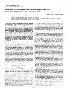
Figure
1
2
4
6
8
10 Fraction
Sephadex G-75 Superfine chromatography
0
2
no.
of ODC from rat brain regms
The crude extracts derived from different rat brain regions were chromatographed as described in the Experimental section. Fractions were assayed in the presence (C1) or absence (U) of 0.1 mM GTP. The results in each panel are the means of duplicate enzyme determinations from 2-4 different experiments.
GTP-activatable rat brain ornithine decarboxylase the kinetic analyses; otherwise the standard L-ornithine concentration was 15 ,tM. Kinetic parameters were determined from Lineweaver-Burk reciprocal plots. To determine the sensitivity to heat inactivation, the extracts were incubated at 55 °C in the presence or absence of 0.1 mM GTP. Aliquots were removed at the indicated times to precooled tubes at 0 °C and subsequently assayed for ODC activity. To measure the effect of DFMO inhibition the samples were preincubated with either L-ornithine (1.25 mM) or DFMO (2.5 mM) for 1 h in 12.5 mM Tris/HCl buffer, pH 7.4, containing 2.5 mM DTT, 0.2 mM PLP and 1 mM EDTA in the presence or absence of 0.1 mM GTP. The incubation mixtures were then dialysed overnight against a 2000fold excess of the same buffer containing 0.5 mM phenylmethanesulphonyl fluoride (PMSF) and 0.020% Brij and assayed for ODC activity both in the presence and in the absence of GTP. The ODC antizyme activities were determined essentially as described by Fong et al. [32]. The principle of the assay is to mix the antizyme sample with an ODC preparation of known activity, and to compare the observed enzyme activity in the mixture with the expected value. The ODC preparation used was a 105000 g supernatant prepared from male mouse kidney or rat brain ODC purified by Sephadex G-75 chromatography.
Table 2 Effect of DFMO on rat brain and kidney ODC activities The samples were preincubated with either L-ornithine (1.25 mM) or DFMO (2.5 mM) for 1 h at 37 OC in 25 mM Tris/HCI, pH 7.4, containing 2.5 mM DTT and 0.1 mM EDTA. The incubation mixtures were then dialysed overnight against a 2000-fold excess of the same buffer containing 0.5 mM PMSF and 0.02% Brij, and assayed for ODC activity in both the presence and the absence of GTP. Samples used were the (NH4)2SO4-precipitated, dialysed extracts for brain region ODCs, and the cytosol for kidney ODC. Inhibition of ODC activity (%) ODC source
-GTP
+ GTP
Cerebellum Cortex Hippocampus Hypothalamus Midbrain Kidney
69 +13 58 +1 65 + 4 61+8 64 + 5 94 + 5
54 +19 54 +1 61+5 65 + 7 64 +13 97 +1
*-GTP
100 - (a)
RESULTS The activity of ODC measured in different regions of rat brain was highest in the hippocampus and lowest in the cerebellum (Table 1). Enzyme assays using cytosol fractions and ammonium sulphate-precipitated dialysates of brain regions in the presence of 0.1 mM GTP indicated that at least cerebellum ODC could be activated by this nucleotide. To further characterize the ODC activities of different brain regions, gel-permeation chromatography in the presence of 0.25 M NaCl was performed in order to dissociate the ODC-antizyme complex and to separate the ODC and the antizyme from each other. As shown in Figure 1, a GTP-activatable ODC activity was found in the eluted fractions from each brain region. The ability of GTP to activate brain ODC was greatest for the enzyme from the cerebellum and least for the ODC from the hippocampus (Table 1). Since it was known from the previous work of Zawia et al. [25] that the basal activity of the ODC in the neocortex and the cerebellum was resistant to inactivation by DFMO, we determined the effect of DFMO on the enzyme activity from the rat brain regions. DFMO inhibited around 60 % of the ODC activity from all brain regions (Table 2). There was no significant difference when activity was assayed in the presence or absence of GTP. As expected, kidney ODC was inhibited totally by DFMO. Because the GTP-activatable ODC could be detected clearly after it had been dissociated from the ODC-antizyme complex, we then asked whether kidney and brain ODCs differed in their sensitivity to the inhibitory protein antizyme. Therefore a constant amount of ODC activity obtained from gel filtration of the cerebrum and from kidney cytosol were mixed with increasing amounts of partially purified brain antizyme and the resulting ODC activity was measured. As shown in Figure 2, there was no difference in the inhibition by antizyme of the kidney ODC in the presence or absence of GTP, although different amounts of ODC activity were used (Figure 2a), whereas the GTP-activatable ODC of the brain seemed to be more sensitive to antizyme inhibition than the non-GTP-activatable ODC (Figure 2b). These data suggest that the GTP-activatable ODC isoform has a greater affinity for antizyme than the non-GTP-activatable ODC isoform.
579
80
a
+GTP
-
o~ 60 r-
60
._
E 40-
(D
o 200 0 100
-
5
10
15
20
25
30
35
(b)
80 6m0
._ E 40 0
o 20 0 0
5
10
15
20
Antizyme (units)
Figure 2 Effect of ODC antizyme on kidney and brain ODCs Rat brain antizyme was added in increasing amounts to a constant amount of ODC activity from kidney (a) and rat cerebrum (b), preincubated for 10 min at 37 °C, and then assayed for ODC activity in the presence (EZ) or absence (-) of 0.1 mM GTP. The amount of ODC activity per tube was for kidney, 23.0 and 452.4 pmol of C02/60 min, and for cerebrum 10.91, 15.06 and 20.27 pmol of C02/60 min assayed in the absence of GTP and 19.63, 31.63 and 39.70 pmol of C02/60 min in the presence of GTP. The antizyme and brain ODCs were from Sephadex chromatography and the kidney ODC from the cytosolic fraction. One antizyme unit inhibits 1 unit (1 pmol of C02) of mouse kidney ODC activity in 1 h. Results are expressed as the ODC activity remaining as a percentage of that in the identical non-incubated aliquots of each extract. Significant differences in the presence and absence of GTP: *P < 0.05, **P < 0.01, ***P < 0.001). mouse
P. T. Kilpelainen and 0. A. Hietala
580 120
100 -
-
E 80
.E
90-
0 a
._
Ei 60E
40'
'a. 20'
CD) o
* GTP a ATP
EE 60-
0, C-
30-
0. . ..
-9 2 6 4 Incubation period (min)
0
8
Figure 3 Sensitivity to heat inactivation of brain and kidney ODCs Aliquots of rat hypothalamic ODC from Sephadex chromatography (O, *) and mouse kidney cytosol (A, A) were incubated at 55 0C in the presence (O., A) or absence (U, A) of 0.1 mM GTP. Aliquots were removed at the indicated times to precooled tubes at 0 0C and subsequently assayed for ODC activity. Results are the means of duplicate enzyme determinations and are expressed as ODC activity remaining as a percentage of that in non-incubated controls.
Table 3 KInetic properties of ODC from rat brain regions Kinetic parameters were determined graphically from Lineweaver-Burk reciprocal plots of data obtained using at least four L-ornithine concentrations (correlation coefficient > 0.9). L-Ornithine Km (mM)
Vm (pmol of C02/ 60 min per mg of
protein)
ODC source
-GTP
+ GTP
-GTP
+ GTP
Cerebellum Cortex Hippocampus Hypothalamus Midbrain
0.058 0.130 0.026 0.089 0.033
0.032 0.052 0.089 0.084 0.096
60.98 84.51 195.65 66.84 86.54
115.85 140.85 478.26 90.91 173.08
Table 4 Effect of nucleotide triphosphates on the ODC activities from midbrain, hypothalamus and kidney Equal amounts of enzyme samples were incubated with the indicated nucleotide at 0.1 mM for 5 min at 0 0C and assayed then for ODC activity. ND, not determined. ODC activity (% of control) Addition
Midbrain
Hypothalamus
Kidney
None ATP dATP CTP GTP dGTP dTTP
100 150 + 3 125 +16 95 + 7 173 +10 178 +14 92 +12
100 158 +16 ND ND 156 +14 195 +15 ND
100 101 +6 103 + 4 ND 96 + 2 102 + 2 105 + 6
To study the possible differences and similarities in brain versus kidney ODCs we determined first the sensitivity of the partially purified brain and kidney enzymes to heat inactivation.
-8
.
.
.
.
.
.
.
.
.
I
.
I
-7 -5 -6 log {([GTPI (M)}
.
I
-4
I.
.
-3
Figure 4 Conc:entratlon-dependence of GTP and ATP activation of rat brain ODC Rat hypothalamic ODC from Sephadex chromatography was preincubated with the indicated concentrations of GTP or ATP for 5 min on ice, and then assayed for ODC activity. Results are the means of duplicate enzyme determinations and are expressed as a percentage of the maximum increment of ODC activity due to the presence of GTP (O) and ATP (a). The arrow indicates the K values.
Kidney ODC lost its activity more rapidly than brain ODC (Figure 3). For example, after 4 min of incubation at 55 °C the kidney ODC retained only few per cent of its activity, whereas brain ODC still retained over 40 % of its activity. Since it is known from previous studies in tumours [26,27,33,34] that GTP decreases the apparent Km values of ODC for Lornithine, we examined the kinetic properties of ODC from different rat brain regions. The effect of GTP on the apparent Km value of the brain enzyme was slight (Table 3), and a reduction in the apparent Km was only found with the cerebellum and cortex enzymes. In contrast, GTP increased the Vmax of the ODC from every brain region (Table 3). The specificity of activation by GTP was tested by incubation of the brain and kidney enzymes with various nucleotide triphosphates, all at 0.1 mM (Table 4). As reported for mammalian ODCs [36,37] the enzyme from kidney was not affected by any of the nucleotides tested (Table 4). However, the ODC of midbrain and hypothalamus was stimulated 1.5-2-fold by GTP, dGTP and ATP, and slightly by dATP (Table 4). The pyrimidine nucleotides tested (CTP, dTTP) were inactive. The KB values for GTP and ATP for activation of hypothalamic ODC were approximately 2 ,uM and 40 ,uM respectively (Figure 4), indicating that GTP is favoured over ATP in activation of brain ODC. When exposed to GTP, the activation of the brain ODC was essentially irreversible after extensive dialysis (Table 5).
DISCUSSION Mammalian ODC from a normal tissue has never been reported, to our knowledge, to be affected by nucleotides such as GTP. Our data provide evidence for the existence of a GTP-activatable ODC isoform in rat brain (Table 1). The ability of GTP to activate brain ODC was greatest for the enzyme from the cerebellum, where the level of activity of the enzyme was lowest, while GTP activation of ODC from the hippocampus was smallest. The absolute level of activity of ODC in the hippocampus, in contrast, was the highest of all brain regions, in agreement with an earlier report from Zawia et al. [25]. A GTPactivatable ODC isoform has been found in mouse skin tumours
GTP-activatable rat brain ornithine decarboxylase
581
Table 5 Irreversibility of the activation of ODC by GTP Identical aliquots of rat midbrain and hypothalamic ODC from Sephadex chromatography were preincubated with L-ornithine (1.25 mM) or with L-ornithine plus GTP (0.1 mM) for 30 min at 37 °C, followed by extensive dialysis (4000 vol. of 25 mM Tris/HCI, pH 7.4, containing 2.5 mM DTT, 0.1 mM EDTA, 0.5 mM PMSF and 0.02% Brij with two changes) for 24 h at 4 °C. The ODC activity was subsequently assayed in the presence and absence of GTP, and the fold stimulation of enzyme activity by GTP was calculated (+GTP/-GTP).
GTP in assay Before preincubation
+
Preincubation with L-ornithine
-
Preincubation with L-ornithine + GTP
+ +
[26,33] and also in human squamous cell carcinomas [34], colorectal adenocarcinomas [27] and gastric carcinomas [35]. In extracts of those tumours studied by gel filtration, multiple species of active ODC are present, and apparently only one of the isoforms present is activated by GTP. However, under the gel filtration conditions that we used, the GTP-activatable ODC isoform could not be separated from the other possible ODC isoforms (Figure 1). The partial resistance of the brain ODC to the potent irreversible inhibitor DFMO is consistent with the observations of other [25]. Furthermore, it could explain why DFMO only inhibits mouse brain ODC at toxic dose levels [8]. The heat sensitivity of brain ODC (Figure 3) was comparable with that of mouse epidermal tumour ODC, which is activated by GTP and is more heat stable than the normal epidermal ODC [33]. However, GTP has a substantial protective effect against heat inactivation of the GTP-activatable ODC of tumours [26], but this effect was not observed with the brain enzyme. The kinetic analyses indicate similarities between the brain and tumour enzymes. The kinetic alterations such as affinity for Lornithine of the brain ODC, in case of the cerebellum and the cortex (Table 3), were modest, as in colon tumour ODCs [27]. The Km values of ODC from different brain regions assayed in the presence or absence of GTP were in the range of earlier reported Km values for brain ODC, i.e. between 40 and 200 ,tM [6,38]. Brain ODC was activated by purine nucleotides (Table 4), in contrast to earlier results with the epidermal tumour enzyme, which was activated only by GTP [26,33]. GTP activated the partially purified brain ODC irreversibly (Table 5), but the exact molecular mechanism involved is not clear. GTP could bind to ODC, directly affecting its catalytic function, or GTP could activate a GTP-binding protein which mediates the activation of ODC, or it could be the substrate of a GTP-dependent kinase which phosphorylates ODC. In order to clarify the mechanism of GTP activation, the ability of GTP to activate brain ODC should be measured during the enzyme purification. If the activation of ODC by GTP is lost, it is likely that an effector molecule is involved. Alternatively, radiolabelled nucleotide could be crosslinked to the brain ODC and the direct binding to ODC could be detected by immunoprecipitation, or the possible phosphorylation could be examined by using the [y-32P]GTP. The K1 for GTP activation of brain ODC (Figure 4) indicates that GTP could be the first activator used in vivo. It is unlikely that GTP is a limiting factor in vivo, and thus the brain ODC may always be in an active state, when/if it is dissociated from the
ODC activity (pmol of C02/60 min)
+ GTP/-GTP
Midbrain
Hypothalamus
Midbrain
Hypothalamus
35.50 60.79 10.91 12.83 25.72 24.67
95.30 217.36 18.26 18.92 26.52 30.31
1.71
2.28
1.18
1.04
0.96
1.14
ODC-antizyme complex. It has been reported that, in the mouse brain, a large amount of ODC is bound to the antizyme as an inactive complex, representing the storage form of the enzyme, which can be rapidly activated by dissociation for local needs [7,9]. To our knowledge there is no information about the histological localization of ODC or antizyme in brain. Therefore it could be possible that the ODC and the antizyme are located in different sites of cell, or even in different cell types of the brain, and that the ODC-antizyme complex is formed during tissue homogenization. It is likely that the presence of the GTPactivatable ODC, which has high affinity for the antizyme (Figure 2), would not be detected in crude brain cytosol. This would give an explanation for the discrepancy between the low ODC activity and the high concentration of putrescine in the brain [5,39,40]. This phenomenon could be further studied immunohistochemically by using highly specific antibodies against the ODC and the antizyme. The existence of a GTP-activatable ODC in the brain raises many questions, which require further study. Is the GTPactivatable ODC tissue-specific? Could it, at least in the brain, partly explain the observed induction of ODC without a change in its mRNA level [25]? The usual explanation for this phenomenon is an increased translational efficiency of mRNA and/or an increased half-life of ODC, neither of which to our knowledge have been studied in the brain. We thank Dr. Thomas G. O'Brien for critical reading of the manuscript. This work received financial support from the Finnish Foundation for Cancer Research, the Finnish Cultural Foundation and the Paulo Foundation.
REFERENCES 1 2 3 4 5 6 7 8 9 10 11 12
Janne, J., Poso, H. and Raina, A. (1978) Biochim. Biophys. Acta 473, 241-293 Tabor, C. W. and Tabor, H. (1984) Annu. Rev. Biochem. 53, 749-790 Pegg, A. E. (1986) Biochem. J. 234, 249-262 Schuber, F. (1989) Biochem. J. 260, 1-10 Raina, A., Pajula, R.-L. and Eloranta, T. (1976) FEBS Lett. 67, 252-255 Laitinen, S. I., Laitinen, P. H., Hietala, 0. A., Pajunen, A. E. I. and Piha, R. S. (1982) Neurochem. Res. 7,1477-1495 Hietala, 0. A. (1983) J. Neurochem. 40, 1174-1177 Hietala, 0. A., Pulkka, A. E., Lapinjoki, S. P., Laitinen, P. H. and Pajunen, A. E. I. (1983) J. Neurochem. 41, 801-808 Laitinen, P. M., Huhtinen, R.-L., Hietala, 0. A. and Pajunen, A. E. I. (1985) J. Neurochem. 44,1885-1891 Seely, J. E. and Pegg, A. E. (1983) Biochem. J. 216, 701-707 Canellakis, E. S., Viceps-Madore, D., Kyriakidis, D. A. and Heller, J. S. (1979) Curr. Top. Cell. Regul. 15, 155-202 Murakami, Y. and Hayashi, S. (1985) Biochem. J. 226, 893-896
582
P. T. Kilpelainen and 0. A. Hietala
13 Murakami, Y., Fujita, K., Kameji, T. and Hayashi, S. (1985) Biochem. J. 225, 689-697 14 Murakami, Y., Nishiyana, M. and Hayashi, S. (1989) Eur. J. Biochem. 180,181-184 15 Murakami, Y., Matsufuji, S., Miyazaki, Y. and Hayashi, S. (1992) J. Biol. Chem. 267, 13138-13141 16 Murakami, Y., Tanaka, K., Matsufuji, S., Miyazaki, Y. and Hayashi, S. (1992) Biochem. J. 283, 661-664 17 Murakami, Y., Matsufuji, S., Kameji, T., Hayashi, S., Igarashi, K., Tanaka, K., Tamura, T. and Ichihara, A. (1992) Nature (London) 360, 597-599 18 Clark, J. L. and Fuller, J. L. (1976) Biochem. Biophys. Res. Commun. 73, 785-790 19 Heller, J. S., Fong, W. F. and Cannellakis, E. S. (1976) Proc. Natl. Acad. Sci. U.S.A. 73,1853-1862 20 McCann, P. P., Tardif, C. and Mamont, P. S. (1977) Biochem. Biophys. Res. Commun. 75, 948-954 21 McCann, P. P., Tardif, C., Duchesne, M. C. and Mamont, P. S. (1977) Biochem. Biophys. Res. Commun. 76, 893-899 22 McCann, P. P., Tardif, C., Hornsperger, J. M. and Bohlen, P. (1979) J. Cell. Physiol. 99, 183-190 23 Metcalf, B. W., Bey, P., Danzin, C., Jung, M. J., Casara, P. and Vevert, J. P. (1978) J. Am. Chem. Soc. 100, 2551-2552 24 Danzin, C., Jung, M. J., Grove, J. and Bey, P. (1979) Life Sci. 24, 519-524 25 Zawia, N. H., Mattia, C. J. and Bondy, S. C. (1991) Neuropharmacology 30, 337-343 26 O'Brien, T. G., Hietala, O., O'Donnell, K. and Holmes, M. (1987) Proc. Natl. Acad. Sci. U.S.A. 64, 8927-8931
Received 27 August 1993/5 January 1994; accepted 27 January 1994
27 Hietala, 0. A., Yum, K. Y., Pilon, J., O'Donnell, K., Holroyde, C. P., Kline, I., Reichard, G. A., Litwin, S., Gilmour, S. K. and O'Brien, T. G. (1990) Cancer Res. 50, 2088-2094 28 Holtta, E., Janne, J. and Pispa, A. (1972) Biochem. Biophys. Res. Commun. 47, 1165-1171 29 Glawinski, J. and Iversen, L. L. (1966) J. Neurochem. 13, 655-669 30 Kallio, A., Lofman, M., P6so, H. and Janne, J. (1979) Biochem. J. 177, 63-69 31 Janne, J. and Williams-Ashman, H. G. (1971) J. Biol. Chem. 246, 1725-1732 32 Fong, W. F., Heller, J. S. and Canellakis, E. S. (1976) Biochim. Biophys. Acta 428, 456-465 33 O'Brien, T. G., Madara, T., Pyle, J. A. and Holmes, M. (1986) Proc. Natl. Acad. Sci. U.S.A. 83, 9448-9452 34 Hietala, O., Dzubow, L., Dlugosz, A. A., Pyle, J. A., Jenney, F., Gilmour, S. K. and O'Brien, T. G. (1988) Cancer Res. 48, 1252-1257 35 Okuzumi, J., Yamove, T., Kitao, Y., Tokiwa, K., Yamaguchi, T., Fugita, Y., Nishino, H., Iwashima, A. and Takahashi, T. (1991) Cancer Res. 51, 1448-1451 36 Friedman, S. J., Halpern, K. V. and Canellakis, E. S. (1971) Biochim. Biophys. Acta 261,181-187 37 Mizoguchi, M., Otani, S., Matsui, J. and Morisawa, S. (1975) Biochem. Biophys. Res. Commun. 66, 328-335 38 Butler, S. R. and Schanberg, S. M. (1977) Life Sci. 21, 877-884 39 Shaw, G. G. (1979) Biochem. Pharmacol. 28, 1-6 40 Shaw, G. G. and Pateman, A. J. (1973) J. Neurochem. 20, 1225-1230