Jul 1, 1974 - 546. F. K. Port, R. D. Wagoner and R. E. Fulton. JULY,. 1974 oligunic renal failure. The importance of early recognition and adequate treatment.
JULY,
ACUTE
RENAL By
FAILURE
FREDERICH
AFTER
ANGIOGRAPHY*
K. PORT, M.D., RICHARD RICHARD E. FULTON,
D. WAGONER, M.D.
and
ROCHESTER,
HE potential nephrotoxic effects of the earlier angiographic contrast media such as acetnizoate (Urokon) are well recognized, and acute renal failure was one reported clinical manifestation of this toxicThe
newer
contrast
media,
puncture Innovar.
in doses
generally
considered
in no patient catheterization
least
Mayo
the years undergoing
Clinic
1968
through
developed
acute
at
renal
*
From
the
Foundation,
Division
of Nephrology Rochester,
and
Internal
8
the
failure
Medicine,
of
angiographic
and
Minnesota.
544
and
for
procedure,
Table naphy
for
failure,
low-output
renal
heart
2
atherosclerotic or 2 had aortography arteniography
at
room. are listed angiocardiog-
unilateral
after an angiographic procedure. These 8 patients quite likely do not represent the total number of complications of this nature because our data were obtained by retrospective review of records of patients for whom nephrologic consultation was requested. During this same period, approximately 7,400 patients underwent angiographic studies. Percutaneous arteniognaphy was performed by a modified Seldinger technique’8 via the femoral or, occasionally, the axillary artery. Cardiac catheterization and coronary arteniography were done by the brachial cut-down technique. The contrast media used were sodium and methyiglucamine diatnizoates (Renografin 76 per cent; Renovist) and sodium iothalamate (Vascoray). The routine preparation included an oral laxative on the previous evening, abstinence from oral intake 8 to io hours before angi-
Mayo
recovery
type
the artery titration of monitored fre-
procedure
in the
aortography for tensive disease,
not ex-
1972,
the
at
and findings I. Two patients had
indications,
was bilateral performed.
evaluation
hour
,
The
such
METHOD
During patients
during
quently
the iothalamate and diatnizoate salts, however, have been considered relatively non-nephrotoxic when used in doses up to 5 mg./kg.”3’ This report describes 8 instances of acute oligunic renal failure occurring after angiography. Currently used contrast media were cessive and renal artery
a local anesthetic site, and intravenous Vital signs were
ography,
as
given
M.D.,
MINNESOTA
T
ity)3’’25
1974
for
in
had
hyperwith
suspected
renal masses, and 2 had selective arteniography of the celiac axis (one for arteniovenous fistulas and the other for hepatic vein obstruction). Angiographic renal findings included benign renal cysts in i patient and bilateral renal artery stenosis in 2 others. Acute renal failure was documented, i to 3 days after angiognaphy, by a marked change in serum creatinine concentration or the occurrence ofoligunia, on both. RESULTS
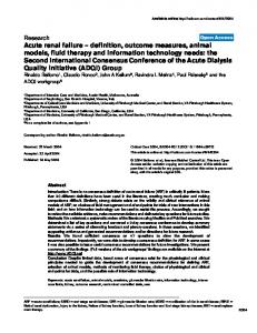
The course of the acute renal failure in our 8 patients is described in terms of serial serum creatinine levels, urinary volumes, and response to diuretics. The severity and duration of renal function impairment, as measured by serial serum creatinine determinations, are shown in Figure I. In all patients, serum creatinine levels increased to more than twice the base-line value, indicating a loss of more than 50 pen cent of renal function and involvement of both kidneys. As early as the first day after angiognaphy, a change in serum creatinine was noted in the 2 patients for whom this information was available. For the remaining patients who did not have serum creatmine determinations until the second or the
Department
of
Diagnostic
Roentgenology,
Mayo
Clinic
and
VOL.
No.
121,
Acute
3
Renal
Failure
after
Case
Age
No.
(yr.)
I
6o
PROCEDURES,
INDICATIONS,
AND
.
Procedure
2
42
3
66
4 5
59 23
Ventriculography
6
8o
Aortography+left
Mitral
.
linding
Mitral insufficiency, artery disease
insufficiency
Budd-Chiari
Hepatic
syndrome
vein
coronary
thrombosis
Hereditary Renovascular
telangiectasia hypertension
Angiomatosis of liver Bilateral renal artery
Congenital
heart
Complete canal
Renal
renal
FINDINGS
.
1ndcation
Ventriculography+coronary arteriography Visceral arteriography Visceral arteriography Aortography
545
I
TABLE ANGIOGRAPHIC
Angiognaphy
disease
mass
Renal
stenosis
atrioventricular cysts,
benign
arteriography 7
49
8
of multiple
Aortography
Occlusion
Aortography+left
arteries Renal mass
renal
Renal artery stenosis, occlusion of multiple arteries Adrenal tumor
arteriography
third day, an average daily increase of more than I mg./dl. was observed. This increase is similar to that seen in acute renal failure from other causes.
The any
duration
output
of
ofoligunia, less
than
defined 20
ml./hn.
as uninon
500
ml./da., is depicted in Figure 2. Low urine volumes were first noted within 12 hours after angiography in 4 patients and before 24 hours in 2 other patients. In the remaining 2 patients, no adequate measurements of urine volume were made until 31 and 3
hours,
respectively,
procedure.
patients was
likely,
shortly not
after
either
in
level
hours
or
The
of diuretics
serum
creatinine
the
oligunia
within
24
of the renal
the
poor
failure
response
and
to
a further
is
large
increase
in
to 4 days after at5 patients. However, of renal function usually comwithin I week; this is earlier than
tempts
at
recovery menced
expected 1
change
nature
by
doses
0
volumes during
angiography.
severe
indicated
urine case
all
but
the 8 patients had in serum creatin-
of
documented
after
in
procedure
every
7
Thus,
a significant
me
angiographic began
the because
measured
hours.
24
the oligunia
documented
not
were
first
after
Most
for
diunesis
for
2
in
most
patients
with
acute
. S
‘2 Serum
creotlifine mg/dI
Pt
0
‘
‘
5
S
6
.#{149}#{149}#{149}#{149}S#{149}#{149}#{149}#{149}#{149}_J
1-
Olguria Furosemlde L0__ManntoI
I0 ...i:
+7
ci)
0
?100mg 12g iv.
v.J
...T)
8
#{163}
02
Days
t
4
I
I
1
2
Angiography
I
I
3
4
Days
Anqiography
Fic.
i.
Serum creatinine values and after angiography.
before
FIG.
2.
Duration
identify
of
curves
oliguria.
in Figure
Symbols i.
5
F. K. Port,
546 oligunic renal early recognition
of this
complication
I, in which kalemia
second during dialysis
fluid
of such
dialysis
The importance adequate treatment
is exemplified
severe were
toneal
ure,
failure. and
R. D. Wagoner
overload
hyper-
that
as early
pen-
as the
day. One death occurred (Case 2) the recovery phase, despite hemoperformed for severe hepatic fail-
and
was
probably
unrelated
severe
R. E. Fulton heart
JULY,
disease
and
in
1974
a newborn
infant.’5”7
by Case and
magnitude
was required
of
and
to renal
failure. In the 7 other patients, gross recovery of renal function to base-line levels occurred before hospital dismissal. The longest recovery time was approximately i6 days.
The literature unexplainable angiography.24’35’37
few reports of failure after
Renovascular
hyperten-
sion was described in 2 patients and diabetes mellitus in a third. In a fourth case, the patient had aortic insufficiency and later developed severe proteinunia; renal biopsy findings in this patient were thought to be compatible with hypersensitivityinduced glomerulonephnitis.4 PATHOPHYS1OLOGIC
There renal
DISCUSSION
contains acute renal
the
are many failure
time
MECHANISMS
possible
after
causes
for acute
angiography.
correlation
Because
between
angiography, renal status prior to angiography, and allergic reactions. Effect of contrast medium dosage and type. The type and amount of contrast medium used in our patients are shown in Table ii.
have
The
occurred
under
specific
An overdose of contrast current multiple myeloma likely cause for several renal damage after
adults.5’24’31’
Two
ally
induced
renal
have
been
documented,
circumstances.
medium or a condisorder was the reported cases of angiography in
cases
of angiographic-
failure
failure
also
in a patient
with
AND
AMOUNT
trast
medium,
established
of
for
dosages
cardiovascular
and
effects
the frequencies
the
concentrations patients
pen cent. In the was the highest,
were
were 70 whose total
the amount
was
MEDIUM
USED
Administered
Type (70-80%)
ml./kg.
I, g./kg.
285
4.6
1.70
210
4.1
1.52
3 4
i8
4.2
1.34
144
3.4
1.26
5
Renovist
230
4.9
1.82
6 7 8
Vascoray
110
1.4
o.6
1.3
0.62
3.4
1.26
Angio-Conray
Renografin-76
simi-
70
310
to
8o
dose
close to
Renografin-76 Renografin-76 Renografin-76 Renografin-76
i
2
pa-
after
II
OF CONTRAST
ml.
renal
our
lar to our total experience for all angiographic studies during this period at the Mayo Clinic. Recorded doses include all test injections and averaged 193 ml. or 3.4
Amount Case
well
onset
tients, certain explanations are possibilities, such as dosage and nephrotoxicity of con-
TABLE TYPE
was
ml./kg.;
children
in
procedure
the
angio-
Currently used angiognaphic contrast media such as the diatnizoate and iothalamate salts have been considered to be safe by many investigators.6”3’ Although acute renal failure has not been reported in animal experiments with these media, sevenal studies have suggested a transient and mild decrease in renal function.27’34’36’38 Few clinical instances of severe impairment of renal function have been reported and most
graphic
and
the
VOL.
121, No.
Acute
3
Renal
Failure
the proposed upper limit of ml./kg. Some workers36 consider this average dose to be too high. The 2 patients who had a relatively small amount of contrast medium had been exposed to contrast agents only I and 3 days previously. Two other patients had had a similar exposure within I week; the others had had no studies with contrast medium for more than I month prior to angiography. Renal function impairment had not been detected after these preceding studies, and all patients had adequate urinary output prior to angiography. The direct mechanism by which contrast medium might cause renal damage is still controversial.’6 Hypertonicity of the medium may cause crenation of enythrocytes with a secondary decrease in renal blood 0 Vasoconstniction and subsequent vasodilatation immediately after exposure to contrast medium and release of histamine, particularly after a repeated study, have also been implicated.2’9’20’2’ In our patients, a relatively high dosage or repeated administration of contrast medium may have been an etiologic factor. However, no patient in this series underwent bilateral selective renal angiography, a procedure that often produces maximal exposure of the kidneys to contrast medium. Usually, aortography utilizing 6o ml. of contrast medium and bilateral selective renal artery injections with 12 to 15 ml. per kidney is used for studies performed because renal
of mass.
contraindication
renovascular
Renal
hypertension
or
a
stenosis
is not
a
artery for
selective
injection
if
occlusion of the renal orifice can be avoided. More than 2,700 such bilateral studies were performed at the Mayo Clinic during the period of review and no renal complications were observed in this group. Cardiovascular effects. A prolonged decrease in blood pressure was not observed in any of our patients despite close monitoring. In only i instance was there a brief, moderate decrease in blood pressure, which responded immediately to a vasopressor agent (Vasoxyl). Decreased peripheral and possibly renal
after
Angiography
perfusion
547
have octhough their blood pressure remained normal. In Case I there was a short episode of atnial fibrillation without associated signs of impaired peripheral perfusion. The congenital heart disease in Case 5 was associated with a chronically low cardiac output. Large arteniovenous fistulas in Case 3 produced a shunt of approximately 5 liters/mm., and curned
this
after
in
may
angiognaphy
several
have
impaired
patients
may
even
perfusion
to other
organs. A suspected pulmonary after venography in Case 2 and
embolus a recurrent gastrointestinal hemorrhage in Case 3 also could have resulted in significantly decreased renal perfusion. However, blood pressures remained stable during these episodes.
Dehydration etiologic factor
has been considered as an in angiographically induced complications.’2 The preparation, with fluid restriction and a laxative, may have induced a mild dehydration in our patients. However, none was prepared for simultaneous renal vein renin determinations-a procedure that often produces marked dehydration. In the same 4 year period, there were no recognized instances of acute renal failure in more than 500 patients who had combined renal vein renin and angiographic study in the salt-depleted and dehydrated state. Some of these salt-depleted patients, however, did receive 500 to I ,000 ml. of saline after the renal venous blood sampling. Dehydration per se apparently did not play a significant role in the development of acute renal failure in our patients. Other causes of decreased renal perfusion cannot be specifically excluded. Renal disease. Renal abnormalities were present prior to arteniography in most of our patients and frequently were related to systemic disorders. Five of the 8 patients had an increased serum creatinine value, which clearly indicated a decreased glomerulan filtration rate, before the angiographic study. The impairment of function was mild to moderate in all cases. Likely conditions responsible for these renal abnormalities included diabetic glomerulopathy, ur-
F. K. Port, R. D. Wagoner
548
ate nephnopathy, bilateral renal artery stenosis, and obstructive uropathy in Cases I 3, 4, and 6, respectively. Numerous patients with more marked impairment of renal function were subjected to arteniography during the same period without apparent complications. In our experience’4 with high-dose urography in patients with decreased renal function, there has not been a deleterious effect of the contrast medium. Diabetic patients may be an exception, however. The combination of renal and hepatic impairment might increase the toxicity of the contrast medium, because of the limitations in both major pathways of excretion.32 Although hyperbilinubinemia has been suggested8 as a predisposing condition for the
,
development
of
acute
failure
renal
expeni-
mentally, clinical experience7 does not support this. Of our 8 patients, 3 had marked abnormalities in liven function tests; 2 of these patients also had renal insufficiency. Perhaps there is a greater susceptibility to renal complications in patients with hepatic insufficiency who undergo arteniography. Proteinunia may produce acute renal failure
after
angiography
by
precipitation
of casts or proteinaceous material. Urinary Tamm-Horsfall protein3 and the panaproteinunia of multiple myeloma,28 particularly in the dehydrated patient, have been proposed as precipitating factors in the development of acute renal failure. Although present in the majority of our patients, proteinunia was only mild to moderate and was not associated with the nephnotic syndrome. Protein electrophoresis (obtained in 6 patients) did not suggest the presence of an abnormal serum protein and in no case was multiple myeloma suspected on subsequently diagnosed. Diabetes mellitus was present in I patient, who also had probable diabetic nephropathy. We have observed the development of acute renal failure after excretory unography in 6 diabetic patients during the years 1968 through 1972. Pillay et al.29 and Teal et al.37 also suggested an
R. E. Fulton
and
association to
of
contrast
renal
JULY,
failure
medium
in
with diabetic this has
1974
exposure patients, not been
but the mechanism for clarified. According to Martin,22 hyperunicemia can delay the excretion ofcontrast medium. Increased urinary uric acid excretion induced
by
Mudge26
and
these and
agents
is a potential
particularly
has
been
Postlethwaite in
and
noted
by
Kelley3#{176}
hazard to renal function, dehydrated patients. Serum were abnormally high in
uric acid levels 2 of our 4 patients in whom this value was available. Hypersensitivity allergic reactions could cause acute renal failure as a consequence of induced vasculitis. An interstitial nephnitis was implicated in i previously reported case.4 The methylglucamine cation present in most media has been thought to release histamine’6 and thus cause bronchospasm.’ In our patients, no overt allergic phenomena were observed, and histories of allergies were negative except for I patient who reported previous reactions. PREVENTION
This review of potential causes for acute renal failure in our patients suggests that multiple factors, alone or in combination, are responsible for this nephnologic complication. The factors that identify the patient at risk are not clean, but all of our
patients
had
severe
medical
disorders.
The trend toward the use of angiography in more severely ill patients and the use of greater amounts of contrast medium should be tempered with an increased awareness of potential renal complications. More frequent consultation between the clinician and the radiologist, particularly when planning a study in severely ill patients, seems warranted. We suggest that consideration be given to the prophylactic administration of mannitol by intravenous infusion prior to and during the procedure and that fluid restriction be avoided whenever possible. Careful monitoring of urinary output and serum creatinine for at least 24 hours after angiography may allow the
VoL.
Acute
3
No.
121,
Renal
Failure
early recognition oF this complication that appropriate therapeutic attempts increase urine output, when impending oliguria is suspected, can be instituted earlier.
after
so to
7.
8.
oligunic
renal
failure
developed
do
Mayo 200
Section
1964,
9. DEAN,
cell
Street
Rochester,
Foundation
BOND,
T.
mental
effects
mi/kg
agents:
12.
hemodynamics
W. E.,
BERDON,
and
BAKER,
uria:
its
in
R. H., Tamm-Horsfall
H.
to
prolonged
children
and
intravenous myeloma.
and
to
urography Radiology,
protein-
renal
16.
of
high
17.
S.,
D.,
HAWKINS,
KAYE, M. Acute renal failure and syndrome after angiocardiography lumine diatrizoate. New England 284,
1971,
5.
Its
Application
Vascular
C
and
nephrotic
with
7.
megMed.,
i8.
by
and
to
Renal
the
Parenchymal
Publisher,
Thomas,
Diagnosis of Renal Lesions. Charles Springfield,
T. A.
A.
J.,
ABRAMS,
extraction
19.
urography
to
renal
in de106,
its
reports Acta
damage.
6I#{231}-
C. G. On and
with
pre-
of
five
radio!.,
473-480.
R. E., WITTEN,
B.
JONES,
D. M., and WAGONER,
Hemorrhagic
pounds.
7. Pediat.,
in renal insuffiRAD. THERAPY 623-634. HOGAN, G. R.,
renal
to ‘970,
R. G. Renal
GRAINGER,
necrosis
in
radiopaque
com-
76, 49-53. toxicity of radiological
media.
Brit.
GRUSKIN,
A. B.,
OETLIKER, N. L.,
M. Bull.,
1972,
28,
KNOX,
ogist, 20.
M.,
0.
H.,
WOLFISH,
191-
Effects histology
JR.
Year
LASSER,
Book
of angiography in infants and
F.
Medical
E. M. Effect on
1965, E.
Angi-
Inc.,
pp.
STIGMAN, plasma
Renal
Publishers,
37-38. G., BUNNELL, I. L., 1966,
J., and
BERNSTEIN,
on renal function and piglets. 7. Pediat., 1970, 76, 41-48. KINCAID, 0. W., and DAVIS, G. D.
angiography
STAMEY,
follow-
dosage
aortography
tests:
relationship
and
III., 1968,
H. L., and of PAH in man
renal
infancy:
renal
Renal
to
severe
47,
Chicago,
Arteriography:
p. 35. 6. DAVIDSON,
of
ography.
592-593.
I. L. Selective
BUNNELL,
W.,
DUGUID,
medium
excretory
due
N. M., GOOTMAN, EDELMANN, C.
722.
4. BORRA,
116,
195.
with 714-
contrast
dose
damage
contrast
fol-
failure
in adults 1969, 92,
on
1968,
hydrated patients. 7. Urol., 1971, 62 I. EDLING, N. P. G., and HELANDER,
and
nephrogram
arteriography
7. Surg.,
15.
J.,
BECKER,
selective
Am.
R. D. Intravenous urography ciency. AM. J. ROENTGENOL., & NUCLEAR MED., 1969, 106, GILBERT, E. I., KHOURY, G. H.,
K.
EKENGREN,
SCHWARTZ,
D.
relationship
infants
lowing multiple
and
during selective angiog1968,3, 389-396.
mnvest. Radio!.,
raphy.
3.
0.,
A., BROBERGER,
APERIA, Renal
and of
FULTON,
374-384. 2.
27-31.
R. W., LIVANEC, G., GUEST, M. M. Experi-
14.
5,
i 970,
situ per-
Radiology, 1972, 104, 557-560. DUDZINSKI, P. J., PETRONE, A. F., PERSOFF, M., and CALLAGHAN, E. E. Acute renal failure fol-
1957,
Radio!.,
P.,
relationship
vention
to contrast
187,
in
complications.
13.
mnvest.
1964,
of acellular
D., and REDMAN, H. C. Myth of in angiography: study to determine
B.
DOUST, I
5oI
reactions
and
angio-
J. R., BROWN,
renal
G. Adverse of problem.
studies
cellular
R. C.
READ, from
7 12-714. II.
REFERENCES
ANSELL, scope
in man. renal damBrit. M. 7.,
damage
perfusion
7.A.M.A.,
DERRICK,
cases I.
anoxic
mannitol.
J. H., and
with
microcirculation.
S.W.
Minnesota
of
in renal
media:
fusates.
M.D. Mayo
factor
kidney
canine
function
810-811.
I,
graphic
10.
effect
R. E., ANDREW,
Red
of Publications
Clinic and First
and hepatic 97, 249-254. J. L. Jaundice and 1970,
protective
lowing
K. Port,
Frederich
renal
DAWSON, age:
in
8 patients after catheterization angiography without bilateral renal arteniography. The cause for this complication remains obscure. Severe dehydration, shock, overdose of contrast medium, or multiple myeloma could not be implicated. From retrospective analysis of these cases, multiple factors seem to be operative in these generally severely ill patients. Early recognition and adequate management may prevent life-threatening effects of this infrequent, but potentially serious angiographic complication.
549
ing abdominal aortography. Radiology, 1965, 8, I043I046. DAVIDSON, A. J., BECKER, J., ROTHFIELD, N., UNGER, G., and PLOCH, D. R. Evaluation of effect of high-dose urography on previously
impaired Radiology,
SUMMARY
Acute
Angiography
glomerular
flow
in
man
ELwooD, of
C. M.,
selective
filtration (abstract).
renal rate
and
Physiol-
8, 210. C.,
WALTERS,
A.,
REUTER,
S. R.,
F. K.
550
LANG, J. Histamine Radiology, 1971,
and media. 21.
C. M.
MARTIN,
trizoate
D.
Wagoner
Myelography
and
by contrast
responses
to
con-
31.
dominal
32.
Survey
of
complications
ab68,
of
Radiology,
1957,
G.,
KUMAR,
33.
J.,
MCEVOY,
MILLER,
Renal
JR.
G. H. agents:
MUDGE,
icity. 933.
New
34.
sodium
of
factor 7. Med.,
England
failure plasma
cholecysto-
1971,
284,
K. G., HUNT, Effects
on kidney function 716-718.
P. E.,
and
35.
of
929-
J. C.,
diatrizoate
36.
7.A.M.A.,
in dogs.
CONN,
H. 0. Acute
renal
after intravenous pyelography cell myeloma. 7.A.M.A., 1958,
V. K. G.,
PILLAY, F.
D.,
and
in
following
R. M. Acute
intravenous
urography
with long_standing azotemia. Radiology,
37.
167,
30.
POSTLETHWAITE, Uricosuric
A. effect
1970,
38.
patients
mellitus
and
and
of
radiocontrast
KELLEY,
and
W. agents:
N.
Radiology.
Williams p. 9. 7. Radiol.,
1972,
Brit.
Urography. 46.
DECTER,
y, selective
renal
suppression
PAH 12,
and
y,
venography
Austra!asian
ratio.
extraction
arteriograph
renal
Radio!.,
252-255.
F. R., and COBURN, J. W. Renal failure following methylglucamine diatrizoate (Renografin) aortography: report of case with unilateral renal artery stenosis. 7. Uro!., 1966, 96, 848-85 I. 1ALNER, L. B., and DAVIDSON, A. J. Effect of STARK,
media
on
renal
Radio!., 1968,3, I’EAL, J. S., RUMBAUGH, BERGERON, R. T., and renal failure following with methylglucamine Radiology,
H. renal
E.,
1972,
H. E.,
WOLLOWICK,
Effect
95, 633-636.
E.,
prepara-
A. Unilateral renal damcontrast dye injection with recovery. 7. Urol., 1967, 97, 30-32. SORBY, W. A., and Hoy, R. J. Renal arteriography and renal function: short-term effect of SIDD,
port.
renal failure in
diabetes
1974
845-852.
Diagnostic Baltimore,
Invest.
P. C., SCHWARTZ,
ROBBINS,
KARK,
J. J.,
M.
used
74,
1971,
Angiography.
Company,
321-3
contrast
2186-2189. 29.
42,
adrenalin
184,
PERILLIE,
& Wilkins SAXTON, H.
on
in nephrotox-
commonly
Med.,
Selective
18.
1968,
action
of four
mt.
L. L. Golden’s
ROBBINS,
aortograph
F.,
aortography.
possible
KINCAID,
1963,
from
Uricosuric
T. F., WAKIM, 0. W.
MULLADY, and
28.
R.
contrast
1954,35,
graphic
27.
and
radiological
complications 885-896.
Surgery, 26.
M.
after
Brit. M. 7., 1970, 4, 717-718. G. M., WYLIE, E. J., and HINMAN,
media. 25.
MCGEOwN,
failure
Renal
JULY,
age due to massive
825-838. 24.
in man Ann.
1969,
aortography.
E. Fulton
Section
1970,
115,
23.
R.
study tions.
683-686.
5, 424-435. with sodium diaCalifornia Med., 197!,
(Hypaque).
57-59. MCAFEE, J. G.
R.
release 100,
P. Hemodynamic media. mnvest. Radio!.,
LINDGREN,
trast 22.
Port,
retraction
of
PAH.
301-317.
C. L., spinal
H. D., R. L. Acute angiography
iothalamate:
case
SEGALL,
SCANLAN,
104,
FOSTER,
re-
561-562.
J. H.,
SNYDER,
YOUNGER, R., and KILLEN, D. A. of abdominal aortography on proximal tubule function. 7. Surg. Res., 1966,
6, 346-353.