MINI REVIEW published: 06 June 2018 doi: 10.3389/fmicb.2018.01184
Advance in Research on Mycobacterium tuberculosis FabG4 and Its Inhibitor Debajyoti Dutta* Department of Biochemistry, Faculty of Medicine and Dentistry, University of Alberta, Edmonton, AB, Canada
Edited by: Noton Kumar Dutta, Johns Hopkins University, United States Reviewed by: Luiz Augusto Basso, Pontifícia Universidade Católica do Rio Grande do Sul, Brazil Li Yuan, Shihezi University, China *Correspondence: Debajyoti Dutta
[email protected];
[email protected]
Increasing evidence from recent reports of drug-resistant mycobacterial strains poses a challenge worldwide. Drug-resistant strains often undergo mutations, adopt alternative pathways, and express drug efflux pumps to reduce or eliminate drug doses. Besides these intrinsic resistance mechanisms, bacteria can evade drug doses by forming biofilms. Biofilms are the concerted growth of adherent microorganisms, which can also be formed at the air-water interface. The growth is supported by the extracellular polymer matrix which is self-produced by the microorganisms. Reduced metabolic activity in a nutrient-deficient environment in the biofilm may cause the microorganisms to take alternative pathways that can make the microorganisms recalcitrant to the drug doses. Recent works have shown that Mycobacterium tuberculosis expresses several proteins during its growth in biofilm, those when deleted, did not show any effect on mycobacterial growth in normal nutrient-sufficient conditions. Studying these unconventional proteins in mycobacterial biofilms is therefore of utmost importance. In this article, I will discuss one such mycobacterial biofilm-related protein FabG4 that is recently shown to be important for mycobacterial survival in the presence of antibiotic stressors and limited nutrient condition. In an attempt to find more effective FabG4 inhibitors and its importance in biofilm forming M. tuberculosis, present knowledge about FabG4 and its known inhibitors are discussed. Based on the existing data, a putative role of FabG4 is also suggested. Keywords: FabG4, Mycobacterium tuberculosis, biofilm, β-oxo acyl-ACP reductase, inhibitor
Specialty section: This article was submitted to Antimicrobials, Resistance and Chemotherapy, a section of the journal Frontiers in Microbiology Received: 26 March 2018 Accepted: 16 May 2018 Published: 06 June 2018 Citation: Dutta D (2018) Advance in Research on Mycobacterium tuberculosis FabG4 and Its Inhibitor. Front. Microbiol. 9:1184. doi: 10.3389/fmicb.2018.01184
INTRODUCTION The ability of Mycobacterium tuberculosis to form biofilms was noted almost 120 years ago (Jones, 1896). However, the physiological and molecular basis of biofilm is only beginning to unravel until recently. The first protein that was found to be involved in M. smegmatis maturation of biofilm is the chaperone GroEL-1 (Ojha et al., 2005). GroEL-1 deficient mutant of M. smegmatis was suggested to lack GroEL-1 interaction with fatty acid synthesis type -II complex thereby reducing the mycolic acid content in biofilms. Mycolic acids are the predominant components of mycobacterial cell envelope that are produced by fatty acid synthesis type-II pathway in mycobacteria. The reason that mycobacterial cell envelope largely contributes to the biofilm attachment endorses mycolic acids may be involved in biofilm formation (Marrakchi et al., 2014). However the physiology of mycobacteria changes as it shifts from planktonic growth to biofilm
Frontiers in Microbiology | www.frontiersin.org
1
June 2018 | Volume 9 | Article 1184
Dutta
Structure and Function of Mycobacterial FabG4
synthesis type-II (Gurvitz et al., 2009). Because yeast ORA1 is a mitochondrial FabG1/FabG4 ortholog (Hiltunen et al., 2009), functional complementation of ORA1 with FabG4 is indicative of an active FabG4 protein that can utilize ACP/Coenzyme A tagged β-oxoacyl substrates and is utilized by mycobacteria during limiting resource or antibiotic stressed condition.
dependent growth and results in several modifications in the expression level of protein and molecules pertaining to the cell envelope (Ojha et al., 2008; Sambandan et al., 2013; Rastogi et al., 2017). Fatty acid synthesis and its associated pathways for mycobacterial cell envelope synthesis are one of the major areas for developing antitubercular drugs (Zumla et al., 2013). Because of this altered phenotypes and a waxy extra-cellular matrix of biofilm, Mycobacteria become resilient to drug doses (Islam et al., 2012). Therefore the conventional TB drugs may not be as effective for biofilm-forming mycobacteria. Membrane and cell envelope-associated biofilm-related proteins are particularly of interest as these proteins are likely to be involved in the making of cellular attachment to the biofilms. For example, recent studies have shown that a lipid transporter MmpL11 is specifically required for biofilm formation (Pacheco et al., 2013). Other works have identified a number of proteins specific to mycobacterial growth in the biofilm at air-water interface (Kerns et al., 2014). One of the proteins that are conserved among mycobacterial species is FabG4. The protein was proposed to possess host antigenic property and has a potential to be a biofilm-specific marker (Kerns et al., 2014). In addition to that, FabG4 was recognized as one of the crucial protein for mycobacterial survival in a stressed condition. This article will discuss the known facts about FabG4, its inhibitors, and discuss its possibility to serve as a candidate to study and treat biofilm-related mycobacteria.
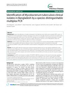
UNIQUE FEATURES OF FabG4 Characterization of recombinantly expressed FabG4 has shown that the protein is highly specific for NADH (Dutta et al., 2011). This is intriguingly different from its close relative (33% homology) FabG1 that utilizes NADPH. Crystal structure of the FabG4 complex with NADH reveals that the specificity comes from a single aspartate residue that reduces the volume of the phosphate binding groove of adenosine ribose phosphate moiety of NADPH (Dutta et al., 2013). Crystal structure further shows that FabG4 contains a “flavodoxin-type” structural domain in its N-terminal and a typical ketoreductase domain in its C-terminal (Figure 1A). The N-terminal structural domain is also found in modular polyketide synthase ketoreductase domain PlmKR1 (Bonnett et al., 2013). PlmKR1 exists as a monomer but FabG4 is different in the sense that the protein is a dimer, where the N-terminal domain of a protomer makes an extensive interaction with the C-terminal domain of the other protomer. Therefore, FabG4 was designated as “High Molecular weight FabG” (HMwFabG) (Dutta et al., 2011). Typical FabG does not contain this structural domain. Many of the N-terminal residues (1–19) of FabG4 cannot be traced in crystal structure suggesting that the region has no rigid secondary structure. Most of the N-terminal residues were traced in FabG4 complex with NAD+ structure (PDB 4FW8) showing a possibility of short helix containing seven residues (21–27) (Figure 1A). Long N-terminal sequences in FabGs are also found among eukaryotes, which were suggested to be the signal sequence (Wickramasinghe et al., 2006). Whether it is true for FabG4 is not known, but sequence alignment of HMwFabGs from actinobacteria and many proteobacteria reveals conserved residues Pro30, Leu33, and Arg35 in N-terminal sequence. Truncation of the N-terminal residues does not have any effect on catalytic activity. On the contrary, the C-terminal residues of FabG4 are apparently involved in catalytic activity since its truncation yields with a defective protein (Dutta et al., 2011). This is because the conserved C-terminal residues are engaged in electrostatic interaction with the active site proximal loops (Dutta et al., 2011).
IMPORTANCE OF FABG4 IN MYCOBACTERIA FabG commits the second step of fatty acid synthesis that is to convert β-oxo acyl-ACP to β-hydroxy acyl-ACP. M. tuberculosis genome contains multiple FabG homologs. Two of them are conserved among all mycobacterial species, FabG1 and FabG4. FabG1 remain at the focus of attention because it takes part in fatty acid synthesis type-II (Marrakchi et al., 2002). On the other hand, several studies have indicated that FabG4 is not an inactive gene in the M. tuberculosis genome. Its expression was first documented in the proteome by using 2-D gel electrophoresis accompanying MALDI-MS analysis (Jungblut et al., 1999) and later verified by others (Rosenkrands et al., 2000; Sinha et al., 2002). Gu et al. (2003), first provide the evidence that the protein is expressed in the mycobacterial membrane fraction. However, it’s requirement in mycobacterial physiology was not shown until the comprehensive work done by McFadden and coworkers (Beste et al., 2009). The authors showed that the protein is uniquely required for mycobacterial growth in Roisin’s minimal media, which contains limited carbon source (Beste et al., 2009). Proteomics studies have further identified that FabG4 is one of the major proteins which expression is induced by the antibiotic stressor (Sharma et al., 2010). The protein expression was also detected in the drug-resistant strains (Singh et al., 2016; Verma et al., 2017). Most interestingly, FabG4 can functionally complement eukaryotic β-oxoacyl-ACP reductase activity. FabG4, when expressed in the knockout of ora1 yeast cells can restore the mitochondrial fatty acid
Frontiers in Microbiology | www.frontiersin.org
INHIBITORS OF FabG4 Structure of FabG4 and its complex with a substrate mimic hexanoyl-CoA (PDB 3V1U) accelerated the work on structurebased inhibitor design against FabG4 (Dutta et al., 2013) (Figure 1B). The particular structure also provides a platform to design FabG specific inhibitors because the structure provided the first evidence of FabG-substrate complex (Table 1). The
2
June 2018 | Volume 9 | Article 1184
Dutta
Structure and Function of Mycobacterial FabG4
two sequential binding sites showing positive cooperativity. Inhibitory effects of β-lactam and Isoniazid-based compounds have shown to be inhibitory toward mycobacterial biofilm formation. Recently, thiophene containing trisubstituted methane compound, S-S006-830 was found to have a substantial inhibitory effect on M. tuberculosis biofilm formation and possesses antitubercular activity (Singh et al., 2017). FabG4 is one of the three membrane-associated proteins identified to strongly interact with S-S006-830. Computational docking study predicts two possible binding sites. One comprises of the region overlapping NADH and CoA substrate binding and the other site is on the N-terminal domain. S-S006-830 is special because apart from the NADH-substrate binding site it also targets to the N-terminal structural domain. Two unique FabG inhibitors are worth mentioning in this regard. The first, Pyridomycin analog specifically targets NADH binding proteins and proposed to exhibit inhibitory effects on FabG4 by bridging both NADH and substrate-binding region (Hartkoorn et al., 2014). The second inhibitor was identified using library screening that uniquely binds to the helical dimeric interface of FabG (Cukier et al., 2013). The helical dimeric interface in FabG is responsible of NAD(P)H binding cooperativity (Dutta et al., 2012). All FabG4 and related inhibitors are summarized in Table 1.
POSSIBLE ROLE OF THE ENZYME High Molecular weight FabG are exclusively found among bacteria. Homologous genes of FabG4 are found in actinobacteria and many proteobacteria (Dutta et al., 2011). Genomic analysis of HMwFabG containing bacteria shows another protein coexisting downstream to FabG4 ORF. In M. tuberculosis this protein is HtdX (Rv0241c). Both the proteins are conserved among mycobacterial species. Even in M. leprae genome where the genomic reduction had happened, retained both fabG4 and its downstream htdX (Cole et al., 2001). However, deletion of htdX or fabG4 does not show any apparent effect on bacterial survival in vivo (Sassetti and Rubin, 2003). Nonetheless, the protein is shown to be essential for survival in minimal media (Beste et al., 2009). Presumably, the conditional dependency of M. tuberculosis on FabG4 indicates an alternative pathway during limiting resource condition. Another clue is the FabG4 dependency on NADH. The NADH utilizing β-ketoacyl reductase activity of FabG4 is four times higher to the reverse reaction that utilizes NAD+ for β-hydroxyacyl dehydrogenase activity (Dutta et al., 2013). Compared to NADPH, NADH is the low energy molecule and mostly associates to the catabolic pathways (Cantó et al., 2015). Shifting the coenzyme specificity toward low energy NADH possibly indicates that the bacteria are rewiring its metabolism toward the energy saving mode. Furthermore, FabG4 is isolated from the membrane fraction, which is indicative of its membrane-associated role (Gu et al., 2003; Singh et al., 2017). The role could be either fatty acid synthesis or modification. Notably, the other gene htdX was found to be responsive to the inhibitors of cell wall synthesis (Boshoff et al., 2004). Since both fabG4 and htdX are situated in a conserved cluster it is tempting to predict that they are involved in the same
FIGURE 1 | Crystal structures of FabG4: (A) N-terminal “flavodoxin type” (orange) and C-terminal ketoreductase (magenta) domains. “N” indicated the N-terminal of the polypeptide chain. The regions those were not be traced in electron density are shown in dotted lines. (B) The substrate mimic hexanoyl coenzyme A (HXC) and NAD+ bound structure shows the putative substrate binding modes.
first reported FabG4 inhibitors are based on the polyphenol compounds (Banerjee et al., 2014, 2015b). Synthesized compounds are also tested against M. smegmatis showing the MIC of 5 µg mL−1 . Docking studies had revealed that the compounds potentially occupy the NADH binding region with a few hydrogen-bonding interactions with the loop-residues responsible for CoA substrate binding. Another study to design and synthesize the inhibitors based on the common pharmacophores like β-lactam and Isoniazid was also successful inhibiting FabG4 with IC50 as low as 15.2 ± 0.5 µM (Banerjee et al., 2015a). Similar to the polyphenol based compounds β-lactam and Isoniazid-based compounds primarily dock on the NADH binding site. Isothermal titration calorimetry indicates
Frontiers in Microbiology | www.frontiersin.org
3
June 2018 | Volume 9 | Article 1184
Dutta
Structure and Function of Mycobacterial FabG4
TABLE 1 | Inhibitors of FabG4. Compound structure
Type of the compounds
General comments
Reference
Triazole-polyphenol hybrid compounds
Possibly compete for NADH binding sites; Cooperative sequential binding with two binding sites IC50 = 34.9 µM Ki = 0.88 µM MIC = 5 µg ml−1
Banerjee et al., 2014, 2015b
Beta-lactam isoniazid based compounds
Possibly compete for NADH binding sites; Cooperative sequential binding with two binding sites; acts as dual inhibitor of HtdX IC50 = 15.2 µM MIC = 15 µg ml−1
Banerjee et al., 2015a
Thiophene containing trisubstituted methane
S-Enantiomer has more specificity toward FabG4; combination with rifampicin has a synergistic effect MIC = 3.125 µg ml−1
Singh et al., 2017
Pyridomycin analogs
An InhA inhibitor that can possibly inhibits FabG4 by binding to the both NADH and substrate binding region
Hartkoorn et al., 2014
Triazol-quinazoline based compound
Inhibitor identified against Pseudomonas aeruginosa FabG from the screening small molecule library, Specifically binds to the helical dimer interface
Cukier et al., 2013
Other inhibitors related to FabG4
manifested antitubercular activity more experiments are needed to call it a drug target. Role of FabG4 and its connection with the membrane in mycobacteria also require further experimentations. To study FabG4 importance in biofilm forming M. tuberculosis it is, therefore, necessary to find out its targeting and interaction with other proteins. Identifying the role of FabG4 in mycobacteria might provide a hint of a new aspect of a survival strategy. The second aspect is the strategy to design more specific inhibitors of FabG4. While the C-terminal domain of FabG4 is common among FabGs, inhibitors against its N-terminal structural domain might be a way to target the protein more specifically. Designing the fluorophore inhibitors against its structural domain could be a way to find out its role in vivo.
pathway. Both sequence analysis and preliminary enzymatic activities are corroborated that HtdX can commit a dehydratase reaction that is to convert β-hydroxyacyl-CoA to enoylacylCoA (Gurvitz, 2009; Sacco et al., 2010; Biswas et al., 2013; Banerjee et al., 2015a). The reaction is theoretically the successive step of FabG4 enzymatic reaction for fatty acid elongation cycle. Altogether the data suggests that during limiting resource condition FabG4 participates into some low-energy requiring pathway that is related to membrane biosynthesis, remodeling or recycling.
FUTURE SCOPE FabG4 is a host antigenic protein reportedly expressed in mycobacterial biofilms. The protein expression was consistently found in membrane fraction and stressor-induced. As a conserved gene in all mycobacterial species, FabG4 can be a biomarker for biofilm. Although, FabG4 inhibitors are
Frontiers in Microbiology | www.frontiersin.org
AUTHOR CONTRIBUTIONS DD is solely responsible for the conception or design, drafting, and revising the work.
4
June 2018 | Volume 9 | Article 1184
Dutta
Structure and Function of Mycobacterial FabG4
Professor Amit Basak, Department of Chemistry, Indian Institute of Technology Kharagpur and Dr. Deb Ranjan Banerjee and Rupam Biswas for their contribution to the FabG4 work from Indian Institute of Technology Kharagpur.
ACKNOWLEDGMENTS The author thanks Professor Amit Kumar Das, Department of Biotechnology, Indian Institute of Technology Kharagpur for his guidance during Ph.D. The author also thanks
REFERENCES
Gurvitz, A. (2009). The essential mycobacterial genes, fabG1 and fabG4, encode 3-oxoacyl-thioester reductases that are functional in yeast mitochondrial fatty acid synthase type 2. Mol. Genet. Genomics 282, 407–416. doi: 10.1007/s00438009-0474-2 Gurvitz, A., Hiltunen, J. K., and Kastaniotis, A. J. (2009). Heterologous expression of mycobacterial proteins in Saccharomyces cerevisiae reveals two physiologically functional 3-hydroxyacyl-thioester dehydratases, HtdX and HtdY, in addition to HadABC and HtdZ. J. Bacteriol. 191, 2683–2690. doi: 10.1128/JB.01046-08 Hartkoorn, R. C., Pojer, F., Read, J. A., Gingell, H., Neres, J., Horlacher, O. P., et al. (2014). Pyridomycin bridges the NADH- and substrate-binding pockets of the enoyl reductase InhA. Nat. Chem. Biol. 10, 96–98. doi: 10.1038/nchembio. 1405 Hiltunen, J. K., Schonauer, M. S., Autio, K. J., Mittelmeier, T. M., Kastaniotis, A. J., and Dieckmann, C. L. (2009). Mitochondrial fatty acid synthesis type II: more than just fatty acids. J. Biol. Chem. 284, 9011–9015. doi: 10.1074/jbc.R80006 8200 Islam, M. S., Richards, J. P., and Ojha, A. K. (2012). Targeting drug tolerance in mycobacteria: a perspective from mycobacterial biofilms. Expert Rev. Anti Infect. Ther. 10, 1055–1066. doi: 10.1586/eri.12.88 Jones, A. C. (1896). On the so-called tubercle bacilli. Report of the 66th Meeting of the British Association for the Advancements of Science. London: John Murray, 1015–1016. Jungblut, P. R., Schaible, U. E., Mollenkopf, H. J., Zimny-Arndt, U., Raupach, B., Mattow, J., et al. (1999). Comparative proteome analysis of Mycobacterium tuberculosis and Mycobacterium bovis BCG strains: towards functional genomics of microbial pathogens. Mol. Microbiol. 33, 1103–1117. doi: 10.1046/ j.1365-2958.1999.01549.x Kerns, P. W., Ackhart, D. F., Basaraba, R. J., Leid, J. G., and Shirtliff, M. E. (2014). Mycobacterium tuberculosis pellicles express unique proteins recognized by the host humoral response. Pathog. Dis. 70, 347–358. doi: 10.1111/2049-632X. 12142 Marrakchi, H., Ducasse, S., Labesse, G., Montrozier, H., Margeat, E., Emorine, L., et al. (2002). MabA (FabG1), a Mycobacterium tuberculosis protein involved in the long-chain fatty acid elongation system FAS-II. Microbiology 148, 951–960. doi: 10.1099/00221287-148-4-951 Marrakchi, H., Lanéelle, M. A., and Daffé, M. (2014). Mycolic acids: structures, biosynthesis, and beyond. Chem. Biol. 21, 67–85. doi: 10.1016/j.chembiol.2013. 11.011 Ojha, A., Anand, M., Bhatt, A., Kremer, L., Jacobs, W. R. Jr., and Hatfull, G. F. (2005). GroEL1: a dedicated chaperone involved in mycolic acid biosynthesis during biofilm formation in mycobacteria. Cell 123, 861–873. doi: 10.1016/j. cell.2005.09.012 Ojha, A. K., Baughn, A. D., Sambandan, D., Hsu, T., Trivelli, X., Guerardel, Y., et al. (2008). Growth of Mycobacterium tuberculosis biofilms containing free mycolic acids and harbouring drug-tolerant bacteria. Mol. Microbiol. 69, 164–174. doi: 10.1111/j.1365-2958.2008.06274.x Pacheco, S. A., Hsu, F. F., Powers, K. M., and Purdy, G. E. (2013). MmpL11 protein transports mycolic acid-containing lipids to the mycobacterial cell wall and contributes to biofilm formation in Mycobacterium smegmatis. J. Biol. Chem. 288, 24213–24222. doi: 10.1074/jbc.M113.473371 Rastogi, S., Singh, A. K., Pant, G., Mitra, K., Sashidhara, K. V., and Krishnan, M. Y. (2017). Down-regulation of PE11, a cell wall associated esterase, enhances the biofilm growth of Mycobacterium tuberculosis and reduces cell wall virulence lipid levels. Microbiology 163, 52–61. doi: 10.1099/mic.0.00 0417 Rosenkrands, I., King, A., Weldingh, K., Moniatte, M., Moertz, E., and Andersen, P. (2000). Towards the proteome of Mycobacterium tuberculosis. Electrophoresis 21, 3740–3756. doi: 10.1002/1522-2683(200011)21:17 3.0.CO;2-3
Banerjee, D. R., Biswas, R., Das, A. K., and Basak, A. (2015a). Design, synthesis and characterization of dual inhibitors against new targets FabG4 and HtdX of Mycobacterium tuberculosis. Eur. J. Med. Chem. 100, 223–234. doi: 10.1016/j. ejmech.2015.06.007 Banerjee, D. R., Dutta, D., Saha, B., Bhattacharyya, S., Senapati, K., Das, A. K., et al. (2014). Design, synthesis and characterization of novel inhibitors against mycobacterial β-ketoacyl CoA reductase FabG4. Org. Biomol. Chem. 12, 73–85. doi: 10.1039/c3ob41676c Banerjee, D. R., Senapati, K., Biswas, R., Das, A. K., and Basak, A. (2015b). Inhibition of M. tuberculosis β-ketoacyl CoA reductase FabG4 (Rv0242c) by triazole linked polyphenol-aminobenzene hybrids: comparison with the corresponding gallate counterparts. Bioorg. Med. Chem. Lett. 25, 1343–1347. doi: 10.1016/j.bmcl.2015.01.014 Beste, D. J., Espasa, M., Bonde, B., Kierzek, A. M., Stewart, G. R., and McFadden, J. (2009). The genetic requirements for fast and slow growth in mycobacteria. PLoS One 4:e5349. doi: 10.1371/journal.pone.000 5349 Biswas, R., Dutta, D., and Das, A. K. (2013). Cloning, overexpression, purification, crystallization and preliminary X-ray diffraction analysis of Rv0241c (HtdX) from Mycobacterium tuberculosis H37Rv. Acta Crystallogr. Sect. F Struct. Biol. Cryst. Commun. 69, 1110–1113. doi: 10.1107/S174430911302 3452 Bonnett, S. A., Whicher, J. R., Papireddy, K., Florova, G., Smith, J. L., and Reynolds, K. A. (2013). Structural and stereochemical analysis of a modular polyketide synthase ketoreductase domain required for the generation of a cis-alkene. Chem. Biol. 20, 772–783. doi: 10.1016/j.chembiol.2013.0 4.014 Boshoff, H. I., Myers, T. G., Copp, B. R., McNeil, M. R., Wilson, M. A., and Barry, C. E. III (2004). The transcriptional responses of Mycobacterium tuberculosis to inhibitors of metabolism: novel insights into drug mechanisms of action. J. Biol. Chem. 279, 40174–40184. doi: 10.1074/jbc.M40679 6200 Cantó, C., Menzies, K. J., and Auwerx, J. (2015). NAD+ metabolism and the control of energy homeostasis: a balancing act between mitochondria and the nucleus. Cell Metab. 22, 31–53. doi: 10.1016/j.cmet.2015.05.023 Cole, S. T., Eiglmeier, K., Parkhill, J., James, K. D., Thomson, N. R., Wheeler, P. R., et al. (2001). Massive gene decay in the leprosy bacillus. Nature 409, 1007–1011. doi: 10.1038/35059006 Cukier, C. D., Hope, A. G., Elamin, A. A., Moynie, L., Schnell, R., Schach, S., et al. (2013). Discovery of an allosteric inhibitor binding site in 3-Oxo-acylACP reductase from Pseudomonas aeruginosa. ACS Chem. Biol. 8, 2518–2527. doi: 10.1021/cb4005063 Dutta, D., Bhattacharyya, S., and Das, A. K. (2012). Crystal structure and fluorescence studies reveal the role of helical dimeric interface of staphylococcal FabG1 in positive cooperativity for NADPH. Proteins 80, 1250–1257. doi: 10.1002/prot.24024 Dutta, D., Bhattacharyya, S., Mukherjee, S., Saha, B., and Das, A. K. (2011). Crystal structure of FabG4 from Mycobacterium tuberculosis reveals the importance of C-terminal residues in ketoreductase activity. J. Struct. Biol. 174, 147–155. doi: 10.1016/j.jsb.2010.11.012 Dutta, D., Bhattacharyya, S., Roychowdhury, A., Biswas, R., and Das, A. K. (2013). Crystal structure of hexanoyl-CoA bound to β-ketoacyl reductase FabG4 of Mycobacterium tuberculosis. Biochem. J. 450, 127–139. doi: 10.1042/BJ2012 1107 Gu, S., Chen, J., Dobos, K. M., Bradbury, E. M., Belisle, J. T., and Chen, X. (2003). Comprehensive proteomic profiling of the membrane constituents of a Mycobacterium tuberculosis strain. Mol. Cell. Proteomics 2, 1284–1296. doi: 10.1074/mcp.M300060-MCP200
Frontiers in Microbiology | www.frontiersin.org
5
June 2018 | Volume 9 | Article 1184
Dutta
Structure and Function of Mycobacterial FabG4
Sacco, E., Slama, N., Bäckbro, K., Parish, T., Laval, F., Daffé, M., et al. (2010). Revisiting the assignment of Rv0241c to fatty acid synthase type II of Mycobacterium tuberculosis. J. Bacteriol. 192, 4037–4044. doi: 10.1128/JB. 00386-10 Sambandan, D., Dao, D. N., Weinrick, B. C., Vilchèze, C., Gurcha, S. S., Ojha, A., et al. (2013). Keto-mycolic acid-dependent pellicle formation confers tolerance to drug-sensitive Mycobacterium tuberculosis. mBio 4:e00222-13. doi: 10.1128/ mBio.00222-13 Sassetti, C. M., and Rubin, E. J. (2003). Genetic requirements for mycobacterial survival during infection. Proc. Natl. Acad. Sci. U.S.A. 100, 12989–12994. doi: 10.1073/pnas.2134250100 Sharma, P., Kumar, B., Singhal, N., Katoch, V. M., Venkatesan, K., Chauhan, D. S., et al. (2010). Streptomycin induced protein expression analysis in Mycobacterium tuberculosis by two-dimensional gel electrophoresis & mass spectrometry. Indian J. Med. Res. 132, 400–408. Singh, A., Gupta, A. K., Gopinath, K., Sharma, D., Sharma, P., Bisht, D., et al. (2016). Comparative proteomic analysis of sequential isolates of Mycobacterium tuberculosis sensitive and resistant Beijing type from a patient with pulmonary tuberculosis. Int. J. Mycobacteriol. 5(Suppl. 1), S123–S124. doi: 10.1016/j. ijmyco.2016.10.028 Singh, P., Kumar, S. K., Maurya, V. K., Mehta, B. K., Ahmad, H., Dwivedi, A. K., et al. (2017). S-Enantiomer of the antitubercular compound S006830 complements activity of frontline TB drugs and targets biogenesis of Mycobacterium tuberculosis cell envelope. ACS Omega 2, 8453–8465. doi: 10.1021/acsomega.7b01281
Frontiers in Microbiology | www.frontiersin.org
Sinha, S., Arora, S., Kosalai, K., Namane, A., Pym, A. S., and Cole, S. T. (2002). Proteome analysis of the plasma membrane of Mycobacterium tuberculosis. Comp. Funct. Genomics. 3, 470–483. doi: 10.1002/cfg.211 Verma, R., Pinto, S. M., Patil, A. H., Advani, J., Subba, P., Kumar, M., et al. (2017). Quantitative proteomic and phosphoproteomic analysis of H37Ra and H37Rv strains of Mycobacterium tuberculosis. J. Proteome Res. 16, 1632–1645. doi: 10.1021/acs.jproteome.6b00983 Wickramasinghe, S. R., Inglis, K. A., Urch, J. E., Müller, S., van Aalten, D. M., and Fairlamb, A. H. (2006). Kinetic, inhibition and structural studies on 3oxoacyl-ACP reductase from Plasmodium falciparum, a key enzyme in fatty acid biosynthesis. Biochem. J. 393, 447–457. doi: 10.1042/BJ20050832 Zumla, A., Nahid, P., and Cole, S. T. (2013). Advances in the development of new tuberculosis drugs and treatment regimens. Nat. Rev. Drug Discov. 12, 388–404. doi: 10.1038/nrd4001 Conflict of Interest Statement: The author declares that the research was conducted in the absence of any commercial or financial relationships that could be construed as a potential conflict of interest. Copyright © 2018 Dutta. This is an open-access article distributed under the terms of the Creative Commons Attribution License (CC BY). The use, distribution or reproduction in other forums is permitted, provided the original author(s) and the copyright owner are credited and that the original publication in this journal is cited, in accordance with accepted academic practice. No use, distribution or reproduction is permitted which does not comply with these terms.
6
June 2018 | Volume 9 | Article 1184