Atherosclerosis 254 (2016) 305e313
Contents lists available at ScienceDirect
Atherosclerosis journal homepage: www.elsevier.com/locate/atherosclerosis
Review article
Advanced glycation end products: An emerging biomarker for adverse outcome in patients with peripheral artery disease Lisanne C. de Vos a, Joop D. Lefrandt a, Robin P.F. Dullaart b, Clark J. Zeebregts c, Andries J. Smit a, * a
Department of Internal Medicine, Division of Vascular Medicine, University of Groningen, University Medical Center Groningen, Groningen, The Netherlands Department of Internal Medicine, Division of Endocrinology, University of Groningen, University Medical Center Groningen, Groningen, The Netherlands c Department of Surgery, Division of Vascular Surgery, University of Groningen, University Medical Center Groningen, Groningen, The Netherlands b
a r t i c l e i n f o
a b s t r a c t
Article history: Received 1 July 2016 Received in revised form 5 October 2016 Accepted 6 October 2016 Available online 6 October 2016
Patients with peripheral artery disease (PAD) suffer from widespread atherosclerosis. Partly due to the growing awareness of cardiovascular disease, the incidence of PAD has increased considerably during the past decade. It is anticipated that algorithms to identify high risk patients for cardiovascular events require being updated, making use of novel biomarkers. Advanced glycation end products (AGEs) are moieties formed non-enzymatically on long-lived proteins under influence of glycemic and oxidative stress reactions. We elaborate about the formation and effects of AGEs, and the methods to measure AGEs. Several studies have been performed with AGEs in PAD. In this review, we evaluate the emerging evidence of AGEs as a clinical biomarker for patients with PAD. © 2016 The Authors. Published by Elsevier Ireland Ltd. This is an open access article under the CC BY license (http://creativecommons.org/licenses/by/4.0/).
Keywords: Advanced glycation end products Peripheral artery disease Biomarker Atherosclerosis
1. Introduction Peripheral artery disease (PAD) has become a global health problem. Partly because of the growing awareness for cardiovascular disease both among the general public and among health care professionals, the reported prevalence of PAD did increase more than 23% within a decade (2000 until 2010) [1]. Patients with PAD do not only suffer from local symptoms, but they are also at increased risk for cardiovascular events, such as myocardial infarction, stroke and cardiovascular death. Therefore patients with PAD are considered to qualify for secondary cardiovascular prevention. Within the group of patients with established cardiovascular disease, including PAD, it remains a challenge to identify the very
Abbreviations: AGEs, advanced glycation end products; ABI, ankle-brachial index; CML, Nε-carboxymethyl-lysine; PAD, peripheral artery disease; ELISA, enzymelinked immuno sorbent assay; SAF, skin autofluorescence; RAGE, receptor for advanced glycation end products. * Corresponding author. University Medical Center Groningen, Department of Internal Medicine, Division of Vascular Medicine, HPC AA41, Hanzeplein 1, 9713 GZ Groningen, The Netherlands. E-mail address:
[email protected] (A.J. Smit).
high risk patient in order to improve stratification for secondary cardiovascular prevention. An important drawback is that the generally proposed cardiovascular risk estimators are designed to identify those patients who classify for primary prevention. In fact, updated algorithms that are employed in the current risk estimators are insufficient for this purpose. The primary prevention Framingham risk score and the Systematic Coronary Risk Evaluation (SCORE) risk chart that are used frequently in the clinic, were developed over 15 years ago [2,3]. In case of secondary cardiovascular prevention, all patients are deemed at high risk, without a generally accepted system for risk differentiation. Due to implementation of cardiovascular preventive medication such as lipidlowering drugs and antihypertensive drugs as secondary prevention strategies, these established risk factors will be incorporated into risk estimating mostly after being modified. For these reasons, there is a clear need for updated and alternative risk prediction models, especially in secondary prevention, which may also include new risk markers. Therefore, an important challenge of current research in cardiovascular disease, and in PAD in particular, is to focus on finding better ways to predict cardiovascular events. A potential way to achieve this is to search for new biomarkers to identify patients with increased cardiovascular risk. One group of biomarkers that are increasingly considered to be
http://dx.doi.org/10.1016/j.atherosclerosis.2016.10.012 0021-9150/© 2016 The Authors. Published by Elsevier Ireland Ltd. This is an open access article under the CC BY license (http://creativecommons.org/licenses/by/4.0/).
306
L.C. de Vos et al. / Atherosclerosis 254 (2016) 305e313
important to better identify high risk patients are advanced glycation end products (AGEs). AGEs are formed by non-enzymatic glycation or by oxidative reactions to form stable structures accumulating on proteins with slow turnover, and are conceivably involved in the process of aging [4]. Negative effects are the formation of cross-links which cause stiffness of interstitial tissue, mitochondrial dysfunction, and binding to the cellular receptors such as receptor for AGE (RAGE) which may lead to cytokine release. These effects modify pathways which contribute to vascular dysfunction and accelerated development of atherosclerotic processes. Increased levels of AGEs have been described in several patient groups at increased cardiovascular risk, specifically those with diabetes mellitus and renal insufficiency [5,6]. In fact, increased AGE levels are associated with cardiovascular endpoints, such as acute myocardial infarction, acute ischemic stroke, and cardiovascular mortality [7e10]. Information on the role of AGEs in patients with PAD is scarce, and this topic has not been reviewed before. In the current review, we evaluate the role of AGEs in PAD. The review first introduces the biochemical background on AGEs, measurement methods, and its association with atherosclerosis. Secondly, we will focus on clinical studies available so far in patients with PAD in which AGEs in plasma, skin and other tissues were measured.
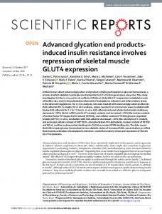
2. Accumulation of AGEs AGEs comprise a heterogeneous group and are formed by a combination of glycation, oxidation, and/or carbonylation, which can be divided into three distinct pathways, as outlined in Fig. 1 [11]. The classical mechanism of AGE formation is the slow Maillard reaction between glucose or reducing sugars and proteins. The interaction between the carbonyl groups of reducing sugars and amino groups of proteins results in the formation of a Schiff base within a few hours. Intramolecular rearrangement of the Schiff base results in more stable Amadori products. Glycated hemoglobin is an example of an Amadori product that is widely used in clinical practice for diagnosis and regulation of diabetes mellitus. The slow process of oxidation of the Amadori products lead to reactive carbonyl compounds and subsequently to the formation of AGEs within weeks to months. The bestknown AGEs derived from this glycoxidation process are pentosidine, Nε-carboxymethyl-lysine (CML) and glucosepane [12]. Other, and much faster evolving, processes involving AGE formation are lipid peroxidation and the glycolysis pathway. In the lipid peroxidation pathway, reactive oxygen species (ROS) alter lipids into reactive carbonyl compounds under influence of oxidation. This formation results into AGEs or advanced lipid end products (ALEs), for example malondialdehyde [11]. This reaction takes place both intracellularly and extracellularly.
Fig. 1. Formation of advanced glycation end products (AGEs). Formation of AGEs by the three different pathways: Maillard reaction, lipid peroxidation and glycolysis pathway. ROS, reactive oxygen species.
L.C. de Vos et al. / Atherosclerosis 254 (2016) 305e313
During the intracellular glycolysis pathway, glucose is altered into reactive carbonyl compounds, of which the best-known is methylglyoxal. The chemical reaction between reactive carbonyl compounds and proteins can result in AGEs. An example of AGEs formed by this pathway is methylglyoxal-derived hydroimidazolone (MG-H1). Besides endogenous formation of AGEs, absorption of exogenous AGEs occurs by two mechanisms. Firstly, accumulation of AGEs occurs by inhalation of tobacco smoke. Tobacco smoke contains highly reactive glycation products which rapidly form AGEs in vitro and in vivo. Serum AGEs are significantly elevated in smokers who smoke at least a package a day as compared to nonsmokers [13]. Secondly, intake of high-AGE food products may lead to an increase of AGEs. The temperature at which food products are prepared is of major importance for the amount of AGEs, with oven frying as most severe inducer [14]. Approximately 10% of the AGEs from food products and beverages are absorbed from the gastrointestinal tract into the blood [15]. For example, serum CML increased after a 6-week high-AGE diet and decreased in low-AGE diet in patients with diabetes mellitus [16].
307
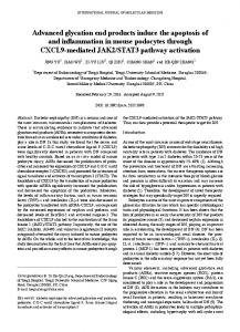
The final mechanism which affects the exposure to and accumulation of AGEs is the clearance of the kidney and metabolism by the liver. Increased level of AGEs can be found in patients with either renal or liver failure [17,18]. This is also in part attributable to increased production of oxidative stress in these diseases, which stimulates formation of AGEs. However, plasma pentosidine decreased with different types of dialysis in renal failure [17]. Furthermore, plasma pentosidine decreased to 80% six months after renal transplantation, and plasma CML decreased to 50% three years after liver transplantation [17,19]. These findings indicate that the accumulation of AGEs in the blood is at least in part reversible in the context of improvement of kidney and liver function. 3. Measurement of AGEs For appropriate assessment of AGEs levels, the biological sample material as well as the method to measure AGEs are important. Firstly, several methods have been developed to measure AGEs in different body compartments, including blood, urine, and tissue (see Fig. 2). Blood and urine samples can obviously be obtained
Fig. 2. Measurements of advanced glycation end products (AGEs).
308
L.C. de Vos et al. / Atherosclerosis 254 (2016) 305e313
easily; however, it is thought that most AGEs are formed intracellularly, but are also bound intracellularly, or in interstitial tissues. For these reasons, circulating AGEs, especially in plasma, do not sufficiently reflect the AGE amount in tissues [20]. In addition, as stated above, the amount of plasma AGEs is affected by its clearance by kidney and liver. Therefore, concentrations of various AGEs in plasma may fluctuate over time. The other possibility is to measure AGEs in tissue. An issue of particular importance is the turnover time of tissues. AGEs crosslinked to collagen or other proteins in interstitial compartments are considered to remain linked during the lifetime of the specific tissue. For example, articular cartilage collagen is thought to have an extremely long half-time, which was calculated to be as high as 117 years [21]. AGEs linked to eye lens proteins remain there lifelong, and accumulate already from the preconceptual period on. Therefore, these AGEs are considered to represent an estimate of long-term metabolic memory. However, most tissue material is not easy to obtain. A relatively easy tissue to acquire is skin tissue, which was calculated to have a half-life of 14.8 years [21]. For this reason, it has been used in early studies on the role of AGE, for example in patients with type 1 diabetes [22]. Several techniques now available to measure AGEs are described in Table 1 [23]. Traditionally, quantitative measurements of AGEs were performed with enzyme-linked immunosorbent assays (ELISA). However, experts in the field state that for quantitative analysis, ELISA has limited specificity and reproducibility [24]. Furthermore, ELISA kits do not measure the difference between protein-bound and free circulating AGEs. Therefore, studies which use this technique, especially when employing older ELISA kits, should possibly be interpreted with caution. High performance and ultra-high performance liquid chromatography methods, most combined with mass spectrometry, have become the technique of choice to measure both free as well as protein-bound AGEs. A method for localization of AGEs is immunohistochemistry, but this technique is not frequently used. In addition, the noninvasive method skin autofluorescence (SAF) has been designed to assess the AGEs of the skin with the so-called AGE Reader™ (DiagnOptics Technologies BV, Groningen, the Netherlands). Eye lens autofluorescence has also recently been proposed for assessing lens AGE accumulation as tool for diabetes screening (Clearpath DS, Freedom Meditech, USA). Some AGEs respond to ultraviolet light by emitting fluorescent light with another wavelength. Meerwaldt et al. showed a strong correlation between SAF and the fluorescent AGE pentosidine as well as the
non-fluorescent AGE CML and Nε-carboxyethyl-lysine (CEL) in the dermal layer of the skin [25]. Since the device uses light to detect AGEs, it is difficult to measure patients with a very dark skin, due to absorption of both the incoming light and the fluorescent light. Another limitation of the AGE Reader™ is the effect of skin cream on SAF measurements. Especially self-browning cream and sun blocker cream block the incident light and cause unreliable SAF measurements [26]. 4. Vascular effects of AGEs AGEs are harmful via two main pathogenic pathways (Fig. 3). Firstly, several AGEs have the potential to form cross-links which results in impaired protein function and turnover, and in increased tissue stiffness as a consequence of collagen and elastin crosslinking. Secondly, AGEs may also bind to cell membrane receptors resulting in release of pro-inflammatory cytokines, thereby enhancing inflammatory reactions. Specific AGEs, such as pentosidine, form cross-links between proteins [27]. Cross-links between and within collagen and elastin fibers result in loss of distensibility and strength, and hence induce arterial stiffness [28]. Results from the Maastricht Study showed a strong association between AGEs and pulse wave velocity as a marker for arterial stiffness. The results showed that plasma protein-bound pentosidine, as well as SAF was positively associated with carotid-femoral pulse wave velocity in 862 patients with normal glucose metabolism, impaired glucose metabolism and type 2 diabetes mellitus [29]. In contrast, plasma CML and CEL were not associated with pulse wave velocity in the latter study. Hofmann et al. showed that tissue AGEs, derived from venous graft material, as well as SAF, were positively associated with pulse wave velocity in patients with coronary artery disease [30]. In addition, stiffness of the heart causes diastolic dysfunction. Several parameter of diastolic dysfunction, measured with echocardiography (e.g. E/A ratio), are associated with both serum AGEs and SAF [31,32]. Secondly, AGEs promote cellular stress responses by engagement to receptors on the cell membrane. The binding of AGEs to RAGE results in intracellular activation of nuclear transcription (NFkB) factor [33]. NF-kB induces the release of several adhesion molecules and pro-inflammatory cytokines such as vascular cell adhesion molecule-1 (VCAM-1), intercellular adhesion molecule-1 (ICAM-1), and interleukin-6. Morita et al. showed that ROS and TNF-a were elevated after stimulation of human aortic endothelial
Table 1 Advantages and disadvantages of techniques to measure AGEs. Technique
Material
Advantages
Disadvantages
Enzyme-linked immunosorbent assay (ELISA)
Liquids including blood and urine and tissue
- Suitable for large number of measurements
(Ultra-)High performance liquid chromatography
Liquids and tissue
Immunohistochemistry
Tissue
-
- Limited specificity, and reproducibility - No differentiation between proteinbound and free circulating AGEs - Lack of standard ELISA kits for AGEs - Expensive due to specific equipment and dedicated personnel
Skin autofluorescence
Skin
- Noninvasive - Fast technique (within seconds) - Point of care testing
Eye lens autofluorescence
Eye lens
- Noninvasive - Fast technique (within seconds)
AGEs, advanced glycation end products.
Accurate and precise measurements Both free as well as protein-bound AGEs Measurement of specific AGEs Localization of AGEs in tissue
- Invasive biopsies needed before analysis - Lack of standardized antibodies for AGEs - Difficult to measure patients with a very dark skin - False elevated levels after use of selfbrowning cream and sun blocker cream block - Limited clinical research
L.C. de Vos et al. / Atherosclerosis 254 (2016) 305e313
309
Fig. 3. Vascular effects of advanced glycation end products (AGEs). CAC, coronary artery calcium; FDG-PET, 18F-fluorodeoxyglucose-positron emission tomography; IMT, intimamedia thickness; PWV; pulse wave velocity; RAGE, receptor for advanced glycation end products.
cells with AGE, which suggests that NF-kB is involved in this reaction [34]. Both TNF-a and ROS induce endothelial dysfunction and, therefore, stimulate the atherosclerotic process. Besides these two pathogenic pathways, there is also evidence in support of local effects of AGEs on plaques in the arterial wall. AGE precursors are associated with macrophage apoptosis, causing rupture-prone atherosclerotic plaques [35]. In 75 carotid artery plaques, increased levels of MG-H1 and CML were found in histological identified rupture-prone plaques versus intermediate and stable plaques [35]. Furthermore, the latter study showed that the AGE precursor dicarbonyl methylglyoxal induced apoptosis of macrophages in vitro. Therefore, AGEs and their precursors are thought to be associated with unstable plaques, which in case of rupture provoke cardiovascular events. The vascular effects of AGE are clinically relevant as shown by the associations between AGEs and atherosclerotic parameters, such as coronary artery calcium (CAC) score, intima-media thickness (IMT), and 18F-fluorodeoxyglucose-positron emission tomography (FDG-PET) (Fig. 3). In a cross-sectional study including 275 Japanese subjects, a positive association was found between glyceraldehyde-derived AGEs, measured with ELISA, and IMT, as well as between glyceraldehyde-derived AGEs and vascular inflammation, measured by FDG-PET [36]. Our research group has found that SAF is associated with IMT in 59 subjects without diabetes mellitus and cardiovascular disease, as well as with CAC score in patients with subclinical atherosclerosis (n ¼ 67) and PAD (n ¼ 60) versus controls (n ¼ 96) [37,38].
5. AGEs in PAD As yet, six observational studies have been published in patients with PAD evaluating levels of AGEs measured in blood or with SAF (Table 2). Lapolla et al. were the first to show elevated serum AGEs including pentosidine in 33 type 2 diabetic patients with PAD versus 66 type 2 diabetic patients without PAD and 20 healthy control subjects [39]. All participants were evaluated by echo Doppler, and in patients with an ankle-brachial index (ABI)