Molecular Biology of the Cell Vol. 19, 5072–5081, December 2008
Aftiphilin and ␥-Synergin Are Required for Secretagogue Sensitivity of Weibel-Palade Bodies in Endothelial Cells Winnie W.Y. Lui-Roberts,* Francesco Ferraro, Thomas D. Nightingale, and Daniel F. Cutler MRC Laboratory of Molecular Cell Biology, Cell Biology Unit and Department of Cell and Developmental Biology, University College London, London WC1E 6BT, United Kingdom Submitted March 20, 2008; Revised September 2, 2008; Accepted September 16, 2008 Monitoring Editor: Francis A. Barr
Formation of secretory organelles requires the coupling of cargo selection to targeting into the correct exocytic pathway. Although the assembly of regulated secretory granules is driven in part by selective aggregation and retention of content, we recently reported that adaptor protein-1 (AP-1) recruitment of clathrin is essential to the initial formation of Weibel-Palade bodies (WPBs) at the trans-Golgi network. A selective co-aggregation process might include recruitment of components required for targeting to the regulated secretory pathway. However, we find that acquisition of the regulated secretory phenotype by WPBs in endothelial cells is coupled to but can be separated from formation of the distinctive granule core by ablation of the AP-1 effectors aftiphilin and ␥-synergin. Their depletion by small interfering RNA leads to WPBs that fail to respond to secretagogue and release their content in an unregulated manner. We find that these non-responsive WPBs have density, markers of maturation, and highly multimerized von Willebrand factor similar to those of wild-type granules. Thus, by also recruiting aftiphilin/␥-synergin in addition to clathrin, AP-1 coordinates formation of WPBs with their acquisition of a regulated secretory phenotype.
INTRODUCTION Cells must rigorously control their release of bioactive proteins. This is reflected in the careful packaging of such material into secretory granules that only undergo exocytosis in response to an external physiological trigger. In contrast, the constitutive secretory pathway is taken by proteins that are not concentrated and stored in secretory granules (Burgess and Kelly, 1987). Many studies of secretory granule formation have focused on the sorting and processing of their secreted contents. Selective aggregation of regulated secretory proteins in response to conditions within the Golgi/trans-Golgi network (TGN) to form the granule core, and the removal of missorted proteins during maturation play prominent roles in models of granule biogenesis (for reviews, see Arvan and Castle, 1998; Dikeakos and Reudelhuber, 2007). For membrane proteins, behavior similar to that of soluble content has been found, i.e., recruitment via direct interaction of their lumenal domains with the core proteins (e.g., Colomer et al., 1996; Rindler, 1998; Michaux et This article was published online ahead of print in MBC in Press (http://www.molbiolcell.org/cgi/doi/10.1091/mbc.E08 – 03– 0301) on September 24, 2008. Present address: * Cambridge Institute for Medical Research, Wellcome Trust/MRC Building, Addenbrooke’s Hospital, University of Cambridge, Hills Rd., Cambridge CB2 0XY, United Kingdom. Address correspondence to: Daniel F. Cutler (
[email protected]). Abbreviations used: GGA, Golgi-localized, ␥-ear-containing, ADP ribosylation factor (ARF)-binding proteins; HEK, human embryonic kidney; HMW, high molecular weight; HUVEC, human umbilical vein endothelial cell; LMW, low molecular weight; PMA, phorbol 12-myristate 13-acetate; ssHRP, signal sequence HRP; VWF, von Willebrand factor; WPB, Weibel-Palade body. 5072
al., 2006b). Granule membrane proteins are also recycled after exocytosis for re-incorporation at the TGN in routing dependent on sequences within their cytoplasmic domains (Milgram et al., 1996; Wasmeier et al., 2005; Harrison-Lavoie et al., 2006). Furthermore, their transmembrane domains may aid in selective partitioning into forming granules (Fleming et al., 1998), although there are examples (e.g., vesicular monoamine transporter 2) where targeting seems solely dependent on elements within their cytoplasmic domain (Waites et al., 2001). In addition, roles for sorting receptors and for protein–lipid interactions have been implicated. All of the processes described above may act together in granule formation (Dikeakos and Reudelhuber, 2007, which references many of the key articles not otherwise cited above). However, in all this work, recruitment of the proteins required for exocytosis of granules has not been much studied, despite the importance of this aspect of their biogenesis. Endothelial cells have a secretory organelle that plays a key role in hemostasis and acute inflammation. These are large cigar-shaped organelles called Weibel-Palade bodies (WPBs) (for recent reviews, see Michaux and Cutler, 2004; Rondaij et al., 2006; Metcalf et al., 2008). Exocytosis of WPBs brings P-selectin and von Willebrand factor (VWF) to the cell surface to recruit leukocytes and platelets, respectively. Freshly released VWF from WPBs, which is highly multimerized and unfurls into platelet-catching filaments that can be hundreds of micrometers long, is particularly active in recruiting platelets (Michaux et al., 2006a). The storage and release of these biologically active contents of WPBs must be tightly controlled. Indeed, defects in the multimerization or expression of VWF lead to von Willebrand’s disease, which is the most common heritable bleeding disorder. Conversely, elevated levels of active VWF have been linked to thrombotic manifestations of various diseases, including © 2008 by The American Society for Cell Biology
Regulation of Weibel-Palade Body Exocytosis
HELLP (hemolysis, elevated liver enzymes, low platelets) syndrome in pregnant women and thrombotic thrombocytopenic purpura (Groot et al., 2007). In addition, atherosclerosis has been linked to high plasma levels of soluble Pselectin (Dong et al., 2000; Molenaar et al., 2003). Conversely, P-selectin knockout mice have defective leukocyte recruitment to sites of inflammation (Wagner, 1995). Since VWF is exceptionally effective at driving the formation of WPBs—most dramatically shown by heterologous expression (Wagner et al., 1991)—-it had been thought to be an example of granule biogenesis driven by core self-assembly. However, we recently found a key role for cytoplasmic machinery in the biogenesis of this granule (Lui-Roberts et al., 2005). We found that adaptor protein-1 (AP-1) controls the initial formation of WPBs at the TGN by recruiting clathrin. Depletion of AP-1 with small interfering RNA (siRNA) or disruption of clathrin function with a dominantnegative AP180 construct leads to the failure of formation of new WPBs at the TGN in addition to an abolition of stimulated secretion and an increase in secretagogue-independent secretion. The extensive AP-1/clathrin coat surrounding forming and immature WPBs is believed to assist the initial formation of these large elongated organelles, which can be up to 5 m in length, by acting as a structural scaffold. Thus, for the biogenesis of WPBs, both content-driven events and cytoplasmic machinery are necessary. Since regulation of release of the pro-thrombotic VWF is so important, we hypothesized that acquisition of the WPB proteins required for their secretagogue response would be coupled to the initial formation of this organelle. If so, is this acquisition dependent on co-aggregation, or on AP-1 and its effectors? Since the removal of AP-1 itself leads to a failure to make WPBs, it was not possible to establish whether AP-1 also contributes to WPB function beyond initial formation by simply depleting it. However, the small VWF-positive vesicular structures found in AP-1– depleted cells do not respond to phorbol 12-myristate 13-acetate (PMA) stimulation (Lui-Roberts et al., 2005), suggesting that AP-1 and its effectors might play a role in the acquisition of the regulated secretory phenotype. We therefore focused on the role of AP-1 effectors in WPB biogenesis. In this article, we used siRNA duplexes to deplete AP-1 effectors, and we analyzed the effect of their removal on formation and secretion of WPBs. We examined the altered WPBs of both human umbilical vein endothelial cells (HUVECs) and those pseudo-WPBs made by the model system of HEK293 cells heterologously expressing VWF. We focused on the AP-1–interacting aftiphilin/␥-synergin/p200 complex, partly because very little was known about its function and partly because although its removal in HeLa cells can phenocopy siRNA-mediated ablation of AP-1, it seems to have additional functions (Hirst et al., 2005). These data made the complex of high interest to us, since we were looking for an AP-1 effector whose loss could give a phenotype distinct from that of AP-1 deficiency. We found that the loss of aftiphilin and to a lesser extent ␥-synergin cause a switch of VWF secretion from the regulated pathway to constitutive release, even though WPBs in aftiphilin-depleted cells are made efficiently and possess all the known hallmarks of maturity. We have therefore managed to uncouple the formation and maturation of a secretory granule from its ability to undergo regulated exocytosis: content packaging and acquisition of the regulated secretory phenotype are coordinated by AP-1 at an early stage of WPB biogenesis. Vol. 19, December 2008
MATERIALS AND METHODS Antibodies and Immunofluorescence Rabbit anti-VWF and its horseradish peroxidase (HRP)-conjugated derivative were purchased from Dako UK (Ely, Cambridgeshire, United Kingdom), and sheep anti-VWF and mouse anti-P-selectin (clone AK6) were from Serotec (Oxford, UK). Mouse anti-green fluorescent protein (GFP) and rabbit-antiactin were supplied by Sigma Chemical (Poole, Dorset, United Kingdom). Aftiphilin (Hirst et al., 2005), ␥-synergin (Page et al., 1999), p200 (Lui et al., 2003), and epsinR (Hirst et al., 2003) antibodies were kind gifts from M. S. Robinson (Cambridge Institute for Medical Research, Cambridge, United Kingdom). Mouse monoclonal anti-CD63 (clone 1B5) was a kind gift of M. Marsh (MRC Laboratory for Molecular Cell Biology, London, United Kingdom). HRP-, fluorescein isothiocyanate (FITC)-, Texas Red-, and Cy5-conjugated secondary antibodies were from Jackson ImmunoResearch Laboratories (West Grove, PA), whereas Alexa Fluor-conjugated secondary antibodies were from Invitrogen (Carlsbad, CA). Immunofluorescence was carried out as described previously (Lui-Roberts et al., 2005). Mounted coverslips were examined at ambient temperature through a 60⫻ oil immersion lens (numerical aperture [NA] 1.4) on an Optiphot 2 microscope (Nikon, Tokyo, Japan), fitted with an MRC 1024 confocal laser scanner (Bio-Rad, Hemel Hempstead, Hertfordshire, United Kingdom), or in Figure 4, through a 63⫻ oil immersion lens (NA 1.3) on a TCS SPE confocal microscope system (Leica, Wetzlar, Germany). For double- and triple-labeling experiments, the channels were scanned sequentially. Adobe Photoshop 6.0.1 and Illustrator 10 (Adobe Systems, Mountain View, CA) were used to generate figures from digital images.
Cell Culture and Transfection HUVECs and HEK293 cells were cultured as described previously (Michaux et al., 2006a). The full-length human VWF construct in pCI-neo (Michaux et al., 2003) and signal sequence (ss)HRP construct (Connolly et al., 1994) have been described previously. GFP-VWF (Romani de Wit et al., 2003) and GFP-Rab27a (Hume et al., 2001) were gifts from J. Voorberg and J. A. Van Mourik (Sanquin Research at CLB, Amsterdam, The Netherlands) and M. C. Seabra (Imperial College London, London, United Kingdom), respectively. DNA and siRNA were transfected into mammalian cells by nucleofection (Amaxa Biosystems, Gaithersburg, MD). Typical transfection rate is 30 –70% for DNA and 90% for siRNA. For microinjection, DNA (0.05– 0.1 g/l) and siRNA (0.05 g/l) were injected together with biotin-dextran into the nuclei of ⬃50 cells over a period of 20 min.
RNA Interference (RNAi) and Secretion Assays All siRNA duplexes were purchased from QIAGEN (Dorking, Surrey, United Kingdom), unless otherwise stated. The sequences were (A)AGCAGUUGCUAGUGGCCAUU for aftiphilin, CAGCAGCUCCUAUUCCAACUU for ␥-synergin (Hirst et al., 2005), AAGGCAUCAAGUAUCGGAAGA for 1A (Hirst et al., 2003), and AAUACAGAUAUGGUCCAGAAA for epsinR (Hirst et al., 2004). For p200, siGENOME SMARTpool XM_042685 (p200a) and siGENOME SMARTpool XM_113763.5 (p200b) from Dharmacon RNA Technologies (Lafayette, CO) were used (Hirst et al., 2005). Cells were transfected with 100 –300 pmol of siRNA by nucleofection (Amaxa Biosystems) by using the nucleofection program A-23 for HEK293 cells and U-01 for HUVECs. Typically, a 15-cm Petri dish of cells that were 70 – 80% confluent were used for six to eight nucleofection reactions. Two reactions were plated onto each 9-cm Petri dish and incubated for 2–3 d at 37°C. The cells were then nucleofected again with 100 –300 pmol of siRNA; and in the HEK293 cells, with 4 g of a full-length VWF DNA construct (Michaux et al., 2003). After plating onto six-well plates, they were incubated for 2–3 d until the cells were ⬃90% confluent before being processed for immunofluorescence and secretion assays. The VWF secretion assay has been described previously (Lui-Roberts et al., 2005). In short, cells were rinsed and incubated in a medium without a secretagogue for 30 min of constitutive secretion. The medium was then collected and replaced with a medium containing PMA as a secretagogue. The stimulated release was collected and the remaining VWF was released by cell lysis. The relative amounts of VWF were quantified by enzyme-linked immunosorbent assay (ELISA) (Blagoveshchenskaya et al., 2002), and the data were normalized using the total VWF signal. The PMA-responsive pool was estimated by subtracting the amount of VWF released by constitutive secretion from that released upon PMA addition. The ssHRP secretion assay was carried out as described in Lui-Roberts et al. (2005).
Subcellular Fractionation For each gradient, two confluent 15-cm dishes of HEK293 cells transiently expressing VWF were homogenized on ice by 10 –12 passages through a ball-bearing homogenizer with a 0.008-mm clearance (European Molecular Biology Laboratory, Heidelberg, Germany) in 1 ml of buffer containing 10 mM HEPES, pH 7.4, 0.25 M sucrose, 1 mM MgCl2, 800 U/ml DNase, and a protease inhibitor cocktail (Sigma Chemical). The postnuclear supernatant was obtained by centrifuging at 600 ⫻ g for 10 min at 4°C in an Avanti 30
5073
W.W.Y. Lui-Roberts et al. centrifuge (Beckman Coulter, Fullerton, CA). It was then loaded onto a preformed 20 – 60% continuous sucrose gradient, made with a Gradient Master (BioComp Instruments, Fredericton, NB, Canada), and centrifuged to equilibrium at 35,000 rpm for 16 h at 4°C in a SW40Ti rotor in an Optima LE-80K ultracentrifuge (Beckman Coulter). Twenty-four fractions of 0.5 ml each were collected from the top using a Fractionator (BioComp Instruments). The relative amounts of VWF were quantified using an ELISA described previously (Blagoveshchenskaya et al., 2002).
Rescue Experiments An expressed sequence tag (EST) encoding full-length aftiphilin (IMAGE Clone I.D. 6014735; gi 21175557) in pCMV-SPORT6 was used for generating an siRNA-resistant aftiphilin construct. The siRNA target sequence was mutated from AGCAGTTGCTAGTGGCCATT to AGCTGTAGCTAGCGG TCATT by using QuikChange site-directed mutagenesis kit (Stratagene, LaJolla, CA). The first round of transfection was carried out as described previously. To prevent unnecessary stress to the cells by introducing too much DNA and siRNA, we have used relatively low amounts of siRNAresistant aftiphilin and VWF DNA in the rescue experiments. One microgram of the mutated aftiphilin and 3 g of full-length VWF plasmids were used in each transfection. VWF secretion assays were carried out 2 d after the second round of transfection.
VWF Multimer Analysis Agarose gels (1.4%) were prepared by dissolving Seakem high gelling temperature agarose (Lonza, Wokingham, United Kingdom) in 0.2 M Tris, 0.1 M glycine, pH 9.0. SDS was then added to a final concentration of 0.1%. Secretion assay samples were concentrated using Vivaspin 500 centrifugal filter units (Sartorius, Goettingen, Germany) before loading onto the SDS-agarose gels. The gels were run at 60 V for 20 min and then 40 V for ⬃3 h in a miniPROTEAN 3 electrophoresis system (Bio-Rad). The proteins transferred to
nitrocellulose membranes were labeled with rabbit anti-VWF (Dako UK) and then HRP-conjugated donkey anti-rabbit, followed by SuperSignal West Pico chemiluminescent substrate (Pierce Chemical, Rockford, IL). The VWF multimer pattern was analyzed using a Molecular Imager GS-800 calibrated densitometer and the Quantity One software (Bio-Rad). The data were normalized to the total signal, after subtraction of the global minimum value. The relative amounts of VWF of different multimeric states were estimated by calculating the areas under different regions of the curve.
RESULTS Aftiphilin and ␥-Synergin Are Critical to Regulated Secretion of VWF Heterologous expression of VWF in HEK293 cells lead to the de novo formation of pseudo-WPBs, which have every characteristic of endothelial WPBs. They have the same elongated shape and incorporate VWF tubules, which have a characteristic striated appearance under the electron microscope; they also recruit all the known components of WPBs, including P-selectin, CD63, and Rab27a. Most importantly, they can undergo regulated secretion triggered by secretagogues (Michaux et al., 2003). The HEK293 system has several advantages in the study of WPB biogenesis and function. RNAi often works more efficiently in HEK293 cells than in the primary HUVECs. In addition, as opposed to HUVECs, the absence of an endogenous pool of VWF within preformed secretory granules makes HEK293 cells a better
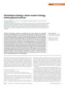
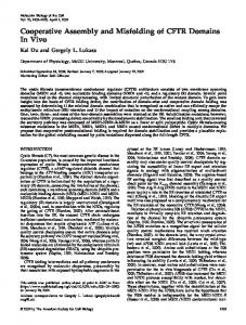
Figure 1. Aftiphilin and ␥-synergin affect the ratio of VWF released in the constitutive and regulated pathways. (A) HEK293 cell lysates from mock and knockdown cells were analyzed by Western blotting. Actin was used as the loading control. The bottom panel shows the protein levels of the p200/aftiphilin/␥-synergin complex when one subunit is targeted by siRNA in the HEK293 system. (B) Effect of siRNAs against AP-1 effectors on VWF secretion assays. HEK293 cells were transfected with siRNA and incubated for 3 d, before the co-transfection of full-length VWF and siRNA. VWF secretion assays were carried out when the cells reached 90% confluence. After 30 min of constitutive secretion (left, blue bars), the medium was collected and fresh medium containing the secretagogue PMA was added. The medium was collected 30 min later. To determine the PMA-responsive pool of VWF (right, red bars), the amount released constitutively was subtracted from the PMA releasate. A representative experiment is shown here. (C) Aftiphilin and ␥-synergin depletion led to significant increase in constitutive secretion and reduction in regulated secretion. Here, the data were normalized by dividing by the mock data from the same experiments, and pooled from multiple secretion assays (n ⫽ 7 for aftiphilin, n ⫽ 6 for ␥-synergin, and n ⫽ 5 for epsinR). The error bars represent SEs (t test results compared with mock: *p ⬍ 0.01; E ⫽ 0.03). (D) Immunofluorescence showing effective knockdown of aftiphilin in siRNA-treated HEK293 cells. Bar, 10 m. (E–L) Partial colocalization of aftiphilin and VWF in the perinuclear region. HUVECs were fixed with 6% paraformaldehyde, permeabilized with saponin, and immunolabeled with sheep anti-VWF (F and H; red in E) and rabbit anti-aftiphilin (G and I; green in E). The secondary antibodies used were Texas Red-anti-sheep and FITC-anti-rabbit. Bar, 10 m. (F–I) Magnified view of the boxed areas in E. (J–L) Partial colocalization of ␥-synergin and VWF in the perinuclear region. HUVECs were fixed and permeabilized using methanol and acetone, followed by immunolabeling with sheep anti-VWF (K; red in J) and rabbit anti-␥-synergin (L; green in J). The secondary antibodies were as above. Bar, 10 m. (K and L) Magnified view of the boxed area in J. 5074
Molecular Biology of the Cell
Regulation of Weibel-Palade Body Exocytosis
option for biochemical studies of WPB biogenesis. Figure 1A shows the efficiency of depletion of the aftiphilin/␥-synergin/p200 complex. Aftiphilin was depleted by 89 ⫾ 5% (n ⫽ 5), whereas ␥-synergin and p200a were knocked down by 63 ⫾ 18% (n ⫽ 3) and 80 ⫾ 8% (n ⫽ 5), respectively. We used an established VWF secretion assay to determine regulated and constitutive exocytosis of VWF (Michaux et al., 2003; Lui-Roberts et al., 2005). Cells were incubated in the absence and then the presence of the secretagogue PMA for the constitutive and stimulated secretions (see Materials and Methods). The PMA-responsive pool was estimated by subtracting the amount of VWF released by constitutive secretion from that released upon PMA addition. Using this assay, we examined the effects of RNA interference of AP-1 effectors by using published siRNA duplexes (see Materials and Methods). The siRNAs were transfected into HEK293 cells and given 3 d to start depletion, before co-transfection with full-length VWF and a further incubation of 2–3 d. Release assays were then carried out to study the effects of siRNA on WPB function. EpsinR (Kalthoff et al., 2002; Wasiak et al., 2002; Hirst et al., 2003; Mills et al., 2003), another AP-1–interacting protein, was depleted by 73% (Figure 1A) and was tested in parallel. Figure 1B shows the results of a VWF secretion assay. Among all the proteins tested, only the depletion of aftiphilin and ␥-synergin had any effect on VWF secretion. Figure 1C shows the normalized results from multiple experiments. The data shown were obtained by dividing the data from secretion assays of the knockdown cells by that of mock-treated cells from the same experiments. Although epsinR siRNA had no significant effect on the regulated secretion of VWF, siRNAs against aftiphilin or ␥-synergin significantly reduced the stimulated release to 20% of mock level (p ⬍ 0.01, t test). Constitutive secretion was increased in both cases (p ⬍ 0.01, t test), with a more dramatic increase to 190% of mock level with aftiphilin depletion and 139% with ␥-synergin depletion. We found that siRNA-mediated depletion of epsinR has a small but significant effect on constitutive release of VWF, but since there is no significant effect on the regulated release of VWF, this is clearly not affecting the formation or behavior of WPBs and is specific to the constitutive secretory pathway. This is in marked contrast to the data obtained on aftiphilin and ␥-synergin and shows that not all AP-1 effectors are involved in WPB biogenesis. The secretory phenotype seen after siRNA-mediated reduction of aftiphilin and ␥-synergin is similar to that seen in experiments where AP-1 has been ablated, i.e., a dramatic reduction of regulated secretion and an increase in constitutive release. Thus losing these AP-1 effectors can phenocopy the loss of AP-1 itself at the level of exocytosis. The similarity in the aftiphilin and ␥-synergin depletion phenotypes is likely to reflect the fact that they belong to the same complex (Hirst et al., 2005), whereas the different degrees of the effects were probably caused by the higher efficiency of aftiphilin knockdown (Figure 1A). p200 is reported to be part of a complex with aftiphilin and ␥-synergin. It was therefore surprising that p200 siRNA did not give the same effect as ablation of the other two members. In HeLa cells, when a subunit of the complex is depleted, the other two components become unstable (Hirst et al., 2005). So, in theory, p200 depletion should at least indirectly cause a phenotype similar to aftiphilin depletion. We determined that p200a was depleted by 80 ⫾ 8% in HEK293 cells. We carried out Western blotting analyses in the HEK293 system and found out that p200 ablation caused a decrease in aftiphilin level to 50% and a milder decrease in ␥-synergin level (Figure 1A)—not to the same extent as in Vol. 19, December 2008
HeLa cells (Hirst et al., 2005). p200b, another isoform of p200, was targeted along with p200a by our Dharmacon siGENOME SMARTpool siRNA mix, but we were unable to determine the level of its knockdown because no antibodies against p200b are available. Therefore, it is currently not possible to distinguish whether there is enough p200b left to contribute to WPB function, or p200 is simply not required. GFP-VWF expression studies can be used to track newly formed secretory granules in HUVECs. This approach showed that immature WPBs are in the perinuclear region of the cell, whereas older mature WPBs are localized in the cell periphery (Hannah et al., 2003). We carried out immunofluorescence studies in HUVECs and observed partial colocalization of endogenous VWF with aftiphilin and also ␥-synergin in the perinuclear region (Figure 1, E–L). This is consistent with the presence of AP-1 on immature WPBs near the TGN (Lui-Roberts et al., 2005). Aftiphilin and ␥-synergin are known to interact with AP-1 (Page et al., 1999; Hirst et al., 2005) and are therefore likely to be recruited to forming WPBs by AP-1 and exert their function at this early stage. Effect of Aftiphilin Depletion on VWF Secretion Is Specific As shown in Figure 1C, the phenotype is stronger with the aftiphilin siRNA than that for ␥-synergin, probably due to a higher efficiency of knockdown (Figure 1A). We have therefore focused this study on aftiphilin, while occasionally referring to our results with ␥-synergin. Figure 1A shows that aftiphilin was efficiently depleted by 89 ⫾ 5% after two rounds of nucleofection in HEK293 cells, and it was further confirmed by immunofluorescence (Figure 1D). To prove that the effect of aftiphilin depletion is not due to off-target effects of the siRNA duplex, we produced an siRNA-resistant clone by introducing four point mutations to a full-length aftiphilin EST clone (IMAGE Clone I.D. 6014735; gi 21175557) from AGCAGTTGCTAGTGGCCATT to AGCTGTAGCTAGCGGTCATT. This mutated clone encodes the same protein sequence, but has four mismatches with the siRNA sequence and hence should be resistant to silencing. HEK293 cells were transfected with aftiphilin siRNA and incubated for 3 d. Then, in the second round of transfection, cells were transfected with the siRNA, fulllength VWF, and the siRNA-resistant aftiphilin construct. It was followed by a VWF secretion assay 2 d later (Figure 2B). To prevent unnecessary stress to the cells by introducing too much DNA and siRNA, we have used relatively low amounts of both DNA constructs; and to optimize expression of the rescue plasmid, we used the cells at low confluence (see Materials and Methods). It has been reported that the number of WPBs is linked to cell confluence (Howell et al., 2004), and this likely explains why the level of regulated VWF secretion in this set of experiments was lower than that in Figure 1B. As shown in Figure 2B, the aftiphilin knockdown phenotype can be partially rescued by mild expression of the siRNA-resistant clone, confirming that the aftiphilin siRNA is specific. The partial nature of the rescue likely arose from two main factors: 1) We used a relatively small amount of siRNA-resistant aftiphilin DNA to prevent any potential side effects of overexpression. Aftiphilin expression in the rescue samples was 61 ⫾ 6% (n ⫽ 6) of that of mock level (Figure 2A). Mild expression of aftiphilin is therefore likely to have contributed to the partial rescue. 2) This was a triple transfection experiment and a complete rescue would be technically difficult to achieve as it relies on co-transfection of all three components into the same cells. That we have a partial but significant rescue with mild expression of the siRNA-resistant aftiphilin clone argues that the effect of aftiphilin depletion is not an off-target effect. 5075
W.W.Y. Lui-Roberts et al.
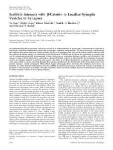
Figure 2. Effect of aftiphilin depletion on VWF secretion is specific. (A and B) Rescue of aftiphilin knockdown by mutant aftiphilin cDNA. HEK293 cells were transfected with aftiphilin siRNA and incubated for 3 d. They were then transfected again with the siRNA and a full-length VWF plasmid, with or without an siRNA-resistant aftiphilin construct. VWF secretion assays were carried out 48 h later. (A) Western blotting showing the expression of siRNA-resistant aftiphilin plasmid restored the expression of aftiphilin to 61 ⫾ 6% of mock level (n ⫽ 6). Actin was used as a loading control. (B) The aftiphilin depletion phenotype was partially rescued by mild expression of the siRNA-resistant aftiphilin plasmid. Error bars represent SEs; n ⫽ 6. (The asterisks, squares and circles show pairs of samples compared using t tests: *p ⫽ 0.015, significantly different; 䡺, p ⫽ 0.059; E, p ⫽ 0.2; F, p ⫽ 0.2, statistically not different). (C) Effect of aftiphilin depletion is specific to VWF. ssHRP, a constitutive secretion marker, was co-transfected into HEK293 cells in the second round of siRNA transfection and left for 1 d. The ratio of the amount of ssHRP released in 6 h to that remained in the cells was plotted in the graph. The triangles and diamonds show pairs of samples compared using t tests. There was no significant difference between mock cells and those transfected with aftiphilin (Œ, p ⬎ 0.05) or epsinR siRNA(〫, p ⬎ 0.05). Error bars represent SEs; n ⫽ 6.
We further tested whether the effect of aftiphilin knockdown is specific to proteins in the regulated secretory pathway. We co-transfected ssHRP (signal sequence HRP), the secretory form of HRP and a marker of the constitutive secretory pathway (Connolly et al., 1994), into HEK293 cells during the second round of siRNA transfection. An ssHRP secretion assay was then carried out 24 h after transfection. Aftiphilin depletion caused no significant difference in ssHRP secretion when compared with mock-treated cells or those transfected with epsinR siRNA (Figure 2C). This shows that the effect of aftiphilin depletion is specific to VWF. It also confirms that there is no general malfunction of the Golgi leading to a blockage in the exit of proteins. Aftiphilin-depleted Cells Can Still Make WPBs As mentioned, WPBs are made upon heterologous expression of VWF in HEK293 cells (Figure 3A). If loss of aftiphilin uncouples the regulated secretory phenotype from core formation, then we should still see recognizable cigar-shaped WPBs after siRNA treatment, in contrast to AP-1– depleted cells, which cannot make the cigar-shaped organelles (Lui-Roberts et al., 2005; Figure 3, M and N). We found that aftiphilin-depleted cells can still make elongated WPBs (Figure 3B). We further tested whether this is also the case for HUVECs. Aftiphilin siRNA was transfected into HUVECs by nucleofection and left for 3 d for the depletion to progress, before introducing GFPVWF in the second round of siRNA transfection to track newly formed granules. As shown in Figure 3, C–F, both mock and aftiphilin knockdown cells are capable of making WPBs. We obtained similar results in ␥-synergindepleted cells (Figure 3, G–J). These data strongly suggest that WPBs core formation can still continue despite loss of secretagogue responsiveness. Another event that is linked to initial formation of WPBs at the TGN and which is lost when core formation is disrupted by AP-1 depletion is the incorporation of the leukocyte receptor P-selectin into the granule (Harrison-Lavoie et al., 2006). To determine whether the VWF-positive structures made in the absence of aftiphilin are capable of normal recruitment of this integral membrane protein, we transfected HUVECs with aftiphilin siRNA and carried out im5076
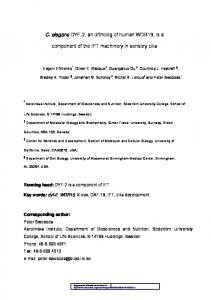
munofluorescence studies using a P-selectin antibody (Figure 4). In marked contrast to AP-1-depleted cells, which have no P-selectin in the VWF-positive structures (LuiRoberts et al., 2005), P-selectin recruitment to WPBs in aftiphilin-depleted cells seems normal, providing further evidence for the formation of WPBs. To quantify whether WPB production is affected by aftiphilin depletion, we carried out subcellular fractionation of HEK293 cells transfected with VWF and aftiphilin siRNA. The postnuclear supernatant was centrifuged in an equilibrium gradient of 20 – 60% sucrose to separate organelles according to density. The relative amount of VWF in the fractions was then measured by ELISA (Blagoveshchenskaya et al., 2002). Figure 3K shows a typical fractionation profile of postnuclear supernatants of mock (blue diamonds) and aftiphilin knockdown (red circles) cells. The light peak is enriched in the endoplasmic reticulum (ER) and Golgi fractions as biosynthetic VWF passes through these organelles, whereas the dense peak is enriched in WPBs. Multimerization occurs as VWF progresses through the secretory pathway (for review, see Michaux and Cutler, 2004). It dimerizes at the ER and further oligomerizes in the Golgi apparatus, and reaches its highest multimeric state in WPBs (Ewenstein et al., 1987). Figure 3L shows the VWF multimer analysis of the light and dense peaks on an SDS-agarose gel. The lowest band is the VWF dimer and the multimeric state is increased by two for each band upwards. The ladder of high molecular weight VWF multimers in fraction 18 confirms that the dense peak is indeed enriched in WPBs. It is important to note that aftiphilin-depleted cells are capable of making WPBs that contain ultra-high molecular weight VWF, which is a hallmark of mature WPBs. In addition, we observed no consistent density shift of the WPB fraction in the subcellular fractionation experiments (Figure 3K), suggesting that there was no major granule condensation defect in aftiphilin-depleted cells. From two independent transfection and fractionation experiments, we found that the amounts of VWF in the dense peak are roughly comparable for mock and aftiphilin knockdown cells, suggesting that aftiphilin depletion does not significantly impair the efficiency of WPB production. Although the high level of dimers in this fraction may represent ER contamination, it is likely that they are true Molecular Biology of the Cell
Regulation of Weibel-Palade Body Exocytosis
Figure 3. WPBs are made in aftiphilin-depleted cells. (A and B) HEK293 cells were nucleofected with aftiphilin siRNA (B) or mocktreated (A), and after 3 d, were further nucleofected with full-length VWF and siRNA. Fixation and immunolabeling with anti-VWF were carried out 2 d later. Insets, magnified view of the boxed areas. Bar, 10 m. (C–F) HUVECs were nucleofected with aftiphilin siRNA (E and F) or mock-treated (C and D). After 3 d, the cells were nucleofected again with siRNA and GFP-VWF and incubated for 1 d, followed by fixation and immunolabeling with anti-aftiphilin and anti-GFP. Secondary antibodies used were Alexa Fluor 594-anti-rabbit and Alexa Fluor 488anti-mouse. Insets, magnified view of the boxed areas. Bar, 10 m. (G–J) ␥-Synergin– depleted cells are capable of making WPBs. HUVECs were nucleofected with ␥-synergin siRNA (I and J) or mock-treated (G and H). After 3 d, the cells were microinjected with siRNA and GFPVWF and incubated for 1 d, followed by methanol/acetone fixation and immunolabeling with anti-␥-synergin (G and I) and anti-GFP (H and J). Insets, magnified view of the boxed areas. Bar, 10 m. (K–L) Subcellular fractionation shows the efficiency of WPB production in aftiphilin-depleted cells. (K) Mock-treated (blue diamonds) and aftiphilin-depleted (red circles) HEK293 cells, co-transfected with fulllength VWF, were homogenized and the post-nuclear supernatants were fractionated using a density gradient of 20 – 60% sucrose. The dense peak corresponds to the WPB fraction. (L) VWF multimer analysis of fractions from the light and dense peaks on a 1.4% SDS-agarose gel. Highly multimerized VWF is found in the WPB-enriched fractions from both mock and aftiphilin-depleted cells. (M–N) AP-1-depleted cells cannot make WPBs. HUVECs were nucleofected with AP-1 siRNA or mock-treated, followed by a 2-d incubation and a second round of nucleofection. Mock-treated and AP-1-depleted cells were plated onto the same dish. After 2 d of incubation, cells were fixed and analyzed by immunofluorescence microscopy using anti-␥-adaptin (M) and anti-VWF (N). Bar, 10 m.
residents in the WPBs in HEK293 cells. Wagner and Marder showed that although the ratio of high-molecular-weight (HMW) multimers to low-molecular-weight (LMW) multimers increases with time, dimers are still prominent even after a 67-h chase— by which time VWF must have left the ER and be within storage organelles (Wagner and Marder, 1984). This is consistent with the idea that multimerization is not yet complete as WPBs leave the TGN. Indeed, short, not fully multimerized VWF tubules are found in budding WPBs at the TGN, and WPBs continue to become more compact as they mature (Zenner et al., 2007). In addition, we suspect that multimerization may be a bit slower in HEK293 cells because they do not make as many WPBs as HUVECs do. As expected, our light fractions contain less HMW multimers than in HUVEC Golgi fractions (Vischer and Wagner, 1994). As a consequence, we would expect more dimers in the WPBs of HEK293 cells. Vol. 19, December 2008
Maturation of WPBs in Aftiphilin-depleted Cells Newly formed WPBs and older mature WPBs are localized in the perinuclear region and the cell periphery, respectively (Hannah et al., 2003; Harrison-Lavoie et al., 2006). The small GTPase Rab27a and the tetraspanin CD63 are absent from the perinuclear WPBs, and their presence is a key feature of mature WPBs. To further investigate whether the switch to constitutive secretion of VWF caused by aftiphilin depletion is related to a failure in WPB maturation, we tested the recruitment of these proteins. HUVECs were transfected with the aftiphilin siRNA duplex by nucleofection and incubated for 3 d (Figure 5, A–H). GFP-Rab27a and the siRNA were then co-microinjected into the nuclei to ensure that both the DNA and siRNA were in the same cell. Figures 5B and 5F show that aftiphilin was effectively depleted by this treatment. As shown in Figure 5, E–H, GFP-Rab27a can be recruited to WPBs even when aftiphilin is depleted. This is 5077
W.W.Y. Lui-Roberts et al.
Figure 4. Recruitment of P-selectin to WPBs in aftiphilin-depleted cells. HUVECs were transfected with aftiphilin siRNA (E–H) or mock-treated (A–D) as described above, fixed and immunolabeled with rabbit anti-aftiphilin (A and E), mouse anti-P-selectin (B and F), and sheep anti-VWF (C and G). D and H are merged images of A, B and C, and E, F and G, respectively. It was then followed by Alexa Fluor 594 donkey antirabbit, Alexa Fluor 488 donkey anti-mouse, and Alexa Fluor 680 donkey anti-sheep. Projected Z-stacks are shown here. Bar, 10 m.
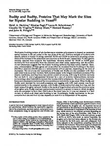
in contrast to the failure of GFP-Rab27a recruitment in AP-1– depleted cells (Lui-Roberts et al., 2005). Similarly, aftiphilin knockdown did not affect CD63 recruitment (Figure 5, I–P). Together with the observations described in the previous section, we conclude that WPBs can undergo maturation in aftiphilin-depleted cells. WPBs in Aftiphilin-depleted Cells Exocytose in an Unregulated Manner We observed a switch from regulated to constitutive secretion in aftiphilin-depleted cells, whereas there is no apparent drop in the quantity of WPBs at steady state. This implies that the increase in constitutive secretion may be caused by uncontrolled WPB exocytosis. To prove that this is the case, we have used the fact that the VWF in WPBs has a higher multimeric state than that in the ER and the Golgi/TGN. As shown in Figure 3L, the VWF from the light fractions enriched in ER and Golgi is only multimerized up to octamers. We therefore defined multimers higher than octamers as HMW multimers that can only be found in WPBs. Figure 6A shows the multimer analysis of constitutively released VWF from a secretion assay, with the data normalized to total signal. The far right peak corresponds to the VWF dimer, and the multimeric state increases from right to left of the graph. Constitutive secretion from mock-treated cells has predominantly LMW VWF multimers (Figure 6A). In contrast, there is a clear increase of HMW multimers in the constitutive release from aftiphilin-depleted cells, suggesting VWF is now released from WPBs even in the absence of stimulation. Figure 6B shows the densitometry traces of constitutive and PMA releasates on the same graph. It should be noted
that, unlike Figure 1B in which the PMA-responsive pool was estimated by subtracting the amount of VWF released by constitutive secretion from that released upon PMA addition, the PMA trace from the VWF multimer gels contains both the constitutively secreted and PMA-responsive pools. The idea that VWF is released from WPBs in aftiphilindepleted cells in the absence of stimulation is further supported by the fact that the densitometry trace of constitutively released VWF from these cells, compared with control cells, bears more resemblance to that of the PMA-stimulated releasate (Figure 6B). ␥-Synergin depletion gives a similar albeit weaker phenotype (data not shown). This is in contrast to AP-1 knockdown cells. These also have an increase in constitutive secretion (Lui-Roberts et al., 2005), but since AP-1– depleted cells do not make WPBs, the proportion of HMW VWF multimers released constitutively is low (Figure 6A). The ratio of HMW to LMW multimers in the PMA releasate from aftiphilin-depleted cells is lower than that from mock cells (Figure 6B). It may reflect a shorter resident time that VWF has in these cells, because WPBs exocytose in an unregulated manner. Consistent with this, in Figure 3L, there is a suggestion of slightly lower multimerization of VWF in the WPB-enriched fraction obtained from the aftiphilin knockdown cells. The relative amounts of HMW and LMW VWF can be estimated using the area under different regions of the curves. By analyzing five VWF multimer gels from two independent experiments, we found that 36% of the constitutive release from aftiphilin-depleted cells was in a high multimeric state, which is 1.8⫻ mock level. We regard this as a conservative estimate, since we have seen a maximum ratio of 2.9 in an experiment with a high efficiency of co-transfection
Figure 5. Recruitment of mature WPB markers Rab27 and CD63 in aftiphilin-depleted cells. (A–H) Rab27 recruitment. HUVECs were nucleofected with aftiphilin siRNA (E–H) or mock-treated (A–D), and after 3 d, were microinjected with GFP-Rab27a and siRNA. The cells were fixed 2 d later and labeled with antiaftiphilin (B and F; red in A and E), anti-GFP (C and G; green in A and E) and anti-VWF (D and H; blue in A and E). The secondary antibodies used were Texas Red-anti-rabbit, FITC-antimouse, and Cy5-anti-sheep. (C, D, G, and H) Magnified view of the boxed areas. Bar, 10 m. (I–P) CD63 recruitment. HUVECs were nucleofected with aftiphilin siRNA (M–P) or mocktreated (I–L), and incubated for 3 d before another round of nucleofection. After 2 d, the cells were fixed and immunolabeled with anti-aftiphilin (J and N; red in I and M), anti-VWF (K and O; green in I and M) and anti-CD63 (L and P; blue in I and M). The secondary antibodies were Texas red-anti-rabbit, FITC-anti-sheep, and Cy5-anti-mouse. (K, L, O, and P) Magnified view of the boxed areas. Bar, 10 m. 5078
Molecular Biology of the Cell
Regulation of Weibel-Palade Body Exocytosis
are now released in an unregulated manner despite being formed and maturing as normal. DISCUSSION
Figure 6. Multimerization analyses of secreted VWF from mock and aftiphilin-depleted cells. (A) WPBs are released in an unregulated manner from aftiphilin-depleted cells, as revealed by VWF multimer analysis of constitutively released VWF. Constitutively secreted VWF was harvested from HEK293 cells transfected with VWF and siRNA duplexes against aftiphilin or AP-1. Samples were analyzed on 1.4% SDS-agarose gels. The figure is a densitometry trace analysis of multimers, with the data normalized to the total signal. The multimeric state of VWF increases from right to left on the multimer profile graph, with the dimer at the far right peak and increments of dimer units leftwards. Octamers or smaller VWF multimers are defined here as LMW multimers; VWF of higher multimeric states are defined as HMW multimers, which can only be found in WPBs. HMW multimers are released from aftiphilindepleted cells constitutively. (B) Comparison of the multimeric states of VWF released in the presence and absence of PMA. Secretion assay samples were collected from aftiphilin-depleted and mock-treated HEK293 cells. The VWF multimers were then analyzed as above. Compared with the control (dark blue line), VWF released constitutively from aftiphilin-depleted cells has more HMW multimers (red line), which is a hallmark of secretion from WPBs (light blue line). The two traces of VWF secreted from aftiphilin-depleted cells are very similar (red and orange lines), reflecting the significant decrease in PMA responsiveness as seen in Figure 1; the slightly higher amount of HMW multimers in the presence of PMA echoes our observation in Figure 1C that there is 20% residual responsiveness to PMA in aftiphilin knockdown.
(Figure 6A). As we have seen in Figure 1C, aftiphilin knockdown has an increased constitutive secretion of 1.9⫻ mock level. By combining the two numbers, we obtained an estimate that aftiphilin-depleted cells release 3.4⫻ the amount of highly multimerized VWF compared with mock cells, even in the absence of a secretagogue. This strongly suggests that WPBs Vol. 19, December 2008
Early investigations of WPB biogenesis revealed that expression of VWF alone was capable of driving the formation of structures that resembled WPBs by both light and electron microscopy, that were secretagogue-sensitive, and that recruited appropriate membrane proteins. Such experiments, suggesting the importance of content and self-assembly in granule biogenesis are paralleled by experiments with neuroendocrine granule content proteins (for review, see Dikeakos and Reudelhuber, 2007). We recently found an equally necessary role for cytoplasmic machinery and have now followed that up by a further analysis of the role of this machinery in WPB biogenesis in relation to acquisition of their secretory phenotype. AP-1 and clathrin are essential for the initial formation of WPBs at the TGN by providing a structural scaffold for membrane buds that accommodate the emerging VWF tubules (Lui-Roberts et al., 2005). In the present study, we probed the involvement of known interactors of this adaptor complex in the biogenesis of this endothelial cell-specific secretory granule. A hallmark of secretory granules is the tight regulation of their exocytosis, occurring only in the presence of the appropriate stimulus. Knockdown of aftiphilin and ␥-synergin revealed that they are required for the acquisition of the regulated secretory phenotype during WPB biogenesis. In fact, aftiphilin and ␥-synergin depletion leads to the release of highly multimerized VWF (a feature unique to mature WPBs) in the absence of secretagogue. Thus, VWF is now released from mature WPBs in an unregulated manner. Raised levels of active VWF, ranging from 2to 15-fold, have been reported under pathological conditions such as hemolysis, elevated liver enzymes, low platelets (HELLP) syndrome, and thrombotic thrombocytopenic purpura (Groot et al., 2007). Our observation that aftiphilindepleted cells release an average of 3.4-fold more active VWF from WPBs may therefore be of pathological significance. One implication of the uncontrolled WPB exocytosis in the aftiphilin knockdown is that VWF ought to transit through the cell rapidly, and we speculate that this may contribute to the lower ratio of HMW multimers to LMW multimers seen in the PMA-stimulated releasate from aftiphilin-depleted cells, compared with that from mocktreated cells (Figure 6B). If maturation of VWF is related to residence-time within a WPB, then uncontrolled release might lead to VWF of slightly lower multimeric state being released. Consistent with this, there may be a slightly lower multimerization within the dense peak in Figure 3L. In previous reports, the loss of regulated secretion was often linked to a failure of formation or maturation of secretory granules (Ciccotosto et al., 1999; Eaton et al., 2000; Kim et al., 2001; Ahras et al., 2006). The increase in constitutive release in these cases is believed to arise from constitutive secretory vesicles budding from either the TGN or immature secretory granules. This is also the case when WPB formation is hindered by AP-1 depletion or disruption of clathrin function with a dominant-negative AP180 construct (LuiRoberts et al., 2005). In contrast, in aftiphilin-depleted HUVEC and HEK293 cells, immunofluorescence and subcellular fractionation showed that WPBs are still made. This is because AP-1 does not require aftiphilin for membrane recruitment (Hirst et al., 2005) and presumably AP-1 and clathrin can still act as a scaffold to assist their initial formation (Lui-Roberts et al., 2005). In striking contrast to the AP-1 5079
W.W.Y. Lui-Roberts et al.
knockdown phenotype, the WPBs in aftiphilin-depleted cells show characteristics of maturation–judged by the recruitment of known mature granule markers, normal density and the presence of ultra-high molecular weight multimers of VWF in the WPB-enriched fractions, and yet they undergo exocytosis in an unregulated manner. Clearly, formation of a WPB, which by many of the established hallmarks seems to be mature, does not guarantee its competence in regulated exocytosis and an early aftiphilin-dependent step is also required. AP-1 thus plays a central role in coordinating the initial formation of WPBs and its entry to the regulated secretory pathway by recruiting clathrin and aftiphilin respectively. Aftiphilin was first identified as a protein with motifs that bind the appendage domain of ␥-adaptin of the AP-1 complex (Mattera et al., 2004). In spite of the structural similarity of the appendage domains of Golgi-localized, ␥-ear-containing, ADP ribosylation factor (ARF)-binding (GGA) proteins and ␥-adaptin (Collins et al., 2003; Miller et al., 2003), aftiphilin preferentially binds ␥-adaptin, whereas its binding to the GGAs and ␣-adaptin of the plasma membrane AP-2 complex is weak (Mattera et al., 2004; Hirst et al., 2005). Although it has been reported that aftiphilin can interact with AP-2 in neuronal tissue (Burman et al., 2005), it is clear that its interaction with AP-1 is essential to its normal localization (Hirst et al., 2005). AP-1 and aftiphilin are both found on newly forming immature WPBs, which leads us to conclude that aftiphilin is most likely recruited by AP-1 during WPB biogenesis. ␥-Synergin, a binding partner of ␥-adaptin (Page et al., 1999), is in a complex with aftiphilin (Hirst et al., 2005). This explains our observation that aftiphilin and ␥-synergin siRNAs led to a similar phenotype in WPB function (Figures 1C and 3, C–J). Pulldowns with glutathione S-transferasetagged ␥-adaptin appendage domain led to the identification of p200 (Lui et al., 2003), which is also part of the aftiphilin/␥-synergin complex (Hirst et al., 2005). It is therefore surprising that we did not observe any effect on VWF secretion upon transfection of siRNA against p200a and p200b. The p200 siRNA mix depleted p200a efficiently and reduced the levels of aftiphilin and ␥-synergin but not to the same extent as in HeLa cells. The remaining levels of aftiphilin and ␥-synergin in our HEK293 system seem sufficient for WPB function. It is possible that aftiphilin and ␥-synergin alone contribute to the regulated secretory phenotype of WPBs. However, since no antibodies to the p200b isoform are available for quantification of knockdown, we cannot formally rule out the possibility that there is enough p200b left to contribute to WPB function. We do not have any explanation as to why loss of epsinR has a small but reproducible effect on constitutive secretion of VWF but not ssHRP. Perhaps the complex folding of VWF within the trans-Golgi (see Metcalf et al., 2008) makes it hypersensitive to a small EpsinR effect. Another possibility is that constitutive VWF and ssHRP secretion occur through separate pathways that are differentially affected by epsinR. What is the mechanism of the action of aftiphilin? We observe aftiphilin only on immature perinuclear WPBs, indicating that aftiphilin is not required on the mature WPBs for regulated secretion to occur, although we cannot formally rule out a later role for the complex. Rather, aftiphilin must somehow cause a permanent change that ensures that WPBs are secretagogue-responsive. Although the AP adaptor complexes are certainly required for efficient coat assembly, there is a growing list of adaptors that regulate the trafficking of specific cargo receptors (for reviews, see Puertollano, 2004; Robinson, 2004; Sorkin, 2004). Aftiphilin might 5080
be another cargo-specific adaptor similar to EpsinR and PACS-1, other AP-1–interacting proteins that function as adaptors for Vti1B and VAMP4 respectively (Hinners et al., 2003; Hirst et al., 2004; Miller et al., 2007). In principle, aftiphilin could act by recruiting a factor required for regulated exocytosis or removal of an inhibitor of regulated exocytosis. We currently have no candidate for such a factor, but we have observed some coated budding profiles that could remove membranes and proteins from immature WPBs (Zenner et al., 2007), consistent with the latter model. The identification of aftiphilin-binding proteins or indeed a WPB proteomic analysis may shed some light on this issue. A recent report suggests that the majority of VWF secreted by HUVECs results from either regulated or basal exocytosis of WPBs, rather than via regulated versus constitutive secretion (Giblin et al., 2008). If so, then our findings here identify machinery likely involved in the choice between these two options. This not only highlights the importance of aftiphilin and ␥-synergin, but also raises the possibility of this machinery controlling the ratio of basal to regulated exocytosis in other cells that carry out regulated exocytosis. In this article, we provide evidence that, at an early stage of WPB biogenesis, there is coordinated packaging of cargo together with factors required for regulated exocytosis, and that these are separable processes. We propose that AP-1 couples the recruitment of clathrin, which acts as a scaffold during initial granule formation, to recruitment of aftiphilin, which directs WPBs into the regulated secretory pathway. It remains to be established whether this extends to other secretory organelles or is unique to WPBs. ACKNOWLEDGMENTS We thank M. S. Robinson, J. Hirst, T. Romani de Wit, J. Voorberg, J. A. Van Mourik, and M. C. Seabra for reagents; and T. A. McKinnon for technical advice on VWF multimer gels. We also thank M. Marsh, M. S. Robinson, J. Burden, C. Futter, and the Cutler laboratory for helpful discussions. This project was funded by the Medical Research Council (UK).
REFERENCES Ahras, M., Otto, G. P., and Tooze, S. A. (2006). Synaptotagmin IV is necessary for the maturation of secretory granules in PC12 cells. J. Cell Biol. 173, 241–251. Arvan, P., and Castle, D. (1998). Sorting and storage during secretory granule biogenesis: looking backward and looking forward. Biochem. J. 332, 593– 610. Blagoveshchenskaya, A. D., Hannah, M. J., Allen, S., and Cutler, D. F. (2002). Selective and signal-dependent recruitment of membrane proteins to secretory granules formed by heterologously expressed von Willebrand factor. Mol. Biol. Cell 13, 1582–1593. Burgess, T. L., and Kelly, R. B. (1987). Constitutive and regulated secretion of proteins. Annu. Rev. Cell Biol. 3, 243–293. Burman, J. L., Wasiak, S., Ritter, B., de Heuvel, E., and McPherson, P. S. (2005). Aftiphilin is a component of the clathrin machinery in neurons. FEBS Lett. 579, 2177–2184. Ciccotosto, G. D., Schiller, M. R., Eipper, B. A., and Mains, R. E. (1999). Induction of integral membrane PAM expression in AtT-20 cells alters the storage and trafficking of POMC and PC1. J. Cell Biol. 144, 459 – 471. Collins, B. M., Praefcke, G. J., Robinson, M. S., and Owen, D. J. (2003). Structural basis for binding of accessory proteins by the appendage domain of GGAs. Nat. Struct. Biol. 10, 607– 613. Colomer, V., Kicska, G. A., and Rindler, M. J. (1996). Secretory granule content proteins and the luminal domains of granule membrane proteins aggregate in vitro at mildly acidic pH. J. Biol. Chem. 271, 48 –55. Connolly, C. N., Futter, C. E., Gibson, A., Hopkins, C. R., and Cutler, D. F. (1994). Transport into and out of the Golgi complex studied by transfecting cells with cDNAs encoding horseradish peroxidase. J. Cell Biol. 127, 641– 652. Dikeakos, J. D., and Reudelhuber, T. L. (2007). Sending proteins to dense core secretory granules: still a lot to sort out. J. Cell Biol. 177, 191–196.
Molecular Biology of the Cell
Regulation of Weibel-Palade Body Exocytosis Dong, Z. M., Brown, A. A., and Wagner, D. D. (2000). Prominent role of P-selectin in the development of advanced atherosclerosis in ApoE-deficient mice. Circulation 101, 2290 –2295. Eaton, B. A., Haugwitz, M., Lau, D., and Moore, H. P. (2000). Biogenesis of regulated exocytotic carriers in neuroendocrine cells. J. Neurosci. 20, 7334 – 7344.
Michaux, G., Hewlett, L. J., Messenger, S. L., Goodeve, A. C., Peake, I. R., Daly, M. E., and Cutler, D. F. (2003). Analysis of intracellular storage and regulated secretion of 3 von Willebrand disease-causing variants of von Willebrand factor. Blood 102, 2452–2458. Michaux, G., Pullen, T. J., Haberichter, S. L., and Cutler, D. F. (2006b). P-selectin binds to the D’-D3 domains of von Willebrand factor in WeibelPalade bodies. Blood 107, 3922–3924.
Ewenstein, B. M., Warhol, M. J., Handin, R. I., and Pober, J. S. (1987). Composition of the von Willebrand factor storage organelle (Weibel-Palade body) isolated from cultured human umbilical vein endothelial cells. J. Cell Biol. 104, 1423–1433.
Milgram, S. L., Mains, R. E., and Eipper, B. A. (1996). Identification of routing determinants in the cytosolic domain of a secretory granule-associated integral membrane protein. J. Biol. Chem. 271, 17526 –17535.
Fleming, J. C., Berger, G., Guichard, J., Cramer, E. M., and Wagner, D. D. (1998). The transmembrane domain enhances granular targeting of P-selectin. Eur. J. Cell Biol. 75, 331–343.
Miller, G. J., Mattera, R., Bonifacino, J. S., and Hurley, J. H. (2003). Recognition of accessory protein motifs by the gamma-adaptin ear domain of GGA3. Nat. Struct. Biol. 10, 599 – 606.
Giblin, J. P., Hewlett, L. J., and Hannah, M. J. (2008). Basal secretion of von Willebrand factor from human endothelial cells. Blood 112, 957–964.
Miller, S. E., Collins, B. M., McCoy, A. J., Robinson, M. S., and Owen, D. J. (2007). A SNARE-adaptor interaction is a new mode of cargo recognition in clathrin-coated vesicles. Nature 450, 570 –574.
Groot, E., de Groot, P. G., Fijnheer, R., and Lenting, P. J. (2007). The presence of active von Willebrand factor under various pathological conditions. Curr. Opin. Hematol. 14, 284 –289. Hannah, M. J., Hume, A. N., Arribas, M., Williams, R., Hewlett, L. J., Seabra, M. C., and Cutler, D. F. (2003). Weibel-Palade bodies recruit Rab27 by a content-driven, maturation-dependent mechanism that is independent of cell type. J. Cell Sci. 116, 3939 –3948. Harrison-Lavoie, K. J., Michaux, G., Hewlett, L., Kaur, J., Hannah, M. J., Lui-Roberts, W. W., Norman, K. E., and Cutler, D. F. (2006). P-selectin and CD63 use different mechanisms for delivery to Weibel-Palade bodies. Traffic 7, 647– 662.
Mills, I. G., Praefcke, G. J., Vallis, Y., Peter, B. J., Olesen, L. E., Gallop, J. L., Butler, P. J., Evans, P. R., and McMahon, H. T. (2003). EpsinR: an AP1/clathrin interacting protein involved in vesicle trafficking. J. Cell Biol. 160, 213–222. Molenaar, T. J., Twisk, J., de Haas, S. A., Peterse, N., Vogelaar, B. J., van Leeuwen, S. H., Michon, I. N., van Berkel, T. J., Kuiper, J., and Biessen, E. A. (2003). P-selectin as a candidate target in atherosclerosis. Biochem. Pharmacol. 66, 859 – 866. Page, L. J., Sowerby, P. J., Lui, W. W., and Robinson, M. S. (1999). Gammasynergin: an EH domain-containing protein that interacts with gamma-adaptin. J. Cell Biol. 146, 993–1004.
Hinners, I., Wendler, F., Fei, H., Thomas, L., Thomas, G., and Tooze, S. A. (2003). AP-1 recruitment to VAMP4 is modulated by phosphorylation-dependent binding of PACS-1. EMBO Rep. 4, 1182–1189.
Puertollano, R. (2004). Clathrin-mediated transport: assembly required. Workshop on Molecular Mechanisms of Vesicle Selectivity. EMBO Rep. 5, 942–946.
Hirst, J., Borner, G. H., Harbour, M., and Robinson, M. S. (2005). The aftiphilin/p200/gamma-synergin complex. Mol. Biol. Cell 16, 2554 –2565.
Rindler, M. J. (1998). Carboxypeptidase E, a peripheral membrane protein implicated in the targeting of hormones to secretory granules, co-aggregates with granule content proteins at acidic pH. J. Biol. Chem. 273, 31180 –31185.
Hirst, J., Miller, S. E., Taylor, M. J., von Mollard, G. F., and Robinson, M. S. (2004). EpsinR is an adaptor for the SNARE protein Vti1b. Mol. Biol. Cell 15, 5593–5602.
Robinson, M. S. (2004). Adaptable adaptors for coated vesicles. Trends Cell Biol. 14, 167–174.
Hirst, J., Motley, A., Harasaki, K., Peak Chew, S. Y., and Robinson, M. S. (2003). EpsinR: an ENTH domain-containing protein that interacts with AP-1. Mol. Biol. Cell 14, 625– 641.
Romani de Wit, T., de Leeuw, H. P., Rondaij, M. G., de Laaf, R. T., Sellink, E., Brinkman, H. J., Voorberg, J., and van Mourik, J. A. (2003). Von Willebrand factor targets IL-8 to Weibel-Palade bodies in an endothelial cell line. Exp. Cell Res. 286, 67–74.
Howell, G. J. et al. (2004). Endothelial cell confluence regulates Weibel-Palade body formation. Mol. Membr. Biol. 21, 413– 421. Hume, A. N., Collinson, L. M., Rapak, A., Gomes, A. Q., Hopkins, C. R., and Seabra, M. C. (2001). Rab27a regulates the peripheral distribution of melanosomes in melanocytes. J. Cell Biol. 152, 795– 808.
Rondaij, M. G., Bierings, R., Kragt, A., van Mourik, J. A., and Voorberg, J. (2006). Dynamics and plasticity of Weibel-Palade bodies in endothelial cells. Arterioscler. Thromb. Vasc. Biol. 26, 1002–1007. Sorkin, A. (2004). Cargo recognition during clathrin-mediated endocytosis: a team effort. Curr. Opin. Cell Biol. 16, 392–399.
Kalthoff, C., Groos, S., Kohl, R., Mahrhold, S., and Ungewickell, E. J. (2002). Clint: a novel clathrin-binding ENTH-domain protein at the Golgi. Mol. Biol. Cell 13, 4060 – 4073.
Vischer, U. M., and Wagner, D. D. (1994). von Willebrand factor proteolytic processing and multimerization precede the formation of Weibel-Palade bodies. Blood 83, 3536 –3544.
Kim, T., Tao-Cheng, J. H., Eiden, L. E., and Loh, Y. P. (2001). Chromogranin A, an “on/off” switch controlling dense-core secretory granule biogenesis. Cell 106, 499 –509.
Wagner, D. D. (1995). P-selectin knockout: a mouse model for various human diseases. Ciba Found Symp. 189, 2–10; discussion 10 –16, 77–78.
Lui, W. W., Collins, B. M., Hirst, J., Motley, A., Millar, C., Schu, P., Owen, D. J., and Robinson, M. S. (2003). Binding partners for the COOH-terminal appendage domains of the GGAs and gamma-adaptin. Mol. Biol. Cell 14, 2385–2398. Lui-Roberts, W. W., Collinson, L. M., Hewlett, L. J., Michaux, G., and Cutler, D. F. (2005). An AP-1/clathrin coat plays a novel and essential role in forming the Weibel-Palade bodies of endothelial cells. J. Cell Biol. 170, 627– 636. Mattera, R., Ritter, B., Sidhu, S. S., McPherson, P. S., and Bonifacino, J. S. (2004). Definition of the consensus motif recognized by gamma-adaptin ear domains. J. Biol. Chem. 279, 8018 – 8028. Metcalf, D. J., Nightingale, T. D., Zenner, H. L., Lui-Roberts, W. W., and Cutler, D. F. (2008). Formation and function of Weibel-Palade bodies. J. Cell Sci. 121, 19 –27. Michaux, G., Abbitt, K. B., Collinson, L. M., Haberichter, S. L., Norman, K. E., and Cutler, D. F. (2006a). The physiological function of von Willebrand’s factor depends on its tubular storage in endothelial Weibel-Palade bodies. Dev. Cell 10, 223–232. Michaux, G., and Cutler, D. F. (2004). How to roll an endothelial cigar: the biogenesis of Weibel-Palade bodies. Traffic 5, 69 –78.
Vol. 19, December 2008
Wagner, D. D., and Marder, V. J. (1984). Biosynthesis of von Willebrand protein by human endothelial cells: processing steps and their intracellular localization. J. Cell Biol. 99, 2123–2130. Wagner, D. D., Saffaripour, S., Bonfanti, R., Sadler, J. E., Cramer, E. M., Chapman, B., and Mayadas, T. N. (1991). Induction of specific storage organelles by von Willebrand factor propolypeptide. Cell 64, 403– 413. Waites, C. L., Mehta, A., Tan, P. K., Thomas, G., Edwards, R. H., and Krantz, D. E. (2001). An acidic motif retains vesicular monoamine transporter 2 on large dense core vesicles. J. Cell Biol. 152, 1159 –1168. Wasiak, S., Legendre-Guillemin, V., Puertollano, R., Blondeau, F., Girard, M., de Heuvel, E., Boismenu, D., Bell, A. W., Bonifacino, J. S., and McPherson, P. S. (2002). Enthoprotin: a novel clathrin-associated protein identified through subcellular proteomics. J. Cell Biol. 158, 855– 862. Wasmeier, C., Burgos, P. V., Trudeau, T., Davidson, H. W., and Hutton, J. C. (2005). An extended tyrosine-targeting motif for endocytosis and recycling of the dense-core vesicle membrane protein phogrin. Traffic 6, 474 – 487. Zenner, H. L., Collinson, L. M., Michaux, G., and Cutler, D. F. (2007). Highpressure freezing provides insights into Weibel-Palade body biogenesis. J. Cell Sci. 120, 2117–2125.
5081