Common causes of neuropathic pain are diabetes mellitus, reactivation of herpes zoster, ..... drome, phantom limb pain, cancer related neuropath- ic pain and ...
suggests that oral cannabis extract is effective for central neuropathic pain, while tetrahydrocannabinol and nabiximols are probably effective; smoked marijua-.
D. Bridges1 2, S. W. N. Thompson3 and A. S. C. Rice1* ..... from complex regional pain syndrome type 1 (CRPS1) can be classified as ...... 96 Mogil JS, Grisel JE.
This article is part of the Topical Collection on Neurologic Manifestations of ..... Special points Tramadol is indicated in the treatment of neuropathic pain as a ...
Jan 7, 2015 - include: tramadol (weak opioid), 8% capsaicin patch, and 5% lidocaine patch (topical agents). There was a positive association between ...
Robert H. Dworkin, PhD; Miroslav Backonja, MD; Michael C. Rowbotham, MD; Robert R. Allen, MD;. Charles R. Argoff ... particular symptoms and signs provides strong support ...... partment of Health and Human Services, Bethesda, Md (Dr.
lidocaine patch, opioid analgesics, tramadol hydrochloride, and tricyclic antidepressants provide ..... tion and response to a brief focal application of topical.
Feb 16, 2017 - Furthermore, methylglyoxal (a by-product of glycolysis) plasma levels are ..... mechanical pain threshold; PPT, pressure pain threshold;.
Oct 10, 2013 - AIDS.11 Complex regional pain syndrome (CRPS) is an important ..... Red ear syndrome and auricular erythromelalgia: the same condition?
Voogt L, Clark J, Moloney N, Meeus M. .... werweeen L, Hermans L, Beckwee D, Voogt L, Clark J, Mo- ... Smart KM, Blake C, Staines A, Doody C. Self-reported.
With great interest we noticed the analgesic effect of palmitoylethanolamide (PEA) in pudendal neuralgia, as described in a recent case report [1]. Recently, at ...
approaches toward imaging of neuropathic pain pathophysi- ology. It includes in .... as neurotrophic growth factor, prostaglandin E2, bradykinin, cytokines, and ...
Kouyoumdjian JA (2006). Peripheral nerve injuries: a retrospective survey of 456 cases. Muscle and Nerve;. 34: 785â787. 3. Ciaramitaro P, Mondelli M, Logullo ...
Estudios Transversales:El dolor mal controlado persiste como 1ª causa de mala calidad de vida. Los efectos 2º de los fármacos pueden contribuir. Muchos ...
Nov 20, 2013 - 3.2 Technical Support Unit (TSU). .... outside of specialist pain management services. ... neuropathic pa
drugs and a lidocaine-containing patch may be effective for peripheral syndromes. Sympathetic ..... HIV-associated neuropathy and chemotherapy-induced.
Oct 10, 2013 - phantom limb pain, postchemotherapy, and in some chronic .... There is a perception that phantom ..... chronic constriction injury (CCI) models.
which makes it difficult to collect large homogenous study cohorts. ... ferent dosages of one opioid were com- pared without a ... nol) because these drugs are not available in Germany. .... dichotomous outcomes, we calculated risk ratios (RR).
Nov 22, 2012 - Krystyna Cegielska-Perun Magdalena Bujalska-Zadrożny. Jan Tatarkiewicz Emilia ..... Cohen J, Del Toro D, Feldman E, Iverson DJ,. Perkins B ...
Levonantradol was found to provide considerable pain relief for patients after operations, but had more intense side effects than. THC (Jain et al., 1981). Another ...
Pain Research and Treatment ... Spinal Nociceptive Circuits: Roles in Neuropathic Pain. Mark S. ...... However, the term psychogenic pain has traditionally meant.
Apr 15, 2015 - including for topical application, such as capsaicin cream and lidocaine patches ... symptoms of diabetic neuropathy, its pathophysiolo- gical mechanisms are ..... side effects include dizziness, somnolence, peripheral edema ...... M.
Dec 17, 2014 - 2000;288:1765-1769. 3. Hans G, Joukes E, Verhulst J, et al. .... Van Harmelen V, Rohrig K, Hauner H. Comparison of proliferation and differen-.
Abstract: Traumatic wounds inflict small- and large-fiber sensory nerve damage, causing neuropathic pain in scar tissue, thus impairing patients' quality of life and leading to the development of psychological disorders. Autologous fat grafting has been clinically shown to improve scar quality, but few studies have explored the effects of this technique on pain. The purpose of this study was to assess the effect of fat grafting on treating neuropathic scar pain. From February 2008 to January 2013, 13 patients who were identified using the Douleur Neuropathique 4 Questions (scores >4/10) were enrolled in this study. The Visual Analog Scale (VAS) and Neuropathic Pain Symptom Inventory (NPSI) were used to evaluate pain preoperatively and 1 week, 4 weeks, and 24 weeks postoperatively. The mechanism of trauma, scar location and size, duration of allodynia, fat graft volume, pharmacologic therapy duration, and total follow-up time were recorded. Thirteen patients experiencing neuropathic pain were enrolled in this study. The mean ± SD age was 33.08 ± 16.35 years. The mean duration of pain was 4.29 ± 2.85 months. The mean VAS score before treatment was 7.54 ± 1.05. The mean VAS scores decreased by 4.38 ± 1.66 after 1 week of treatment (P = 0.009), 5.38 ± 2.06 after 4 weeks of treatment, and 5.62 ± 2.18 after 24 weeks of treatment. The mean NPSI scores were 49.38 ± 13.25 before treatment, 25 ± 14.4 after 1 week of treatment (P = 0.004), 21 ± 17.78 after 4 weeks of treatment, and 14.62 ± 16.88 after 24 weeks of treatment. The 13 patients followed a mean of 24 weeks; 10 (77%) of the patients had improvement of 5 or greater on the VAS score. The mean follow-up period was 19.3 ± 12.26 months (range, 6–38 months). No surgical complications were noted in this series. In our study, both VAS and NPSI scores decreased significantly, revealing that the autologous fat grafting can alleviate neuropathic scar pain 1 week after operation and in the long term. Key Words: scar, neuropathic pain, fat graft (Ann Plast Surg 2015;74: S99–S104)
N
europathic scar pain is directly caused by traumatic injury to the afferent nerve, leading to aberrant somatosensory processing and the sensitization of central neurons.1–3 Symptoms of neuropathic pain include allodynia, hypoesthesia, and burning, stabbing, or paroxysmal
pain. These symptoms affect quality of life.4,5 Patients with neuropathic pain are identified using the Douleur Neuropathique 4 Questions (DN4) (Supplementary Table 1, http://links.lww.com/SAP/A132), a questionnaire recommended by Isoardo et al6 and Bouhassira et al.7 The DN4 is a patient interview comprising 7 items regarding symptoms and a clinical examination in which hypoesthesia and allodynia are measured. Scores of DN4 4/10 or greater indicate neuropathic pain. Neuropathic pain may respond to antidepressants, anticonvulsants, opioids, and N-methyl-D-aspartate antagonists8 and to novel treatments such as lidocaine 5% patches,3,9 capsaicin patches,10 and botulinum toxin type A.11–13 Treatment often consists of a combination of these agents because of the difficulty of treating and eliminating pain as well as the lack of a cure.3,8,12,14,15 Furthermore, some patients are unable to tolerate the adverse effects of certain treatments and experience only shortterm relief. Autologous fat grafting has been used in several fields. Klinger et al16 reported that fat injection can significantly improve the texture and thickness of burn scars and that treated skin exhibited new collagen deposition, local hypervascularity, dermal hyperplasia at the histologic level, and a reduction in pain during clinical observation. Fat grafting has been applied in treating some types of neuropathic pain, achieving favorable results.17–19 Vaienti et al19 used fat grafting in treating painful upper limb end-neuromas, yielding a 22% improvement in the Visual Analogue Scale (VAS) score at the 12-month follow-up. Klinger et al18 demonstrated that fat injection can improve occipital neuralgia, a type of neuropathic pain. Caviggioli et al17,20 used autologous fat grafting to treat postmastectomy pain syndrome successfully, with the effectiveness of a single treatment lasting over 13 months. These clinical results suggest that fat tissue grafting can be used to alleviate posttraumatic neuropathic scar pain effectively, but research on this topic has been scant. In this study, we investigated the effect of autologous fat grafting on posttraumatic neuropathic scar pain.
Annals of Plastic Surgery • Volume 74, Supplement 2, May 2015
An institutional review board approved this chart review study. Between February 2008 and January 2013, we used the DN4, VAS, and Neuropathic Pain Symptom Inventory (NPSI) (Supplementary Table 2, http://links.lww.com/SAP/A133)21 scores to survey patients' neuropathic scar pain. The exclusion criteria included psychological problems, peripheral arterial occlusion disease, motor deficits, and a history of stroke. In addition, patients with other confounding conditions such as radiculopathy, carpal tunnel syndrome, sciatica, and alcoholism; those with a DN4 score of 3/10 or less; and those with symptoms that persisted for fewer than 3 months were excluded from this study. Thirteen patients with painful neuropathic scars resulting from crush injuries, surgery, or other types of trauma were enrolled in this study. In some cases, opioids or local intralesional steroid injections were necessary for pain control, but symptoms still persisted (Table 1). The age, sex, scar location, duration of scar pain, scar size, DN4 score, VAS score, NPSI score, pharmacologic therapy duration, and Vancouver www.annalsplasticsurgery.com
S99
Annals of Plastic Surgery • Volume 74, Supplement 2, May 2015
Huang et al
TABLE 1. Patients' Profiles and VAS Score at Follow-Up Patient No.
1 2 3 4 5 6* 7 8 9 10* 11* 12† 13
Sex
Age
Male Male Male Female Male Male male Male Male Female Female Female Male
38 39 19 75 20 20 23 24 34 24 22 54 38
Duration (mos) VAS
3 3 4 3.5 4.5 6 3.75 3.25 3.5 6 8 3 13
DN4
Trauma Mechanism/Scar Location
Scar Size (cm)
Fat Graft Vol (mL)
VAS 1 wk
7 6 7 7 6 7 5 6 7 6 7 9 6
Crushing injury/ring finger pulp Crushing injury/ring finger PIPJ Surgery/left sole Surgery/right sole Soft tissue defect s/p ALT flap/left heel Friction burn /left knee keloid Avulsion injury/left foot dorsal area Crushing injury/right index finger pulp Crushing injury/right index finger pulp Dog bite/left palm scar Avulsion injury/knee hypertrophy scar Degloving injury/left leg and heel Crushing injury/right ring finger
Duration means the period when symptoms were persistent. *Patients received intralesional steroid injections before autologous fat graft. ALT, anterolateral thigh; DN4, Douleur Neuropathique en 4 questions; PIPJ, proximal interphalangeal joint; mos, months; wk, week; VAS, visual analogue scale; vol, volume; F/U, follow-up. †The patient had taken Ultracet and carbapetin for pain control before operation. She also received another 2 sessions of fat graft at 12 and 16 months within 18 months F/U. Visual Analog Score: 2 NPSI:6.16 and stopped pharmacologic therapy at 13 months, that is, 1 month after the second operation.
Scar Scale score (data not shown) of each patient were recorded before and after the operation.
was assessed using a one-way analysis of variance. The VAS scores and the sum of the NPSI subscores were compared before and after treatment. P < 0.05 indicated significance.
Autologous Fat Grafting Procedure The Coleman procedure with some modifications22,23 was applied in fat graft harvesting, refinement, transfer, and placement. One surgeon performed all the procedures. Local anesthesia or general anesthesia was used according to scar size, scar location, and patients' preferences. Fat was harvested from the lower abdominal area because of the high availability of fatty tissue. After centrifugation was performed at 3000 rpm for 3 minutes, middle-layer fat tissue was obtained for grafting. The adipose tissue was transferred to a 1-mL Becton, Dickinson and Company Luer-Lok syringe with a 19-gauge needle. This needle enables percutaneous release of the scar and fibrotic tissue before injection. The fat graft was injected into painful scars at the dermal-hypodermal junction and in subcutaneous areas beneath scars in a parallel-tunnel and multiple-layer manner (Fig. 1). The volume of each injection was determined according to the size and depth of the scar. After we performed parallel-tunnel and multiple-layer injection and once the fat graft effused from the puncture site, we stopped administering the injection.
RESULTS Patients' Demographics The patients' demographic information is listed in Table 1. Four of the 13 patients were women and 9 were men. The patients' ages
Outcome Assessment The VAS and NPSI scores were measured preoperatively and at 1, 4, and 24 weeks postoperatively. The VAS scores were measured by asking patients to grade their current pain severity on a scale of zero (no pain) to 10 (unbearable pain). The NPSI scores were measured by asking patients to grade 5 aspects of neuropathic pain, namely, superficial, burning spontaneous pain; deep, pressing spontaneous pain; paroxysmal pain; evoked pain; and paresthesia and dysesthesia, on a scale of zero to 10 and according to a total intensity score of 100.
Statistical Analysis All analyses were performed using the SPSS Package version 12 (SPSS Inc, Chicago, IL). Reduction in VAS pain scores after treatment S100
Annals of Plastic Surgery • Volume 74, Supplement 2, May 2015
Fat Graft for Neuropathic Scar Pain
ranged between 19 and 75 years, and the mean age was 33.08 ± 16.35 years. The mean duration of pain was 4.29 ± 2.85 months. Before treatment, the mean VAS score was 7.54 ± 1.05 and the mean DN4 score was 6.54 ± 0.78. The mechanisms and locations of trauma are listed in Table 1. The scar size ranged from 0.12 to 10.5 cm2. The mean fat graft volume was 2.25 mL, and the volume ranged from 0.3 mL to 14 mL. Only one patient (case 12) received pharmacologic therapy for pain control. This patient was administered 4 tablets of Ultracet (acetaminophen, 325 mg, and tramadol, 37.5 mg) per day and 300 mg of carbapetin per day and did not exhibit considerable improvement after the first operation. However, she discontinued pharmacologic treatment 1 month after the second fat graft operation. We observed no adverse effects or complications after the operation.
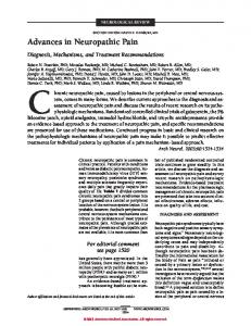
Change in VAS Scores The VAS scores decreased by 4.38 ± 1.66, more than 50%, 1 week after treatment (P = 0.009). In addition, the VAS scores decreased by 5.38 ± 2.06 after 4 weeks (P = 0.008) and 5.62 ± 2.18 after 24 weeks (P = 0.007), as shown in Figure 2. Autologous fat grafting yielded improvement after 1 week, and the effects persisted for at least 6 months, as long as the mean follow-up time of 19.31 ± 12.26 months. Two patients (cases 4 and 12) exhibited little improvement in neuropathic pain 1 week after treatment, but these patients also exhibited no increase in VAS scores after 6 months.
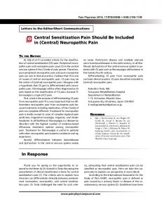
Change in Neuropathic Pain Symptom Inventory The sum of NPSI subscores indicates the efficacy of neuropathic pain treatment.24 The mean NPSI score was 49.38 ± 13.25 before treatment and 25 ± 14 after 1 week of treatment (P = 0.004). The mean NPSI score continued to decrease to 21 ± 17.78 after 4 weeks (P = 0.0009) and to 14.62 ± 16.88 after 24 weeks (P = 0.0008) as shown in Figure 3. Figure 4 shows that the subscore categories exhibiting the most significant improvement were evoked pain and paresthesia and dysesthesia (P < 0.001). Table 2 shows a detailed summary of the VAS scores, total NPSI intensity scores, and preoperative and postoperative NPSI subscores.
FIGURE 3. Preoperative NPSI score and those 1, 4, and 24 weeks after operation. The NPSI scores significantly decreased after the operation (before operation and 1 week after operation, P = 0.004; 4 weeks after operation, P = 0.0009; and 24 weeks after operation, P = 0.0008).
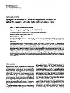
Case Report One 20-year-old man (case 6) sustained a friction burn over the left knee area. After 3 months of wound healing, the patient complained of a painful hypertrophic scar, and the VAS score was 7. After 3 sessions of intralesional injection of 10-mg/mL triamcinolone acetonide, the hypertrophic scar faded, but the neuropathic scar pain persisted. Before the operation, the patient's VAS and NPSI scores were 6 and 23.66, respectively. One month after a 1.8-mL fat graft injection, the VAS and NPSI scores improved to 1 and 8.16, respectively, and the effect persisted even until a 21-month follow-up telephone call. However, 6 months after the operation, the pigmentation score of the Vancouver Scar Scale was decreased from 3 (hyperpigmentation) to 2 (mixed pigmentation), but the height score increased from zero to 1; the pliability and vascularity scores did not change (Fig. 5).
Surgery as well as crushing and avulsion injuries can cause neuropathic pain in scar areas. Symptoms of neuropathic pain include hyperalgesia, allodynia, burning, tingling, and electric shock. Hyperalgesia is an elevated sensation of pain in response to a painful stimulus, and allodynia is a sensation of pain in response to an innocuous stimulus. These conditions are thought to result from damage to nociceptors or peripheral nerves,25 leading to a robust inflammatory response accompanied by excessive fibrosis and causing neuropathic scar pain.26,27 Treatment of the inflammatory response by using corticosteroids has been reported to be successful in suppressing inflammation and fibroblast activity.28,29 Subcutaneous fatty tissue, which secretes the anti-inflammatory cytokine interleukin (IL)-10, can also suppress inflammation.30 Fat grafts are a rich source of mesenchymal stem cells (MSCs),31–33 and adiposederived MSCs (AD-MSCs) can induce the production of IL-1034 as well as inhibit the production of various inflammatory mediators.35,36 Recent studies have indicated that intrathecal injection of IL-10 gene therapy can suppress chronic peripheral neuropathic pain.37 Sacerdote et al reported that AD-MSCs can reverse nociceptive hypersensitivity in neuropathic pain, thus serving as an anti-inflammatory therapy.38 Adipose tissue contains an extracellular matrix of collagen, laminin, and www.annalsplasticsurgery.com
S101
Annals of Plastic Surgery • Volume 74, Supplement 2, May 2015
Huang et al
FIGURE 4. Comparison of preoperative and 24-week postoperative NPSI subscores. The subscore categories that exhibited the most significant improvement were evoked pain and paresthesia and dysesthesia. P < 0.001; **P < 0.01 and ***P < 0.001.
fibronectin39 as well as cellular components such as adipocytes and stem cells.31 Mesenchymal stem cells can inhibit the proliferation of cluster of differentiation 4 (CD4) and CD8 T lymphocytes,40 and scar pain relief might be related to the suppression of inflammation by MSCs in adipose tissue. Therefore, autologous fat grafts may reduce scar inflammation and relieve neuropathic pain because AD-MSCsecreting cytokines such as IL-10 inhibit the production of inflammatory cytokines. Cheville et al41 asserted that treatment strategies should reduce neuropathic pain by stabilizing neuronal membranes and blocking pain signals and that fat grafting can block nociceptive impulses and reduce persistent signal input to the central nerve system. Similarly, Vaienti et al19 reported that fat grafts act as a cushion around the nerve stump to prevent stimulation and reduce local inflammation. Functionally, this cushioning effect enables fat grafts to reduce neuropathic pain. The NPSI is a questionnaire designed to evaluate neuropathic pain. The NPSI includes 10 descriptors and 2 temporal items that enable the quantification of 5 distinct factors. These clinically relevant dimensions of neuropathic pain syndromes are used to verify whether patients respond to therapeutic intervention.24,42,43 The most critical feature of the NPSI is its sensitivity to the effects of treatment24; a decrease in the total NPSI score indicates subjective improvement. This study
evaluated patients' preoperative and postoperative VAS and NPSI scores to determine the effectiveness of fat grafts in treating neuropathic scar pain. Comparing preoperative and 24-week postoperative NPSI subscores revealed that the most significant improvement occurred in evoked pain and in paresthesia and dysesthesia (P < 0.001; Fig. 4). These results indicated that autologous fat acts as cushion or insulation to prevent stimulation and block abnormal sensation. The rapid significant decrease in VAS and NPSI scores 1 week after treatment observed in our study differs from the results reported in other studies. Vaienti et al observed pain alleviation 2 months after treatment; this alleviation persisted to 6 months but deteriorated after 12 months. Caviggioli et al did not indicate when fat grafting initially became effective in treating postmastectomy pain but reported that VAS scores decreased by 3.23 points 13 months after treatment.17 Ulrich et al used lipofilling to treat the neuropathic pain in episiotomy and perineal laceration scars and observed that short-form McGill Pain Questionnaire scores significantly decreased 1 month after treatment and continued to decrease over the subsequent 6 months. In addition, 8 of 20 patients exhibited immediate, subjective pain improvement after the release of their severe scar contractures.44 According to these results, the positive effects of fat grafting may be observed as early as 1 week after treatment.
TABLE 2. Summary of VAS, Total Intensity Score of NPSI and Subscores Preoperative and Postoperative Follow-Up Pain Evaluation
VAS, mean ± SD NPSI symptoms, mean ± SD Superficial pain (0–10) Deep pain (0–10) Paroxysmal pain (0–10) Evoke pain (0–10) Paresthesia and dysesthesia (0–10) Sum of NPSI score (0–100)
Analysis of differences between preoperative and follow-up scores was performed by one-way analysis of variance. *P < 0.05, **P < 0.01, and ***P < 0.001.
Annals of Plastic Surgery • Volume 74, Supplement 2, May 2015
Fat Graft for Neuropathic Scar Pain
FIGURE 5. Case 6. A, Photograph of the painful scar 3 months after trauma. B, After intralesional injection of 10-mg/mL triamcinolone acetonide every month for 3 sessions: VSS score, 3 points; pigmentation, 3; vascularity, zero; pliability, zero; and height, zero. C, Immediately after a 1.8-mL fat graft injection. D, Six months after operation: VSS score, 3 points; pigmentation, 2; vascularity, zero; pliability, zero; and height, 1.
This study had some limitations. In 2 cases (cases 4 and 12), fat grafting failed to alleviate neuropathic pain, and patients were unsatisfied with this result. The failure to respond to treatment may be attributed to age (older than 50 years). Ding et al45 harvested abdominal subcutaneous fat from 3 age groups (30–39, 40–49, and 50–60 years) and observed that older patients exhibited a lower adipogenetic differentiation capacity. Van Harmelen et al46 and Alt et al47 determined that age reduces adipose-derived stem cell proliferation and adipogenetic capacity as well as increases cellular senescence. Further research is necessary to determine whether patients older than 50 years either require more fat grafting to reach the required amount for cell therapy or should not be recommended for autologous fat grafting. Another limitation is that the percentage of volume retention is difficult to measure. In addition, the scar areas addressed in this study were relatively small, and the effects of fat grafting on large scars require further research. Recent studies have demonstrated the high survival rate, longterm stability, and safety of autologous fat grafting in cosmetic filler and reconstructive procedures.22,48–50 In this study, no relapse in neuropathic pain was observed 6 months after treatment, and the mean follow-up period was 19.3 ± 12.26 months (range, 6-38 months). Autologous fat grafting is a promising long-term treatment for traumatic neuropathic scar pain. Complications such as donor site seroma, hematoma, and wound infection were not observed in this study. Autologous fat grafting is a safe technique for treating neuropathic scar pain.
This differentiates fat grafting from pharmacological courses of treatment that must be followed continually. Further long-term clinical observation and basic study are necessary. ACKNOWLEDGMENTS The authors thank Yen-Hsin Kuo for assistance in preparing figures and tables. This study was supported by a grant provided by the National Science Council (NSC 102-2314-B-037-014-MY2) and Kaohsiung Medical University (KMU-Q103002), (KMU-TP103G02), (KMU-TP103G04) & (KMU-TP103G05). REFERENCES 1. Treede RD, Jensen TS, Campbell JN, et al. Neuropathic pain: redefinition and a grading system for clinical and research purposes. Neurology. 2008;70: 1630–1635. 2. Woolf CJ, Salter MW. Neuronal plasticity: increasing the gain in pain. Science. 2000;288:1765–1769. 3. Hans G, Joukes E, Verhulst J, et al. Management of neuropathic pain after surgical and non-surgical trauma with lidocaine 5% patches: study of 40 consecutive cases. Curr Med Res Opin. 2009;25:2737–2743. 4. de Andrade DC, Ferreira KA, Nishimura CM, et al. Psychometric validation of the Portuguese version of the Neuropathic Pain Symptoms Inventory. Health Qual Life Outcomes. 2011;9:107. 5. Attal N, Fermanian C, Fermanian J, et al. Neuropathic pain: are there distinct subtypes depending on the aetiology or anatomical lesion? Pain. 2008;138: 343–353. 6. Isoardo G, Stella M, Cocito D, et al. Neuropathic pain in post-burn hypertrophic scars: a psychophysical and neurophysiological study. Muscle Nerve. 2012;45: 883–890. 7. Bouhassira D, Attal N, Alchaar H, et al. Comparison of pain syndromes associated with nervous or somatic lesions and development of a new neuropathic pain diagnostic questionnaire (DN4). Pain. 2005;114:29–36.
www.annalsplasticsurgery.com
S103
Huang et al
8. Attal N, Cruccu G, Haanpaa M, et al. EFNS guidelines on pharmacological treatment of neuropathic pain. Eur J Neurol. 2006;13:1153–1169. 9. Nayak S, Cunliffe M. Lidocaine 5% patch for localized chronic neuropathic pain in adolescents: report of five cases. Paediatr Anaesth. 2008;18:554–558. 10. Mou J, Paillard F, Turnbull B, et al. Qutenza (Capsaicin) 8% Patch Onset and Duration of Response and Effects of Multiple Treatments in Neuropathic Pain Patients. Clin J Pain. 2014;30:286–294. 11. Fabregat G, Asensio-Samper JM, Palmisani S, et al. Subcutaneous botulinum toxin for chronic post-thoracotomy pain. Pain Pract. 2013;13:231–234. 12. Ranoux D, Attal N, Morain F, et al. Botulinum toxin type A induces direct analgesic effects in chronic neuropathic pain. Ann Neurol. 2008;64:274–283. 13. Fabregat G, De Andres J, Villanueva-Perez VL, et al. Subcutaneous and perineural botulinum toxin type A for neuropathic pain: a descriptive review. Clin J Pain. 2013;29:1006–1012. 14. Finnerup NB, Otto M, McQuay HJ, et al. Algorithm for neuropathic pain treatment: an evidence based proposal. Pain. 2005;118:289–305. 15. O'Connor AB, Dworkin RH. Treatment of neuropathic pain: an overview of recent guidelines. Am J Med. 2009;122:S22–32. 16. Klinger M, Marazzi M, Vigo D, et al. Fat injection for cases of severe burn outcomes: a new perspective of scar remodeling and reduction. Aesthet Plast Surg. 2008;32:465–469. 17. Caviggioli F, Maione L, Forcellini D, et al. Autologous fat graft in postmastectomy pain syndrome. Plast Reconstruct Surg. 2011;128:349–352. 18. Klinger M, Villani F, Klinger F, et al. Anatomical variations of the occipital nerves: implications for the treatment of chronic headaches. Plast Reconstruct Surg. 2009; 124:1727–1728; author reply 1728. 19. Vaienti L, Gazzola R, Villani F, et al. Perineural fat grafting in the treatment of painful neuromas. Tech Hand U Extrem Surg. 2012;16:52–55. 20. Caviggioli F, Vinci V, Codolini L. Autologous fat grafting: an innovative solution for the treatment of post-mastectomy pain syndrome. Breast Cancer. 2013;20: 281–282. 21. Calmels P, Mick G, Perrouin-Verbe B, et al. Neuropathic pain in spinal cord injury: identification, classification, evaluation. Ann Phys Rehabil Med. 2009;52:83–102. 22. Coleman SR. Structural fat grafting: more than a permanent filler. Plast Reconstr Surg. 2006;118:108S–120S. 23. Pu LL, Coleman SR, Cui X, et al. Autologous fat grafts harvested and refined by the Coleman technique: a comparative study. Plast Reconstr Surg. 2008;122: 932–937. 24. Bouhassira D, Attal N, Fermanian J, et al. Development and validation of the Neuropathic Pain Symptom Inventory. Pain. 2004;108:248–257. 25. Vanegas H, Schaible H. Effects of antagonists to high-threshold calcium channels upon spinal mechanisms of pain, hyperalgesia and allodynia. Pain. 2000;85:9–18. 26. Singer AJ, Clark RA. Cutaneous wound healing. N Engl J Med. 1999;341: 738–746. 27. Aarabi S, Longaker MT, Gurtner GC. Hypertrophic scar formation following burns and trauma: new approaches to treatment. PLoS Med. 2007;4:e234. 28. Fitzpatrick RE. Treatment of inflamed hypertrophic scars using intralesional 5-FU. Dermatol Surg. 1999;25:224–232. 29. Yang JY, Huang CY. The effect of combined steroid and calcium channel blocker injection on human hypertrophic scars in animal model: a new strategy for the treatment of hypertrophic scars. Dermatol Surg. 2010;36:1942–1949. 30. Juge-Aubry CE, Henrichot E, Meier CA. Adipose tissue: a regulator of inflammation. Best Pract Res Clin Endocrinol Metab. 2005;19:547–566. 31. Zuk PA, Zhu M, Ashjian P, et al. Human adipose tissue is a source of multipotent stem cells. Mol Biol Cell. 2002;13:4279–4295.
S104
www.annalsplasticsurgery.com
Annals of Plastic Surgery • Volume 74, Supplement 2, May 2015
32. Gimble J, Guilak F. Adipose-derived adult stem cells: isolation, characterization, and differentiation potential. Cytotherapy. 2003;5:362–369. 33. Lin TM, Tsai JL, Lin SD, et al. Accelerated growth and prolonged lifespan of adipose tissue-derived human mesenchymal stem cells in a medium using reduced calcium and antioxidants. Stem Cells Dev. 2005;14:92–102. 34. Zhou B, Yuan J, Zhou Y, et al. Administering human adipose-derived mesenchymal stem cells to prevent and treat experimental arthritis. Clin Immunol. 2011;141: 328–337. 35. Gonzalez-Rey E, Gonzalez MA, Varela N, et al. Human adipose-derived mesenchymal stem cells reduce inflammatory and T cell responses and induce regulatory T cells in vitro in rheumatoid arthritis. Ann Rheum Dis. 2010;69:241–248. 36. Gonzalez MA, Gonzalez-Rey E, Rico L, et al. Adipose-derived mesenchymal stem cells alleviate experimental colitis by inhibiting inflammatory and autoimmune responses. Gastroenterology. 2009;136:978–989. 37. Milligan ED, Penzkover KR, Soderquist RG, et al. Spinal interleukin-10 therapy to treat peripheral neuropathic pain. Neuromodulation. 2012;15: 520–526; discussion 526. 38. Sacerdote P, Niada S, Franchi S, et al. Systemic administration of human adiposederived stem cells reverts nociceptive hypersensitivity in an experimental model of neuropathy. Stem Cells Dev. 2013;22:1252–1263. 39. Gregoire FM, Smas CM, Sul HS. Understanding adipocyte differentiation. Physiol Rev. 1998;78:783–809. 40. Le Blanc K. Mesenchymal stromal cells: tissue repair and immune modulation. Cytotherapy. 2006;8:559–561. 41. Cheville AL, Sloan JA, Northfelt DW, et al. Use of a lidocaine patch in the management of postsurgical neuropathic pain in patients with cancer: a phase III double-blind crossover study (N01CB). Support Care Cancer. 2009;17: 451–460. 42. Truini A, Padua L, Biasiotta A, et al. Differential involvement of A-delta and A-beta fibres in neuropathic pain related to carpal tunnel syndrome. Pain. 2009; 145:105–109. 43. Ducreux D, Attal N, Parker F, et al. Mechanisms of central neuropathic pain: a combined psychophysical and f MRI study in syringomyelia. Brain. 2006;129: 963–976. 44. Ulrich D, Ulrich F, van Doorn L, et al. Lipofilling of perineal and vaginal scars: a new method for improvement of pain after episiotomy and perineal laceration. Plast Reconstr Surg. 2012;129:593e–594e. 45. Ding DC, Chou HL, Hung WT, et al. Human adipose-derived stem cells cultured in keratinocyte serum free medium: donor's age does not affect the proliferation and differentiation capacities. J Biomed Sci. 2013;20:59. 46. Van Harmelen V, Rohrig K, Hauner H. Comparison of proliferation and differentiation capacity of human adipocyte precursor cells from the omental and subcutaneous adipose tissue depot of obese subjects. Metabolism. 2004;53:632–637. 47. Alt EU, Senst C, Murthy SN, et al. Aging alters tissue resident mesenchymal stem cell properties. Stem Cell Res. 2012;8:215–225. 48. Nguyen PS, Desouches C, Gay AM, et al. Development of micro-injection as an innovative autologous fat graft technique: the use of adipose tissue as dermal filler. J Plast Reconstr Aesthet Surgery. 2012;65:1692–1699. 49. Clauser LC, Tieghi R, Galie M, et al. Structural fat grafting: facial volumetric restoration in complex reconstructive surgery. J Craniofac Surg. 2011;22: 1695–1701. 50. Phulpin B, Gangloff P, Tran N, et al. Rehabilitation of irradiated head and neck tissues by autologous fat transplantation. Plast Reconstr Surg. 2009;123: 1187–1197.