www.nature.com/scientificreports
OPEN
received: 27 April 2015 accepted: 09 September 2015 Published: 07 October 2015
An improved chloride-conducting channelrhodopsin for light-induced inhibition of neuronal activity in vivo Jonas Wietek1, Riccardo Beltramo2,3, Massimo Scanziani2,3, Peter Hegemann1, Thomas G. Oertner4 & J. Simon Wiegert4 Channelrhodopsins are light-gated cation channels that have been widely used for optogenetic stimulation of electrically excitable cells. Replacement of a glutamic acid in the central gate with a positively charged amino acid residue reverses the ion selectivity and produces chloride-conducting ChRs (ChloCs). Expressed in neurons, published ChloCs produced a strong shunting effect but also a small, yet significant depolarization from the resting potential. Depending on the state of the neuron, the net result of illumination might therefore be inhibitory or excitatory with respect to action potential generation. Here we report two additional amino acid substitutions that significantly shift the reversal potential of improved ChloC (iChloC) to the reversal potential of endogenous GABAA receptors. As a result, light-evoked membrane depolarization was strongly reduced and spike initiation after current injection or synaptic stimulation was reliably inhibited in iChloC-transfected neurons in vitro. In the primary visual cortex of anesthetized mice, activation of iChloC suppressed spiking activity evoked by visual stimulation. Due to its high operational light sensitivity, iChloC makes it possible to inhibit neurons in a large volume of brain tissue from a small, point-like light source.
Optogenetic stimulation of neurons with channelrhodopsin variants has been applied to many neurobiological questions in a wide variety of organisms and model systems. It has been especially useful to dissect and manipulate the brain circuitry of small rodents and to derive neuronal correlates of behavioral adaptations and disorders1–4. While optogenetic activation is widely used, better tools for optogenetic inhibition of neuronal activity are wanted, as loss-of-function experiments are often more specific and easier to interpret in the context of a complex system like the brain. Inhibition of neuronal activity, however, has proven to be technically difficult. So far, the only inhibitory tools that were successfully applied in vivo are the light-driven chloride pump Halorhodopsin5 and the proton pump Archaerhodopsin6. Both pumps require dense expression in the plasma membrane and high light intensities for reliable inhibition of neuronal activity as only a single ion is transported per absorbed photon. During prolonged activation, pumps affect the intracellular ion composition and can severely change the effects of endogenous GABAergic inhibition7. Recently engineered light-gated chloride channels8,9 (here commonly referred to as ChloCs) are in principle much more efficient than pumps since thousands of ions can pass the channel per absorbed photon. The operational light sensitivity is further increased by the slow 1
Institute for Biology, Experimental Biophysics, Humboldt-Universität zu Berlin, D-10115 Berlin, Germany. 2Center for Neural Circuits and Behavior, University of California San Diego, La Jolla, CA 92093-0634, USA. 3Howard Hughes Medical Institute, University of California San Diego, La Jolla, CA 92093-0634, USA. 4Institute for Synaptic Physiology, Center for Molecular Neurobiology Hamburg, University Medical Center Hamburg-Eppendorf, D-20251 Hamburg, Germany. Correspondence and requests for materials should be addressed to P.H. (email: hegemann@ rz.hu-berlin.de) or T.G.O. (email:
[email protected])
Scientific Reports | 5:14807 | DOI: 10.1038/srep14807
1
www.nature.com/scientificreports/
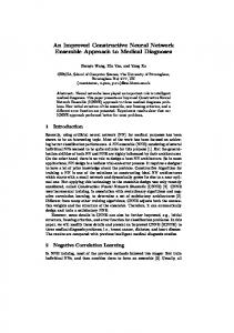
Figure 1. Strategy for improving the chloride selectivity of ChloC. (A) Position of key negative (blue) and positive charges (red) in wt Channelrhodopsin, slowChloC, and iChloC with respect to the aqueous pore (light blue)14. (B) Structural model of ChR2-E90R8; view from the intracellular side. Residues having a stake in the inner gate are highlighted in blue and cyan (TM: transmembrane helix). The negatively charged E83 carboxylate is located in the center of the inner gate pore. (C) Closer look at the inner gate residues (side view). Hydrogen bonds may form between E82 and R268 as well as between E83 and H134. Atomic distances (black dotted lines) are shown in Å. The retinal (RET) is shown in orange.
off-kinetics of some ChloC variants, sustaining inhibition for several seconds. High light sensitivity is an important feature if large volumes of neuronal tissue are to be addressed from a localized light source such as a fiber-coupled laser or LED. The reversal potential (Erev) of published ChloCs, however, is not quite as negative as Erev of endogenous chloride channels (e.g. GABAA receptors), indicating a residual conductance for cations8. Given the dramatic developmental changes in [Cl−]10 i and its dependence on the local concentration of impermeant anions11, the net effect of published ChloC variants on neuronal excitability in vivo is difficult to predict. Here, in an attempt to improve the ion selectivity of ChloC, we combine the three point mutations of slowChloC8 with two additional amino acid substitutions to reduce the number of negative charges protruding into the water pore. In neurons, photocurrents of our improved ChloC (iChloC) reverse at nearly identical membrane potentials than GABAergic IPSCs, suggesting a high selectivity for Cl− ions. iChloC no longer shows depolarizing activity in patch-clamped neurons and reliably inhibits synaptically evoked spikes in undisturbed CA1 pyramidal cells. When expressed in primary visual cortex, iChloC strongly suppressed visually evoked spiking of pyramidal neurons in vivo. Thus, due to its high Cl−-selectivity and operational light sensitivity, iChloC is an ideal tool to silence neurons in vivo with very low light exposure.
Results
The previously published slowChloC carries a Glutamate-to-Arginine substitution at position 90 (E90R), introducing a positive charge to the central gate of the protein. In addition, Threonine 159 was mutated to Cysteine (T159C) to increase photocurrents12 and Aspartic acid 156 was substituted with Asparagine (D156N) to stabilize the open state of the channel and increase its operational light sensitivity13. To further increase the selectivity of the channel for Cl− and to suppress cation conductance, we attempted to render the inner gate as well as the9 extracellular access channel more permissive for anions. Residue E83 is a key component of the inner gate, forming a hydrogen bond to H134 in ChR214,15. In our ChR2-E90R model8, E83 extends its negatively charged carboxylate group into the pore, thus forming a potential diffusion barrier for anions (Fig. 1A & B). In addition, E83 is part of the proton transfer chain and its replacement reduces cation conductance in ChR2 [15, 16]. To eliminate the negative charge at this position without Scientific Reports | 5:14807 | DOI: 10.1038/srep14807
2
www.nature.com/scientificreports/
Figure 2. Characterization of slowChloC variants in HEK293 cells. (A) Photocurrent example traces of slowChloC, slowChloC (E101S), slowChloC (E83Q) and slowChloC (E83Q, E101S) (iChloC) in HEK 293 cells at different holding potentials (20 mV steps). (B) I-E curves for the 4 ChloC variants. (C) Photocurrent amplitudes for the 4 ChloC variants measured at a holding potential of 0 mV. (D) Reversal potential in HEK 293 cells (slowChloC, − 52.5 ± 1.3 mV; slowChloC (E101S), − 59.2 ± 0.3 mV; slowChloC (E83Q), − 60.8 ± 1.5 mV; iChloC, − 65.6 ± 1.1 mV). Dashed line indicates calculated Nernst potential for Cl− (− 69.6 mV). n = 9 for slowChloC, n = 8 for slowChloC (E101S), n = 6 for other two variants.
disturbing the overall geometry, we replaced E83 with a Glutamine (Q) residue (Fig. 1A, B and C). The resulting ChloC variants were characterized in HEK 293 cells. Compared to slowChloC, the E83Q replacement shifted the reversal potential by approx. − 8 mV (Erev slowChloC[E83Q] = − 60.8 ± 1.5 mV), but reduced current amplitudes from 188 ± 38 pA to 80 ± 27 pA (Fig. 2). E101, which is positioned at the collar of the vestibule facing the extracellular space14, also carries a negative charge that may hinder anion diffusion. In addition, E101, like E83, is a constituent of the proton transfer chain and deletion of its side chain (E101A) reduced cation conductance16,17. When we replaced E101 in the access channel with a neutral Serine (S), we obtained very large photocurrents (475 ± 98 pA) with a reversal potential of –59.2 ± 0.3 mV, suggesting that anion conductance was increased (Fig. 2). When we combined the two mutations, we measured a current amplitude of 210 ± 32 pA and an additional shift of the reversal potential (Erev = − 65. 6 ± 1.1 mV) to a value 13 mV more negative than slowChloC (Erev = − 52.5 ± 1.3 mV) and close to the calculated Nernst potential for Cl− (− 69.6 mV, Fig. 2D). The improved chloride-conducting channelrhodopsin ChR2(E83Q,E90R,E101S,D156N,T159C) we termed iChloC. For neuron-specific expression, we assembled an expression plasmid driven by the human CaMKII-promotor, including a red florescent protein (tdimer2) behind a ribosomal skip sequence (CaMKII -iChloC-2A-tdimer2). The cytoplasmic tdimer2 allows straightforward identification of transfected neurons and evaluation of their morphology. Four to five days after single cell electroporation, expressing CA1 pyramidal cells could be readily identified by their red fluorescence and were indistinguishable from non-transfected neurons under Dodt contrast18 (Fig. 3B,C). Dendritic morphology and electrophysiological properties showed no abnormalities (Fig. 3A, Table 1). Brief light pulses (476 nm, 5 ms, 1 mW/mm2) induced large photocurrents in transfected neurons with a decay time constant of 5.3 ± 0.4 s (Fig. 3D). Holding potentials were corrected for a liquid junction potential of − 10.6 mV. The measured reversal potential of a chloride-conducting channel depends on the chloride concentration gradient and is therefore very sensitive to the composition of the recording solutions. To compare iChloC and slowChloC under near-physiological conditions we used artificial cerebrospinal fluid (ACSF) containing 2 mM Ca2+ and 1 mM Mg2+. Compared to our previous measurements in ACSF with a higher concentration of divalent ions8, the measured reversal potential of slowChloC was 8 mV more positive. To provide a biological calibration value, we stimulated inhibitory inputs to iChloC expressing neurons at various holding potentials (5–10 mV increments) while blocking glutamatergic transmission (10 μ M NBQX, 10 μ M CPPene). This allowed us to precisely determine the membrane voltage under which GABAergic currents reverse and to directly compare it to the reversal potential of iChloC (Fig. 4A). On average, IPSCs reversed at − 77.3 ± 1.4 mV (Fig. 4B), close to the calculated Nernst potential for Cl− (− 75.9 mV). Light-induced iChloC currents reversed at − 76.7 ± 1.6 mV, statistically not different Scientific Reports | 5:14807 | DOI: 10.1038/srep14807
3
www.nature.com/scientificreports/
Figure 3. Expression of iChloC in CA1 pyramidal cells in organotypic hippocampal slice culture. (A) Overview of neuronal morphology 5 days after electroporation (maximum intensity projection of two-photon images). (B) Fluorescence of co-expressed tdimer2 was used to target transfected neurons for electrophysiological recordings (same cells as in (A)). (C) Dodt contrast image of cells shown in (B). (D) Photocurrents in response to a single light pulse (476 nm, 5 ms, 1 mW/mm2) at different holding potentials. Photocurrents reversed at very negative holding potentials and were large at depolarized holding potentials, where the Cl− inward driving force was highest (same neuron as in (A–C)). The inset shows the onset of the photocurrents at higher temporal resolution. Indicated holding potentials were rounded to full numbers after subtraction of the liquid junction potential (− 10.6 mV). Indicated tau value was derived from 9 independent measurements in 9 slice cultures.
iChloC (n = 9)
S.D.
wt (n = 9)
S.D.
P
− 38.77
5.37
− 43.42
7.08
0.16
AP peak voltage (mV)
36.19
3.70
35.81
2.22
0.81
AP amplitude (mV)
117.52
4.04
111.06
9.94
0.11
n AP’s
11.22
4.47
14.44
1.83
0.08
AP threshold (mV)
Erest (mV)
− 81.33
3.39
− 75.25
10.52
0.14
RM (Mohm)
181.01
39.75
144.90
54.81
0.20
Table 1. Neuronal membrane parameters. Electrical parameters of untransfected pyramidal CA1 neurons and neurons expressing iChloC together with tdimer2. Action potentials (APs) were evoked by a square current pulse (500 ms, 500 pA) in current clamp (IC) mode. Threshold, peak voltage and amplitude were calculated for the first AP. Membrane resistance (RM) was measured in voltage clamp mode in response to a square voltage pulse (− 5 mV, 100 ms). Erest = resting membrane potential, S.D. = standard deviation. Right column indicates P values from unpaired t-test for each parameter. No significant differences were detected. All measurements were liquid junction potential corrected.
from endogenous GABAergic inhibition (P = 0.6). SlowChloC photocurrents, in contrast, had a reversal potential of − 59.7 ± 1.0 mV, significantly different from GABAergic inhibition in the same cells (− 75.7 ± 1.8 mV, P = 0.0013), indicating a residual cation conductance (Fig. 4C,D). In contrast to the photocurrent reversal potentials, which significantly differed (P