MOLECULAR AND CELLULAR BIOLOGY, Dec. 2004, p. 10397–10405 0270-7306/04/$08.00⫹0 DOI: 10.1128/MCB.24.23.10397–10405.2004 Copyright © 2004, American Society for Microbiology. All Rights Reserved.
Vol. 24, No. 23
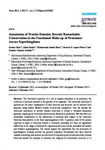
An Intramolecular Association between Two Domains of the Protein Kinase Fused Is Necessary for Hedgehog Signaling Manuel Ascano, Jr.,1,2 and David J. Robbins1,3* Department of Pharmacology and Toxicology, Dartmouth Medical School, Hanover,1 and Norris Cotton Cancer Center, Dartmouth-Hitchcock Medical Center, Lebanon,3 New Hampshire, and Department of Molecular Genetics, Graduate Program, University of Cincinnati Medical Center, Cincinnati, Ohio2 Received 21 July 2004/Returned for modification 3 September 2004/Accepted 14 September 2004
The protein kinase Fused (Fu) is an integral member of the Hedgehog (Hh) signaling pathway. Although genetic studies demonstrate that Fu is required for the regulation of the Hh pathway, the mechanistic role that it plays remains largely unknown. Given our difficulty in developing an in vitro kinase assay for Fu, we reasoned that the catalytic activity of Fu might be highly regulated. Several mechanisms are known to regulate protein kinases, including self-association in either an intra- or an intermolecular fashion. Here, we provide evidence that Hh regulates Fu through intramolecular association between its kinase domain (⌬Fu) and its carboxyl-terminal domain (Fu-tail). We show that ⌬Fu and Fu-tail can interact in trans, with or without the kinesin-related protein Costal 2 (Cos2). However, since the majority of Fu is found associated with Cos2 in vivo, we hypothesized that Fu-tail, which binds Cos2 directly, would be able to tether ⌬Fu to Cos2. We demonstrate that ⌬Fu colocalizes with Cos2 in the presence of Fu-tail and that this colocalization occurs on a subset of membrane vesicles previously characterized to be important for Hh signal transduction. Additionally, expression of Fu-tail in fu mutant flies that normally express only the kinase domain rescues the fu wing phenotype. Therefore, reestablishing the association between these two domains of Fu in trans is sufficient to restore Hh signal transduction in vivo. In such a manner we validate our hypothesis, demonstrating that Fu self-associates and is functional in an Hh-dependent manner. Our results here enhance our understanding of one of the least characterized, yet critical, components of Hh signal transduction. Hedgehog (Hh) is a morphogen required for the proper development of numerous organisms (13). Cells interpret the level of Hh that they receive by means of a large multiprotein signaling complex, termed the Hedgehog signaling complex (HSC) (17, 28). In Drosophila, the HSC includes the Ser/Thr protein kinase Fused (Fu) (34), the kinesin-related protein Costal 2 (Cos2) (36, 39), and the transcription factor Cubitus interruptus (Ci) (31). The HSC also associates with Su(fu) (21, 42), an additional component of the Hh pathway originally identified as an extragenic suppressor of fu mutations (33). In a manner regulated by Hh, the HSC, via Cos2, binds specifically with microtubules and membrane vesicles, suggesting that these two associations are necessary for Ci regulation (36, 41). The primary role of the HSC is to regulate Ci processing: to the transcriptional repressor Ci75 in the absence of Hh and to at least two transcriptional activators in the presence of Hh (1, 3, 6, 16, 19, 30, 45). Fu is a critical component of the HSC, since mutations of fu result in deregulation of all forms of Ci processing, ultimately leading to a loss of Hh signaling (1, 16, 30, 45). fu flies, which reach adulthood through a maternal rescue (26, 34), exhibit a number of phenotypes, including a fusion of longitudinal veins 3 and 4 (LV3 and LV4) in the wing blade and a posterior expansion of the double-row wing margin bristles (7). The development of these two wing structures depends on at least two different Hh target genes. The LV3-LV4 intervein region * Corresponding author. Mailing address: Dartmouth Medical School, Department of Pharmacology and Toxicology, 10 North College St., 7650 Remsen Hall, Hanover, NH 03755. Phone: (603) 6501716. Fax: (603) 650-1129. E-mail:
[email protected].
requires decapentaplegic (dpp) expression, whereas the margin bristles in this same region are specified by late third-instar anterior engrailed (en) expression (4, 23, 38). These downstream target genes require two distinct transcriptional activator forms of Ci, Ciact and Ciⴱ, respectively. Loss of Fu kinase activity results in the failure to convert Ci to either Ciact or Ciⴱ (1, 16, 20, 30, 45). Two major classes of fu alleles, class I and class II, were identified based on genetic interactions with Su(fu) (35, 43). In the presence of Su(fu), the phenotype of class I fu mutants is indistinguishable from that of class II mutants. However, while loss of Su(fu) rescues the fu phenotype of both classes of alleles, the resulting phenotype of the double-mutant animal differs depending on the class of the rescued fu allele. In a Su(fu) mutant background, class I fu flies appear to be phenotypically wild type, while class II fu mutant flies exhibit a neomorphic cos2-like phenotype. The two classes of mutations either affect the amino-terminal protein kinase domain, class I, or truncate the large carboxyl-terminal domain, class II. Based on these studies, it was proposed that the kinase domain of Fu functions, primarily, in direct opposition to Su(fu) function. Additionally, the combined loss of Su(fu) and the carboxylterminal domain of Fu (Fu-tail) appeared to overcome the ability of Cos2 to regulate Ci. The carboxyl-terminal domain of Fu has now been shown to associate directly with Cos2 (2, 22). This interaction is absolutely required for HSC function, since its disruption leads to a loss of Hh signaling (2). The carboxylterminal domain is the only domain of Fu necessary for processing Ci to Ci75, since loss of Fu kinase activity does not appear to affect production of Ci75 (1, 2, 16, 20). Hence, the carboxyl-terminal domain of Fu is required to tether the pro-
10397
10398
ASCANO AND ROBBINS
tein to the HSC and facilitates the production of Ci75 in a kinase-independent manner. The kinase domain of Fu is less well characterized. Although Cos2 and Su(fu) are phosphorylated in an Hh- and Fu-dependent manner, it has not been established that this phosphorylation is direct (18, 27, 44). Taken together, it is clear that a central role of Fu is to regulate Cos2 and Su(fu) function. Despite this, the mechanism that leads to Fu activation is not understood. Part of the difficulty in identifying the substrates of Fu stems from the inability to develop an in vitro kinase assay for it (8, 24, 27, 44) (unpublished data). Thus, to characterize the role that Fu plays in Hh signaling, we decided to explore possible regulatory mechanisms that might control Fu kinase activity. A number of protein kinases are regulated through selfassociation, in either an inter- or an intramolecular fashion (40). We hypothesized that Fu may be regulated through such a mechanism. Here, we report that Fu can associate with itself through an intramolecular association between its kinase and carboxyl-terminal domains. We demonstrate that the two domains of Fu, when expressed separately, stably interact. Additionally, we show that they colocalize with Cos2 in Drosophila cells in a manner consistent with vesicular localization. To substantiate the functional significance of Fu self-association, we rescue the wing phenotype of fu mutant flies lacking the carboxyl-terminal domain by transgenically providing it. Thus, by expressing the exogenous Fu carboxyl-terminal domain in these flies, we restore patterning to regions of the wing which requires Hh-dependent Fu kinase activity. MATERIALS AND METHODS DNA constructs. Full-length Fu (amino acids [aa] 1 to 805) cDNA was subcloned into the pFastBac baculovirus vector containing a 5⬘ Flag epitope tag (Fig. 1) or a similar vector that was left untagged (Fig. 2). Construction of the ⌬Fu (aa 1 to 305) (see Fig. 1) baculovirus was described previously (2). ⌬Fu cDNA (aa 1 to 305) (Fig. 2 and 3) was also subcloned into the pIZT insect expression vector, where it is in frame with a 3⬘ V5-–six-His epitope tag. Generation of the six-Histagged Fu-tail (aa 421 to 805 of Fu) (see Fig. 1 and 2) construct and baculovirus was described previously (2). Fu-tail was also subcloned into the pIZT expression vector containing a 5⬘ Flag epitope tag and used in Fig. 3. A larger Fu-tail construct (aa 270 to 805) was used to generate the Fu-tail transgenic fly strain (Fig. 4) (2). Full-length Flag-tagged Cos2 (aa 1 to 1201) baculovirus (Fig. 2) (2) and pAct eGFP-Cos2⌬N (Fig. 3) (41) have been described previously. Enhanced green fluorescent protein (eGFP)-Cos2⌬N is a fusion construct containing amino acids 500 to 1201 of Cos2 fused to the cDNA of eGFP. Generation of glutathione S-transferase (GST) baculovirus was described previously (2). Preparation of baculovirus. Baculoviruses were produced and titers were determined as previously described (42). Infections were carried with Sf21 cells at a total multiplicity of infection (MOI) of 1 to 10. Wild-type (wt) baculovirus was used to normalize the coinfections done in this study. The infected Sf21 cells were allowed to incubate postinfection for 40 to 44 h. Cell culture and transfection. Sf21 cells were cultured in Grace’s insect medium (Invitrogen, Inc.). S2 cells were cultured in Schneider’s Drosophila medium (Invitrogen, Inc.). Both cell lines were also supplemented with 10% fetal bovine serum and 1% penicillin-streptomycin. Sf21 and S2 cells were transfected by using Cellfectin (Invitrogen, Inc.) according to the manufacturer’s instructions. pIZT-⌬Fu V5-HIS, pIZT-Flag-Fu-tail, and pAct-eGFP-Cos2⌬N were used in various amounts depending on the experiment. Further details are available upon request. Where appropriate, empty pIZT or pAc 5.1A vectors (Invitrogen) were used to normalize the total amount of DNA. Cellular lysates. Sf21 or S2 cells were lysed in a Nonidet P-40 (NP-40) buffer as previously described (2). The lysates were centrifuged at 6,000 ⫻ g for 20 min at 4°C. The resulting supernatants were either subjected to immunoprecipitation analysis or resolved directly by sodium dodecyl sulfate-polyacrylamide gel electrophoresis (SDS-PAGE) and immunoblotted for the appropriate proteins. Batch purification of Fu. Twenty million Sf21 cells were infected with Flag-Fu baculovirus at a MOI of 10. The cells were lysed 44 h postinfection, using an
MOL. CELL. BIOL. NP-40 lysis buffer as previously described (2). The postnuclear supernatant was diluted 10-fold, using the NP-40 lysis buffer, and 0.5 ml of a 50:50 slurry of anti-Flag M2 resin was added. The M2 resin was rocked in the diluted supernatant for 16 h at 4°C. The Flag resin was then collected by low-speed centrifugation and washed three times using 15 ml of lysis buffer. The Flag resin was then incubated in 2 ml of elution buffer (NP-40 lysis buffer containing 0.65 M NaCl and 200 mM Flag peptide [Sigma]) overnight with rocking at 4°C. Purified Fu was dialyzed in NP-40 lysis buffer containing 10% glycerol. Isolation of the trimolecular complex by Flag purification. Fifty million Sf21 cells were infected with wt, Flag-Cos2, and His-Fu-tail baculovirus in various combinations at a MOI of 5 each for a total of 10 (see Results and Discussion). Cells were lysed 40 to 44 h postinfection in an NP-40 lysis buffer as before. Anti-Flag M2 resin (0.25 ml) was then applied to each lysate and incubated overnight as described above. The beads were then washed with a total of 25 ml of lysis buffer. Meanwhile, Sf21 cells transfected with pIZT-⌬Fu V5-HIS were lysed in the same lysis buffer, aliquoted evenly to the three pools of washed beads, and then allowed to incubate overnight as before. The beads were then washed with 2.5 ml of lysis buffer and then 5 ml of lysis buffer supplemented with NaCl for a final concentration of 0.5 M. Flag complexes were then eluted in the 0.5 M NaCl lysis buffer with 0.2 mg of 3⫻ Flag peptide (Sigma)/ml, collecting 10 0.25-ml fractions. The first three eluted fractions are shown in Fig 2; the first eluted fraction contained none of the three proteins of interest, whereas fractions following the third did not have ⌬Fu. Antibodies. The following antibodies were used to immunoblot the appropriate proteins: affinity-purified polyclonal rabbit anti-Fu kinase domain (antiFuKD) (0.1 g/ml) (44), affinity-purified polyclonal rabbit anti-Fu hinge domain (anti-FuH) (0.044 g/ml) (36), mouse monoclonal antibody (MAb) 5D6 directed against Cos2 (0.073 g/ml) (2), mouse MAb M2 directed against the Flag epitope (anti-Flag) (0.2 g/ml) (Sigma), mouse MAb Penta-His directed against the six-His epitope (0.2 g/ml) (QIAGEN), mouse MAb directed against the V5 epitope (0.2 g/ml) (Invitrogen), and mouse MAb directed against the GST epitope (anti-GST) (0.1 g/ml) (Santa Cruz Biotechnology). Anti-FuH antibodies were used to immunoblot Fu proteins unless otherwise noted. Immunoprecipitation of cytosolic lysates. Immunoprecipitations of baculovirus-infected Sf21 lysates and S2 cellular lysates were carried out essentially as described previously (2). The appropriate antibody was used for each immunoprecipitation reaction at 2.0 g. Mouse immunoglobulin G1 (IgG1) (Jackson ImmunoResearch) was used as the isotype-matched control for the various MAbs used in this work. Rabbit serum IgG was used as an immunoprecipitation control when using either of the polyclonal rabbit anti-Fu antibodies. Size exclusion chromatography. Gel filtration analyses were carried out by using a Superose 12 gel filtration column installed on an AKTA fast-performance liquid chromatography system (Amersham-Pharmacia). Affinity-purified Flag-Fu was fractionated over the column equilibrated in isotonic running buffer (150 mM NaCl, 40 mM HEPES [pH 7.6], 1 mM EDTA, 10% glycerol, 0.001% NP-40, 1 mM dithiothreitol, 1:1,000 protease inhibitor cocktail [2]). The following molecular mass standards (Amersham-Pharmacia) were used to calibrate the column: ferritin, 440 kDa; catalase, 232 kDa; aldolase, 158 kDa; albumin, 67 kDa; ovalbumin, 48 kDa; chymotrypsinogen A, 25 kDa; and RNase A, 13.7 kDa. A standard curve was constructed as described in the calibration kit manual. Drosophila strains and generation of transgenic flies. Drosophila strains were maintained as previously described (9). Upstream activation sequence (UAS)6xHis fu-tail transgenic flies were generated as previously described (2). The fuG3 (class II fu) (43) flies were kindly provided by P. Therond (Nice, France). The fuG3 mutant allele is missing aa 407 to 805 of Fu but adds 3 aa due to a frameshift mutation. ptc-GAL4 (12) was kindly provided by the Kornberg laboratory (University of California, San Francisco). ap-GAL4 (11) was a kind gift from X. Lin (Cincinnati, Ohio). Crosses were conducted at 18, 25, and 29°C; the crosses performed at 25°C provided the best results. The wings which corresponded to the genotype fuG3/Y; GAL4/UAS-fu-tail were examined for their vein and wing margin development. Adult wings were mounted using Euparal (Asco Laboratories) for observation. A more detailed description of the mating scheme is available upon request. Immunofluorescence of Drosophila S2 cells. Indirect immunofluorescence analysis of S2 cells was performed essentially as described before (29). Polyclonal rabbit anti-FuH (2.2 g/ml) and monoclonal mouse anti-V5 (Invitrogen) (2 g/ml) primary antibodies were used. Goat anti-mouse Alexa-647 (4 g/ml) and goat anti-rabbit Alexa-594 (4 g/ml) (Molecular Probes) secondary antibodies were used. Green fluorescent protein signal was detected by the appropriate excitation and filter parameters. Confocal images were collected by using a Leica Confocal laser scanning microscope. Images were processed with Adobe Photoshop 7.0. Colocalization analyses were performed essentially as described previously except with use of Zeiss LSM colocalization analysis software (29).
VOL. 24, 2004
PROTEIN KINASE Fu SELF-ASSOCIATES
10399
FIG. 1. The protein kinase Fu associates with itself. (A) Purified recombinant Fu appears dimeric. Flag-tagged Fu was affinity purified and then fractionated on a Superose 12 gel filtration column. Samples obtained during the purification were resolved by SDS-PAGE and silver stained (left panel). The flow-through lane represents proteins which did not bind the anti-Flag resin. A single enriched protein was detected at a size consistent with it being Fu (arrow, elution lane). The eluted fractions from the Superose 12 column were resolved by SDS-PAGE and then immunoblotted using anti-Fu antibodies (right panel). Pure Fu elutes with a peak at fraction 55. Standard proteins were used to generate a calibration curve (see Materials and Methods); their elution peaks are marked (arrows). Based on this curve, the apparent molecular mass of Fu is 150 to 200 kDa, approximately twice the size predicted by its primary sequence. (B) Full-length Fu associates with two distinct Fu domains. Baculovirus expressing various Fu constructs or GST (as a negative control) were expressed in Sf21 cells, singly or in combination. The cellular lysates were resolved by SDS-PAGE and immunoblotted with the appropriate antibodies (left panel). The lysates were also immunoprecipitated with anti-Flag (␣-Flag), anti-GST (␣-GST), or mouse IgG1 isotype-matched antibodies. The immunoprecipitates were resolved by SDS-PAGE followed by immunoblotting with the appropriate antisera (right panel). GST did not significantly associate with any of the Fu proteins tested. (C) Fu-tail associates with ⌬Fu. Fu-tail and ⌬Fu were coexpressed, and the resulting lysates were resolved and immunoblotted (left panel) or immunoprecipitated (right panel) by using two different Fu antibodies. Fu-tail, specifically immunoprecipitated by anti-FuH (␣-FuH) antibodies, is able to coimmunoprecipitate ⌬Fu. A similar result is observed when ⌬Fu is specifically immunoprecipitated with anti-FuKD (␣-FuKD) antibodies. Control (Ctrl.) lanes represent normal rabbit serum, which was used as an antibody control. GST was also coexpressed with each of the Fu domains or full-length Fu, and no significant coimmunoprecipitation was observed. The bands which appear in the IgG and ␣-GST immunoprecipitation lanes represent mouse antibody cross-reactivity. Fu-tail consistently migrates faster than these antibody-cross-reacting proteins.
RESULTS AND DISCUSSION The catalytic activities of a growing list of protein kinases are found to be regulated by various autoinhibition strategies. We hypothesized that Fu kinase activity may also be regulated by an analogous inhibitory mechanism, occurring via an inter- or intramolecular association. To begin testing this hypothesis, we purified Fu to assess its native molecular size. Flag epitopetagged Fu was affinity purified from lysates of Sf21 cells in-
fected with baculovirus containing recombinant Fu cDNA (Fig. 1A, left panel). We subjected the affinity-purified protein to size exclusion chromatography and found that pure Fu elutes with a molecular size larger than its predicted mass of 89 kDa (Fig. 1A, right panel). Using known molecular size protein standards, we generated a calibration curve and determined that purified Fu elutes with an apparent molecular mass between 150 and 200 kDa. Our result suggests that highly purified
10400
ASCANO AND ROBBINS
MOL. CELL. BIOL.
FIG. 2. A head-to-tail monomer of Fu binds Cos2. (A) Two models that are consistent with the data presented in Fig. 1. Fu is either a head-to-tail dimer (left panel) or a monomer (right panel), where the kinase domain associates with an adjacent (dimer) or its own (monomer) carboxyl-terminal domain. (B) A Fu monomer binds to Cos2 at the expense of Fu dimer formation. Full-length Fu, six-His-tagged Fu-tail (6XHis Fu-tail) (which is monomeric), and increasing amounts of Cos2 (MOI of 2 and 4 of Cos2 baculovirus, respectively), were expressed in Sf21 cells in various combinations (left panel). The resulting lysates were immunoprecipitated specifically for Fu-tail using anti-His (␣-His) or isotypematched IgG antibodies (right panel). (C) Fu-tail can tether ⌬Fu to Cos2. After Flag-Cos2 (lanes C) or Flag-Cos2 with Fu-tail (lanes C/F) were immobilized on a Flag affinity resin, Sf21 lysate containing ⌬Fu was passed through each column and then specifically eluted with Flag peptide (see the text). The mock column (M) contained lysate-infected wt baculovirus and was treated as described above. ⌬Fu specifically associated with Cos2 from the C/F column compared to results with the C or M column.
recombinant Fu is predominantly dimeric. Alternately, Fu may migrate through a sizing column as a nonglobular protein, appearing to have a larger mass. To isolate the region of Fu responsible for self-association, we coexpressed full-length Flag-Fu with either of two truncated forms of Fu, ⌬Fu or Fu-tail. ⌬Fu contains only the kinase domain of Fu, whereas Fu-tail contains the carboxyl-terminal domain. We found that both ⌬Fu and Fu-tail coimmunoprecipitate with full-length Fu (Fig. 1B). These interactions were specific, since neither ⌬Fu nor Fu-tail is found in isotype matched IgG control immunoprecipitates. Additionally, none of the Fu proteins were coimmunoprecipitated, using anti-GST antibodies, from lysates where they were coexpressed with GST (Fig. 1B). Since both domains of Fu bind full-length Fu, we tested whether the two distinct domains of Fu could associate with each other. We prepared lysates from Sf21 cells expressing ⌬Fu or Fu-tail on their own, with each other, or with a control baculovirus expressing GST. Immunoprecipitations from these lysates were performed, using antibodies that specifically recognize ⌬Fu, Fu-tail, or GST (as a negative control). We found that ⌬Fu and Fu-tail associated with each other but not with GST (Fig. 1C). Our results clearly demonstrate that Fu can associate with itself. However, while our size exclusion analysis of purified Fu is consistent with Fu behaving like a dimer, our results do not rule out that Fu might be monomeric in vivo. We hypothesized
two models of how Fu might exist in vivo, as a dimer or a monomer (Fig. 2A). In this first case, a dimerization interface would exist between the carboxyl-terminal and kinase domains of adjacent Fu proteins (head-to-tail dimer). The second case predicts that Fu is normally monomeric, with the kinase domain interacting with its own carboxyl-terminal domain (headto-tail monomer). In this scenario, purifying overexpressed Fu or coexpressing it with truncated domains of Fu would force the normally monomeric protein to interact with a neighboring Fu molecule. To distinguish between these models, we decided to identify the form of Fu that associates with Cos2. In Drosophila cell lysates, Fu and Cos2 are predominantly found associated with each other (36). The Fu-Cos2 association is direct and absolutely necessary for proper Hh signaling. Therefore, the form of Fu bound to Cos2 would represent the physiologically relevant form of the protein kinase. We expressed full-length Fu and six-His-tagged Fu-tail, with increasing levels of Cos2, in Sf21 cells (Fig. 2B, left panel). In this experiment Fu-tail represents a monomeric form of Fu, since it cannot form a dimer with itself (data not shown), which is capable of binding directly to Cos2. Since Fu and Fu-tail associate with each other quantitatively (Fig. 1B), we reasoned that we could utilize their interaction to determine what form of Fu binds Cos2. If a dimer of Fu interacts with Cos2, then immunoprecipitating Fu-tail should coimmunoprecipitate full-length Fu
VOL. 24, 2004
equally well in the absence or presence of increasing amounts of Cos2. That is, the Fu-tail–Fu association should not be affected by Cos2. If a Fu monomer, in this case Fu-tail, normally associates with Cos2, then Cos2 would bind to Fu-tail at the expense of the Fu-tail–Fu association. We find that the association between Fu-tail and Fu is significantly reduced in the presence of increasing levels of Cos2 (Fig. 2B, right panel), with a corresponding increase in the Fu-tail–Cos2 association. Therefore, our results indicate that a Fu monomer preferentially binds to Cos2. We have previously shown that truncated Fu proteins which lack the carboxyl-terminal domain do not significantly interact with Cos2 (2, 36). Since Fu-tail can bind to both ⌬Fu and Cos2, we tested whether the two truncated domains of Fu could associate with Cos2 as a trimolecular complex. However, when we coexpressed all three proteins in Sf21 or Drosophila S2 cells, we did not observe a significant interaction by coimmunoprecipitation assays. This may be due to a change in the affinity between Fu-tail and ⌬Fu once Fu-tail binds Cos2, destabilizing the trimolecular complex under the conditions used. To overcome this difficulty, we subjected lysates containing either Flag-Cos2 or Flag-Cos2 and Fu-tail to affinity purification with anti-Flag M2 chromatography resin. We also took lysate from Sf21 cells infected with wt baculovirus and mock purified it as a negative control. In this manner we immobilized Flag-Cos2, with or without associated Fu-tail, in separate affinity columns to act as bait. We then made a lysate from Sf21 cells expressing ⌬Fu and distributed it equally between the Flag-Cos2, FlagCos2–Fu-tail, and mock affinity columns. After successive washes, we selectively eluted the Flag complexes using Flag peptide and immunoblotted the resulting fractions (Fig. 2C). We found that ⌬Fu enriched specifically on the Flag-Cos2–Futail column, compared to results with the Flag-Cos2 or control affinity columns. Taken together, our results are consistent with a model where the kinase and carboxyl-terminal domain of Fu bind each other in an intramolecular head-to-tail manner in the presence of Cos2. Since ⌬Fu and Fu-tail associate in a heterologous expression system, we next wanted to determine if these proteins could interact in a Drosophila cell line and do so in subcellular locations important for Hh signaling. An important aspect of the Hh pathway is the subcellular localization of the various signaling components and how these locations affect Hh activation (14, 17, 18, 28, 29, 36, 37, 41, 42). We found that when ⌬Fu was expressed alone in S2 cells, it localized within the cytoplasm of cells in a diffuse manner (Fig. 3A). When ⌬Fu was coexpressed with Fu-tail, both proteins colocalized with each other and have a punctate appearance, consistent with localizing to various membrane vesicles (Fig. 3E to G). Previously, we found that Cos2 colocalized with the vesicle marker, Rab11, giving a similar punctate staining pattern (41). Vesicular localization of Cos2 and Fu was found to be biologically relevant, since this pool of the HSC is responsive to Hh activation. We hypothesized that Fu-tail-dependent redistribution of ⌬Fu might be representative of the kinase domain regaining the ability to interact with Cos2 and hence the HSC. To test this hypothesis, we expressed eGFP-Cos2⌬N (41), Fu-tail, and ⌬Fu singly or in combination and prepared S2 cells for immunofluorescence analysis. eGFP-Cos2⌬N localizes in cells in a punctate manner similar to that of full-length Cos2 (41), and Fu
PROTEIN KINASE Fu SELF-ASSOCIATES
10401
binds Cos2 within this region (22). When all three proteins are coexpressed, they colocalize in regions of the cytoplasm consistent with vesicular association (Fig. 3H to K, compare to panels B to D). Our results here indicate that we can recapitulate a trimolecular complex between the two domains of Fu and Cos2 in a biologically relevant cell line. More importantly, we show that this trimolecular complex localizes in a manner similar to that of the endogenous HSC. To address if Fu self-association is important to its function in the Hh pathway, we transgenically expressed Fu-tail in a class II fu mutant fly. fuG3 is a particularly strong class II fu allele that is missing the last 398 amino acids of Fu but still encodes an intact kinase domain (43). fuG3 mutants exhibit a severe fusion of LV3 and LV4 across the entire wing blade and an abnormal extension of double-row bristles from LV3 to LV4 (Fig. 4B; compare to panel A). We expressed Fu-tail by using the GAL4/UAS system (5), under the control of either the ptc-GAL4 driver (Fig. 4C) or the ap-GAL4 driver (data not shown). We observed a rescue of the fu wing vein fusion phenotype in 15% of the flies expressing Fu-tail in the fuG3 background, under the control of the ptc-GAL4 driver. The other 85% of the flies of the same genotype exhibited a phenotype indistinguishable from that of the fuG3 mutant flies. Using the ap-GAL4 promoter, only 1% of the fu wing phenotype was rescued. The wings of the rescued transgenic flies, under the control of either GAL4 driver, show only minor fusions near the cross vein between LV3 and LV4 or small ectopic veins in the distal intervein region. However, the transgenic flies still developed a double-row bristle extension (Fig. 4C⬘; compare to panels A⬘ and B⬘). Thus, by providing Fu-tail in trans, we were able to restore the majority of Fu function to a fu mutant fly expressing an endogenous ⌬Fu-like molecule. Our in vivo results indicate that expression of Fu-tail in the fuG3 mutant background allows the proper development of the LV3-LV4 intervein space, thus rescuing one of the regions disrupted by the loss of fu function. Although the fuG3 allele encodes an intact kinase domain, it is not sufficient for Fu function. Since the addition of Fu-tail rescues the LV3-LV4 intervein region, we propose that recapitulating the interaction between Fu-tail and the kinase domain in vivo allows the conversion of Ci to Ciact. We therefore predict that this interaction must restore some functional kinase activity, since kinase-inactive Fu mutants cannot convert Ci to Ciact. Although the association between the two domains of Fu is able to produce Ciact, it does not appear to do so in a constitutive manner. When Fu-tail is expressed using the ap-GAL4 driver, it is present throughout the ventral-anterior compartment of the wing disk (2; also data not shown). This would afford FuG3 and Fu-tail the opportunity to interact in a large field of cells that receive the full spectrum of Hh activation. If their interaction is the only event necessary to promote Ci to Ciact, then Ciact would be ectopically produced in far-anterior sections of the wing disk. This would lead to constitutive activation of the Hh pathway, a prediction inconsistent with the rescued phenotype of the transgenic flies. While we only observed a small number of flies rescued using our ap-GAL4, none of the nonrescued flies exhibited any anterior ectopic wing growth. Thus, when Fu-tail and FuG3 interact, they appear capable of correctly regulating Ciact production and do so in a manner that depends on Hh signaling. However, the rescue of functional Fu kinase
10402
ASCANO AND ROBBINS
MOL. CELL. BIOL.
FIG. 3. ⌬Fu colocalizes with Cos2 in a Fu-tail-dependent manner. ⌬Fu was coexpressed with Fu-tail and/or eGFP-Cos2⌬N in S2 cells and prepared for immunofluorescence analysis. (A) ⌬Fu (red) singly expressed in S2 cells appears diffuse. (B and C) The ⌬Fu (B, red) staining pattern is unchanged upon coexpression with eGFP-Cos2⌬N (C, blue). (D) Merged image of panels B and C. (E to F) ⌬Fu (E, red) and Fu-tail (F, green) colocalize in punctate structures (arrows). (G) Merged image of panels E and F. (H to J) ⌬Fu (H, red), Fu-tail (I, green), and eGFP-Cos2⌬N (J, blue) all colocalize in punctate structures, consistent with vesicular localization (arrows). (K) Merged image of panels H to J.
activity does not appear sufficient to produce Ciⴱ, since the wing margin bristles still develop abnormally. Our inability to rescue margin bristle development may be due to incomplete recapitulation of all Fu kinase activities. Alternately, high levels of overexpressed Fu-tail may prevent Ciⴱ formation by competing with the recapitulated Fu monomer for binding of Cos2. Overexpression of Fu-tail in wt flies causes marked fusion of LV3 and LV4, indicating that it has the capacity to shut down the Hh pathway (2). Thus, Fu-tail appears capable of disrupting or promoting Hh signaling under different genetic contexts. Therefore, a fine balance might exist between providing enough Fu-tail to allow the kinase domain reentry into the HSC and too much Fu-tail, which would bind Cos2 and the kinase domain independently of each other. We propose a model in which Fu, associated with Cos2, exists in equilibrium between kinase-active and kinase-inactive states, with the activity states being dependent on the interaction between its catalytic and carboxyl-terminal domains. We
suggest that in the inactive state, the carboxyl-terminal domain associates stably with the kinase domain, thereby preventing substrate access (Fig. 5). In the absence of Hh, Fu bound to Cos2 is held in an inactive, but poised, state. Hh signaling would promote dissociation of the Fu carboxyl-terminal domain from its kinase domain, thereby activating the protein kinase. In a wild-type setting, dissociation of the kinase domain from the carboxyl-terminal domain would not completely remove it from the HSC, allowing the kinase domain access to all its various substrates. While we present a model where Fu is a monomeric self-associating molecule, we cannot rule out the possibility that Fu might also exist as a dimer in vivo. Given that Fu associates with Cos2 preferentially as a monomer, a Fu dimer would likely represent a minor inactive fraction. Purified Fu migrates at a size consistent with it being a dimer, at least by size exclusion chromatography. In agreement with this possibility, size fractionation of endogenous HSC complexes from Drosophila extracts reveal a minor peak (peak C), absent of all
VOL. 24, 2004
PROTEIN KINASE Fu SELF-ASSOCIATES
10403
FIG. 4. Transgenic expression of Fu-tail rescues the wing vein phenotype of a fu class II mutant fly. (A) A wt wing showing the five distinct longitudinal veins (LV1 to LV5) of an adult Drosophila wing. (B) fuG3 is a strong class II fu allele that causes a fusion of LV3 and LV4. (C) Expression of Fu-tail, in a fuG3 mutant background, rescues the wing vein phenotype in 15% of the flies. (A⬘ to C⬘) A higher magnification of the wings in A to C. (A⬘) The double row of bristles (DR) does not normally extend past LV3 into the single-row (SR) domain between LV3 and LV4. (B⬘) fu mutant wings, like fuG3, exhibit a posterior extension of DR bristles into the SR domain. (C⬘) Expression of Fu-tail does not fully rescue the wing margin bristle phenotype, since some DR bristles can still be seen. The wings shown were taken from flies that were crossed at 25°C.
HSC components except Fu, that migrates with a molecular size consistent with it being dimeric (36). Self-association is not a novel regulatory mechanism, since many protein kinases are regulated in this fashion. In most cases, self-association regulates the catalytic activation of the protein kinase. Activation of the tyrosine protein kinase c-Src is regulated by its own SH3 and SH2 domains (10, 46). In the
inactive state, the SH3 domain binds to a linker region between the SH2 domain and the catalytic domain, inactivating the protein kinase. Dissociation of the SH3 domain from the linker region allows substrate access and kinase activation. Additionally, many members of the PKC family of protein kinases contain pseudosubstrate regions within their regulatory domains, which must be dissociated from the active site prior to
FIG. 5. A model of Fu activation. In low to no Hh activation, Fu is held in a repressed inactive state. In the repressed state, the kinase and carboxyl-terminal domains of Fu are associated, preventing kinase activity and Cos2 activation. This state appears sufficient to facilitate processing of Ci to Ci75. Hh activation promotes dissociation of the kinase domain of Fu from its carboxyl-terminal domain, perhaps by phosphorylation, thereby activating the kinase domain of Fu. The HSC is then further activated by Fu phosphorylation of Cos2 and/or Su(fu), which leads to Ci maturation into its transcriptional activator forms, Ciact and Ciⴱ.
10404
ASCANO AND ROBBINS
the proteins’ activation (32, 40). More recently, the cell cycleregulated kinases Chk1 and Nek11 have both been found to contain an autoregulatory domain that binds to and inactivates the catalytic domain (15, 25). In each case the mechanism to relieve the autoinhibitory effect varies, ranging from phosphorylation to additional protein-protein interactions. These examples underscore the important role autoregulatory domains play in regulating protein kinases and illustrate the diversity of mechanisms used to overcome this autoregulation. In future work, we intend to identify the Hh-dependent mechanisms that allow Fu to overcome its own autoregulation, facilitating our efforts to identify direct substrates of Fu kinase activity. ACKNOWLEDGMENTS We thank the members of the Robbins laboratory, Y. Sanchez (University of Cincinnati), and R. Craig (Dartmouth Medical School) for their helpful discussions and/or critical review of the manuscript. We thank the members of the Cartwright laboratory (University of Cincinnati) and the Lin laboratory (University of Cincinnati) for their assistance in our transgenic Drosophila experiments. We thank K. Nybakken (Harvard) for providing the Fu-tail transgenic Drosophila and for useful discussions. We also thank A. Lavanway (Dartmouth College) and K. Orndorff (Dartmouth-Hitchcock Medical Center) for expert assistance in confocal microscopy and colocalization analysis. This work was supported by National Institutes of Health grant CA82628 (to D.J.R.), NCI training grant 5T32 ES07250 (to M.A.), and the Albert J. Ryan Foundation (to M.A.). REFERENCES 1. Alves, G., B. Limbourg-Bouchon, H. Tricoire, J. Brissard-Zahraoui, C. Lamour-Isnard, and D. Busson. 1998. Modulation of Hedgehog target gene expression by the Fused serine-threonine kinase in wing imaginal discs. Mech. Dev. 78:17–31. 2. Ascano, M., Jr., K. E. Nybakken, J. Sosinski, M. A. Stegman, and D. J. Robbins. 2002. The carboxyl-terminal domain of the protein kinase fused can function as a dominant inhibitor of hedgehog signaling. Mol. Cell. Biol. 22:1555–1566. 3. Aza-Blanc, P., F. A. Ramirez-Weber, M. P. Laget, C. Schwartz, and T. B. Kornberg. 1997. Proteolysis that is inhibited by hedgehog targets Cubitus interruptus protein to the nucleus and converts it to a repressor. Cell 89: 1043–1053. 4. Blair, S. S. 1992. Engrailed expression in the anterior lineage compartment of the developing wing blade of Drosophila. Development 115:21–33. 5. Brand, A. H., and N. Perrimon. 1993. Targeted gene expression as a means of altering cell fates and generating dominant phenotypes. Development 118:401–415. 6. Chen, C. H., D. P. von Kessler, W. Park, B. Wang, Y. Ma, and P. A. Beachy. 1999. Nuclear trafficking of Cubitus interruptus in the transcriptional regulation of Hedgehog target gene expression. Cell 98:305–316. 7. Fausto-Sterling, A. 1978. Pattern formation in the wing veins of the fused mutant (Drosophila melanogaster). Dev. Biol. 63:358–369. 8. Fukumoto, T., R. Watanabe-Fukunaga, K. Fujisawa, S. Nagata, and R. Fukunaga. 2001. The Fused protein kinase regulates Hedgehog-stimulated transcriptional activation in Drosophila Schneider 2 cells. J. Biol. Chem. 276:38441–38448. 9. Gans, M., C. Audit, and M. Masson. 1975. Isolation and characterization of sex-linked female-sterile mutants in Drosophila melanogaster. Genetics 81: 683–704. 10. Gonfloni, S., J. C. Williams, K. Hattula, A. Weijland, R. K. Wierenga, and G. Superti-Furga. 1997. The role of the linker between the SH2 domain and catalytic domain in the regulation and function of Src. EMBO J. 16:7261– 7271. 11. Greco, V., M. Hannus, and S. Eaton. 2001. Argosomes: a potential vehicle for the spread of morphogens through epithelia. Cell 106:633–645. 12. Hinz, U., B. Giebel, and J. A. Campos-Ortega. 1994. The basic-helix-loophelix domain of Drosophila lethal of scute protein is sufficient for proneural function and activates neurogenic genes. Cell 76:77–87. 13. Ingham, P. W., and A. P. McMahon. 2001. Hedgehog signaling in animal development: paradigms and principles. Genes Dev. 15:3059–3087. 14. Jia, J., C. Tong, and J. Jiang. 2003. Smoothened transduces Hedgehog signal by physically interacting with Costal2/Fused complex through its C-terminal tail. Genes Dev. 17:2709–2720. 15. Katsuragi, Y., and N. Sagata. 2004. Regulation of Chk1 kinase by autoinhibition and ATR-mediated phosphorylation. Mol. Biol. Cell 15:1680–1689.
MOL. CELL. BIOL. 16. Lefers, M. A., Q. T. Wang, and R. A. Holmgren. 2001. Genetic dissection of the Drosophila Cubitus interruptus signaling complex. Dev. Biol. 236:411–420. 17. Lum, L., and P. A. Beachy. 2004. The Hedgehog response network: sensors, switches, and routers. Science 304:1755–1759. 18. Lum, L., C. Zhang, S. Oh, R. K. Mann, D. P. von Kessler, J. Taipale, F. Weis-Garcia, R. Gong, B. Wang, and P. A. Beachy. 2003. Hedgehog signal transduction via Smoothened association with a cytoplasmic complex scaffolded by the atypical kinesin, Costal-2. Mol. Cell 12:1261–1274. 19. Methot, N., and K. Basler. 1999. Hedgehog controls limb development by regulating the activities of distinct transcriptional activator and repressor forms of Cubitus interruptus. Cell 96:819–831. 20. Methot, N., and K. Basler. 2000. Suppressor of fused opposes hedgehog signal transduction by impeding nuclear accumulation of the activator form of Cubitus interruptus. Development 127:4001–4010. 21. Monnier, V., F. Dussillol, G. Alves, C. Lamour-Isnard, and A. Plessis. 1998. Suppressor of Fused links Fused and Cubitus interruptus on the Hedgehog signalling pathway. Curr. Biol. 8:583–586. 22. Monnier, V., K. S. Ho, M. Sanial, M. P. Scott, and A. Plessis. 2002. Hedgehog signal transduction proteins: contacts of the Fused kinase and Ci transcription factor with the kinesin-related protein Costal2. BMC Dev. Biol. 2:4. 23. Muller, B., and K. Basler. 2000. The repressor and activator forms of Cubitus interruptus control Hedgehog target genes through common generic gli-binding sites. Development 127:2999–3007. 24. Murone, M., S. M. Luoh, D. Stone, W. Li, A. Gurney, M. Armanini, C. Grey, A. Rosenthal, and F. J. de Sauvage. 2000. Gli regulation by the opposing activities of fused and suppressor of fused. Nat. Cell Biol. 2:310–312. 25. Noguchi, K., H. Fukazawa, Y. Murakami, and Y. Uehara. 2004. Nucleolar Nek11 is a novel target of Nek2A in G1/S-arrested cells. J. Biol. Chem. 279:32716–32727. 26. Nusslein-Volhard, C., and E. Wieschaus. 1980. Mutations affecting segment number and polarity in Drosophila. Nature 287:795–801. 27. Nybakken, K. E., C. W. Turck, D. J. Robbins, and J. M. Bishop. 2002. Hedgehog-stimulated phosphorylation of the kinesin-related protein Costal2 is mediated by the serine/threonine kinase Fused. J. Biol. Chem. 277:24638– 24647. 28. Ogden, S. K., M. Ascano, Jr., M. A. Stegman, and D. J. Robbins. 2004. Regulation of Hedgehog signaling: a complex story. Biochem. Pharmacol. 67:805–814. 29. Ogden, S. K., M. Ascano, Jr., M. A. Stegman, L. M. Suber, J. E. Hooper, and D. J. Robbins. 2003. Identification of a functional interaction between the transmembrane protein Smoothened and the kinesin-related protein Costal2. Curr. Biol. 13:1998–2003. 30. Ohlmeyer, J. T., and D. Kalderon. 1998. Hedgehog stimulates maturation of Cubitus interruptus into a labile transcriptional activator. Nature 396:749–753. 31. Orenic, T. V., D. C. Slusarski, K. L. Kroll, and R. A. Holmgren. 1990. Cloning and characterization of the segment polarity gene cubitus interruptus Dominant of Drosophila. Genes Dev. 4:1053–1067. 32. Orr, J. W., L. M. Keranen, and A. C. Newton. 1992. Reversible exposure of the pseudosubstrate domain of protein kinase C by phosphatidylserine and diacylglycerol. J. Biol. Chem. 267:15263–15266. 33. Preat, T. 1992. Characterization of Suppressor of fused, a complete suppressor of the fused segment polarity gene of Drosophila melanogaster. Genetics 132:725–736. 34. Preat, T., P. Therond, C. Lamour-Isnard, B. Limbourg-Bouchon, H. Tricoire, I. Erk, M. C. Mariol, and D. Busson. 1990. A putative serine/threonine protein kinase encoded by the segment-polarity fused gene of Drosophila. Nature 347:87–89. 35. Preat, T., P. Therond, B. Limbourg-Bouchon, A. Pham, H. Tricoire, D. Busson, and C. Lamour-Isnard. 1993. Segmental polarity in Drosophila melanogaster: genetic dissection of fused in a Suppressor of fused background reveals interaction with costal-2. Genetics 135:1047–1062. 36. Robbins, D. J., K. E. Nybakken, R. Kobayashi, J. C. Sisson, J. M. Bishop, and P. P. Therond. 1997. Hedgehog elicits signal transduction by means of a large complex containing the kinesin-related protein costal2. Cell 90:225–234. 37. Ruel, L., R. Rodriguez, A. Gallet, L. Lavenant-Staccini, and P. P. Therond. 2003. Stability and association of Smoothened, Costal2 and Fused with Cubitus interruptus are regulated by Hedgehog. Nat. Cell Biol. 5:907–913. 38. Sanchez-Herrero, E., J. P. Couso, J. Capdevila, and I. Guerrero. 1996. The fu gene discriminates between pathways to control dpp expression in Drosophila imaginal discs. Mech. Dev. 55:159–170. 39. Sisson, J. C., K. S. Ho, K. Suyama, and M. P. Scott. 1997. Costal2, a novel kinesin-related protein in the Hedgehog signaling pathway. Cell 90:235–245. 40. Soderling, T. R. 1990. Protein kinases. Regulation by autoinhibitory domains. J. Biol. Chem. 265:1823–1826. 41. Stegman, M. A., J. A. Goetz, M. Ascano, Jr., S. K. Ogden, K. E. Nybakken, and D. J. Robbins. 2004. The Kinesin-related protein Costal2 associates with membranes in a Hedgehog-sensitive, Smoothened-independent manner. J. Biol. Chem. 279:7064–7071. 42. Stegman, M. A., J. E. Vallance, G. Elangovan, J. Sosinski, Y. Cheng, and D. J. Robbins. 2000. Identification of a tetrameric hedgehog signaling complex. J. Biol. Chem. 275:21809–21812. 43. Therond, P., G. Alves, B. Limbourg-Bouchon, H. Tricoire, E. Guillemet, J.
VOL. 24, 2004 Brissard-Zahraoui, C. Lamour-Isnard, and D. Busson. 1996. Functional domains of fused, a serine-threonine kinase required for signaling in Drosophila. Genetics 142:1181–1198. 44. Therond, P. P., J. D. Knight, T. B. Kornberg, and J. M. Bishop. 1996. Phosphorylation of the fused protein kinase in response to signaling from hedgehog. Proc. Natl. Acad. Sci. USA 93:4224–4228.
PROTEIN KINASE Fu SELF-ASSOCIATES
10405
45. Wang, G., K. Amanai, B. Wang, and J. Jiang. 2000. Interactions with Costal2 and suppressor of fused regulate nuclear translocation and activity of cubitus interruptus. Genes Dev. 14:2893–2905. 46. Weijland, A., J. C. Williams, G. Neubauer, S. A. Courtneidge, R. K. Wierenga, and G. Superti-Furga. 1997. Src regulated by C-terminal phosphorylation is monomeric. Proc. Natl. Acad. Sci. USA 94:3590–3595.