| FLYBOOK DEVELOPMENT AND GROWTH
Anatomy and Physiology of the Digestive Tract of Drosophila melanogaster Irene Miguel-Aliaga,*,1 Heinrich Jasper,†,‡ and Bruno Lemaitre§ *Medical Research Council London Institute of Medical Sciences, Imperial College London, W12 0NN, United Kingdom, yBuck Institute for Research on Aging, Novato, California 94945-1400, ‡Immunology Discovery, Genentech, Inc., San Francisco, California 94080, and xGlobal Health Institute, School of Life Sciences, École polytechnique fédérale de Lausanne, CH-1015 Lausanne, Switzerland ORCID ID: 0000-0002-1082-5108 (I.M.-A.)
ABSTRACT The gastrointestinal tract has recently come to the forefront of multiple research fields. It is now recognized as a major source of signals modulating food intake, insulin secretion and energy balance. It is also a key player in immunity and, through its interaction with microbiota, can shape our physiology and behavior in complex and sometimes unexpected ways. The insect intestine had remained, by comparison, relatively unexplored until the identification of adult somatic stem cells in the Drosophila intestine over a decade ago. Since then, a growing scientific community has exploited the genetic amenability of this insect organ in powerful and creative ways. By doing so, we have shed light on a broad range of biological questions revolving around stem cells and their niches, interorgan signaling and immunity. Despite their relatively recent discovery, some of the mechanisms active in the intestine of flies have already been shown to be more widely applicable to other gastrointestinal systems, and may therefore become relevant in the context of human pathologies such as gastrointestinal cancers, aging, or obesity. This review summarizes our current knowledge of both the formation and function of the Drosophila melanogaster digestive tract, with a major focus on its main digestive/absorptive portion: the strikingly adaptable adult midgut. KEYWORDS FlyBook; Drosophila; intestine; midgut; enteric nervous system; microbiota; immunity; metals; aging; digestion; absorption; enteroendocrine; stem cells
TABLE OF CONTENTS Abstract
357
Introduction
358
Structure of the Digestive Tract Embryonic and larval development The adult gut and its cell types: genetic and anatomical compartmentalization
358 358 359
Organ Plasticity Stem cells: signals and niches Intestinal plasticity during aging Nutritional and metabolic plasticity Sex differences and reproductive plasticity
363 364 364 365 366 Continued
Copyright © 2018 Miguel-Aliaga et al. doi: https://doi.org/10.1534/genetics.118.300224 Manuscript received May 31, 2018; accepted for publication July 26, 2018 Available freely online through the author-supported open access option. This is an open-access article distributed under the terms of the Creative Commons Attribution 4.0 International License (http://creativecommons.org/licenses/by/4.0/), which permits unrestricted use, distribution, and reproduction in any medium, provided the original work is properly cited. 1 Corresponding author: Level 2, Room 232, ICTEM Bldg., MRC London Institute of Medical Sciences, Hammersmith Campus, Du Cane Rd., London W12 0NN, United Kingdom. E-mail:
[email protected]
Genetics, Vol. 210, 357–396 October 2018
357
CONTENTS, continued
Functions Digestion and absorption
367 367
Digestive enzymes and their regulation Absorption of carbohydrates Absorption of proteins Absorption of lipids and sterols Intestinal pH Absorption of water and osmolytes Absorption of metal ions Copper Iron Zinc Maintaining metal homeostasis Transit and excretion
367 369 369 369 370 371 372 373 373 374 374 375
Gut microbiota and immunity
376
Intestinal microbiota Drosophila and its microbial communities The effect of microbiota on host traits Mucosal immunity
376 376 376 377
Interorgan signaling
378
Gut-innervating neurons Anatomy of enteric innervation Intestinal and nonintestinal roles of enteric neurons
378 378 380
Systemic and enteroendocrine signals
381
Enteroendocrine hormones Interorgan signaling involving other intestinal cell types
381 382
Conclusions and Outlook
C
ONTROL mechanisms are key to animal survival; they ensure stability and can also drive adaptive change. It is relatively straightforward for animals to keep their internal environment under check, but their homeostasis is also critically dependent on a fluctuating external environment over which they have much less control. By capturing part of their immediate external environment inside the lumen of their digestive tract, however, animals were provided with an excellent opportunity to sense and react to their (now ingested) outside world; to transform and extract what they need from it, and to mount defense responses against it if necessary. It is therefore not surprising that digestive tracts, including those of insects, are complex and remarkably plastic organs. It is somewhat more surprising that it took so long for the community of Drosophila researchers to “discover” the digestive tract of their fruit flies. Once they did, however, they exploited its genetic amenability in powerful and creative ways that have shed light on broader biological questions around stem cells and their niches, interorgan signaling and immunity. In the following sections, we summarize our current knowledge of the development and physiology of the Drosophila melanogaster digestive tract, with a major focus on its main digestive/absorptive portion: the strikingly adaptable adult midgut.
358
I. Miguel-Aliaga, H. Jasper, and B. Lemaitre
383
Structure of the Digestive Tract The Drosophila intestine is a complex organ consisting of multiple cell types of heterogeneous developmental origin. While it may be unsurprising that its muscles, neurons, and tracheal supply arise from cell clusters located in different embryonic territories, even its epithelial lining originates from two different germ layers and three distinct sites in the embryo. The behavior of its different cell types can also differ quite dramatically during the transition from larval to adult life (ranging from apoptosis to persistence without remodeling). Partly as a result of these heterogeneous origins and complex developmental trajectory, the adult intestine is a regionalized and plastic organ, and some of its portions can undergo striking remodeling throughout adult life. This section describes both the development and adult structure of the Drosophila intestine, with a focus on the midgut: the major site of digestion and absorption, as well as the main focus of scientific interest in the past decade. Embryonic and larval development
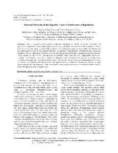
Figure 1 illustrates key developmental transitions and mediators. As opposed to the foregut and hindgut, which are of ectodermal origin, the Drosophila midgut originates from the endoderm and is thus established during gastrulation. After
induction of the endodermal fate by maternal factors, endoderm is further specified by several transcription factors that are widely conserved in evolution, including the GATA transcription factor Serpent (Srp) and the HNF/Fork Head (Fkh) transcription factors (Takashima et al. 2013). Endodermal cells will then undergo specification into either enterocyte (EC)-like or enteroendocrine (EE)-like cells through the action of proneural proteins (such as Lethal of scute, which promotes endocrine fates) and Notch signaling (activation of Notch promotes EC fates) (Takashima et al. 2011a, 2013). The balance between proneural protein activity and Notch signaling activity will thus ultimately determine the cellular composition of the midgut, yet the upstream regulators of proneural gene expression (in addition to GATA and Fkh transcription factors) remain largely unknown (Takashima et al. 2011a, 2013). Extracellular signals derived from the adhering visceral mesoderm then promote differentiation of the midgut endoderm around stage 16 [for reviews see Bienz (1997), Nakagoshi (2005)]. The four posterior Homeobox (Hox) genes in the visceral mesoderm promote the expression of signaling molecules that specify the subdivision of the midgut endoderm along its anterior-posterior axis [for reviews see Bienz (1997), Miller et al. (2001a,b)]. These factors include Decapentaplegic (Dpp), a member of the Bone morphogenetic protein (BMP)/Transforming growth factor b (Tgfb) superfamily, and Wingless/Wnt (Wg), which in turn induce the expression of Vein, a ligand for the EGF receptor, in the visceral mesoderm (Immerglück et al. 1990; Reuter and Scott 1990). All three signaling molecules are involved in the induction of morphogenetic events that subdivide the midgut (Immerglück et al. 1990; Reuter and Scott 1990; Casas-Tinto et al. 2008). In parasegment 7 of the endoderm, they induce, for example, labial (lab): a gene coding for a Hox protein required for endoderm differentiation (Immerglück et al. 1990; Reuter and Scott 1990; Casas-Tinto et al. 2008). Complex interactions between Lab and other transcription factors induced by Dpp and Wg further shape the midgut. teashirt (tsh) negatively regulates lab and is required for interstitial cell precursors (Mathies et al. 1994), whereas defective proventriculus (dve) is broadly expressed in midgut precursor cells and is later repressed by lab (Nakagoshi et al. 1998). Dpp is believed to form a morphogenetic gradient that induces the high-threshold target lab and the lowthreshold target dve in different fields of the gradient, resulting in the specification of two different types of ECs: copper cells (Lab-positive) and interstitial cells (Dve-positive), respectively (Nakagoshi 2005). In addition to the formation of the larval midgut, endodermal progenitors for the adult midgut are also formed in the early embryo. These cells, adult midgut progenitors (AMPs), form small clusters of proliferating, undifferentiated cells that are attached to the basal surface of the larval gut epithelium. During metamorphosis, AMPs form the adult midgut by dispersing and proliferating within distinct islands, in a process that is regulated by Epidermal growth factor receptor (Egfr)
signaling (Jiang and Edgar 2009). AMPs are regulated by a transient niche in the larval midgut that is established through Notch signaling and maintains AMPs in an undifferentiated state through Dpp signaling (Mathur et al. 2010). Niche cells go on to differentiate during metamorphosis, spreading out between the newly forming adult gut and the degenerating larval midgut and forming a transient pupal epithelium. At that stage, AMPs form large cell clusters that eventually fuse to make the adult midgut epithelium (Takashima et al. 2011b). Degeneration of the larval midgut requires activation of autophagy rather apoptosis (Denton et al. 2009), and is modulated by Dpp, the class I phosphoinositide-3-kinase pathway and ecdysone (Denton et al. 2012, 2018). The physiological role of the transient pupal epithelium remains to be established and will be of interest for future work. The adult gut and its cell types: genetic and anatomical compartmentalization
As shown in Figure 2C, the “ground plan” of the adult Drosophila gut consists of a tube lined by an epithelial monolayer consisting of four cell types: intestinal stem cells (ISCs), absorptive ECs, secretory EE cells, and enteroblasts (EBs): a postmitotic, immature cell type which will differentiate as an EC (or, possibly, as an EE, see below for current view of lineage relationships). Of note, midgut epithelial cells have a reverse arrangement of junctions compared to other Drosophila epithelia, with occluding junctions above adherens junctions, as in vertebrates (Chen et al. 2018a). This epithelium is surrounded by visceral muscle and protected toward the lumen by secreted mucus and, posterior to the foregut, by a chitinous layer: the peritrophic matrix (Hegedus et al. 2009). There are, however, substantial variations of this common theme, both at the gross anatomy and cellular levels. These are primarily determined by the developmental origin of a given gut region, as well as its specific location along the antero-posterior axis. Anatomical specializations and regional compartmentalization both enable sequential ingestion, storage, digestion, absorption, and defecation (Karasov et al. 2011). Anteriorly, the ectodermally derived foregut is subdivided into esophagus, crop, and cardia (Figure 2B). The crop is a diverticulated structure unique to Diptera, consisting of a complex array of valves and sphincters ensuring transit of intestinal contents in and out of the crop into the main alimentary canal. Although its functions in Drosophila remain to be investigated, work in other insects suggests that it may function in early digestion, detoxification, microbial control, and/or food storage (Stoffolano and Haselton 2013). The cardia (also known as proventriculus) is a complex bulbshaped organ composed of three epithelial layers. It produces the peritrophic matrix, is a major site of antimicrobial peptide production (King 1988; Tzou et al. 2000) and may also act as a valve, regulating the entry of ingested food into the midgut. Posterior to the cardia, the endodermally derived midgut, with an average length of 6 mm in adult flies, occupies a large
The Drosophila melanogaster Intestine
359
Figure 1 Developmental transitions and key factors in intestinal cell fate decisions. See section Embryonic and larval development for details.
part of the abdomen and is commonly regarded as the main digestive/absorptive portion (Demerec 1950; Douglas 2013) (Figure 2, A and B). The Malpighian tubules, tubular excretory organs, discharge at the junction between the midgut and the ectodermally derived hindgut. The hindgut is further subdivided into pylorus (a second valve-like structure), ileum, and rectum, where water/ion exchange may occur (Demerec 1950; Douglas 2013). The muscles surrounding the epithelium are striated, in contrast to the smooth muscles found in mammalian intestines (Sandborn et al. 1967). Circular muscles are present throughout the tract, and an outer layer of longitudinal muscles surrounds the midgut. Physiology of the intestine is regulated by autonomic innervation and by hormones (Figure 2C and Figure 5, see Interorgan signaling for details of their functions). The gut is further
360
I. Miguel-Aliaga, H. Jasper, and B. Lemaitre
influenced by the tracheal system (Figure 2C), which forms a branched structure surrounding the gut during development (Linneweber et al. 2014) and may influence epithelial regeneration in the adult, although the mechanism(s) mediating such interactions remain controversial and provide interesting ground for future work (Guo et al. 2013; Z. Li et al. 2013). Ectodermally derived regions of the intestinal epithelium are relatively poorly understood compared to the midgut, which has been characterized in exquisite detail in recent years. The midgut is grossly subdivided into the anterior midgut, the middle midgut and the posterior midgut, but has been morphologically and molecularly subdivided into 10–14 regions (Murakami et al. 1994; Buchon et al. 2013b; Marianes and Spradling 2013) (Figure 2B and Figure 3B).
Figure 2 The adult intestine and its cell types. (A) The digestive tract is highlighted in gray inside an adult fly. (B) Main anatomical features of the adult digestive tract. (C) General cellular composition of the digestive tract. See section The adult gut and its cell types: genetic and anatomical compartmentalization for details.
Indeed, each midgut region is characterized by specific histological and cellular features (villi size, lumen width), stem cell proliferation rates, physical properties (e.g., luminal pH), and gene expression profiles (Murakami et al. 1994; Strand and Micchelli 2011, 2013; Buchon et al. 2013b; Marianes and Spradling 2013). The middle midgut (R3) contains a copper cell region in R3ab, which produces gastric acid, followed by a large flat cell region (R3c) with unclear function. Two boundaries flanking this region are inflection points where the midgut folds stereotypically inside the body cavity. Regionalization is not confined to the epithelium—it is also apparent in the muscles, trachea and neurons that surround it (Cognigni et al. 2011; Buchon et al. 2013b; Marianes and Spradling 2013; Linneweber et al. 2014). Although our genetic knowledge of midgut compartmentalization is far from comprehensive, the genes involved in its establishment during development may also play important roles in their adult maintenance. A case in point is the role of the transcription factor Lab, involved in both specification and later maintenance of the copper cell region of the R3 region (Hoppler and Bienz 1994; Buchon et al. 2013b; H. Li et al. 2013). Graded activities of the Wnt ligand Wingless are observed at several compartment boundaries and may determine their position
and identity (Buchon et al. 2013b; Tian et al. 2016). These boundaries may act as tissue-organizing centers from which Wingless may signal as a morphogen, akin to its roles in development (Buchon et al. 2013b; Tian et al. 2016). Extensive future studies are needed to gain a detailed understanding of maintenance and plasticity of midgut compartmentalization in the adult. While significant differences in cellular composition and function exist in the different regions of the adult intestinal epithelium, all regions of the midgut contain ISCs able to regenerate all cell types of their particular region (Buchon and Osman 2015). The ISC lineage was first characterized in the posterior midgut by two groups simultaneously (Micchelli and Perrimon 2006; Ohlstein and Spradling 2006) and is depicted in Figure 3A. Since then, a large number of studies have characterized the regulation of ISCs and their lineages. Midgut ISCs are uniformly interspersed among their differentiated progeny, and are located basally in close proximity to visceral muscles (Figure 2C and Figure 3A). They are, however, heterogeneous in both their cellular behavior and gene expression, which may contribute to specifying compartment differences (Marianes and Spradling 2013; Dutta et al. 2015b) (Figure 3, B and C). Consistent with this idea, mosaic
The Drosophila melanogaster Intestine
361
Figure 3 Regional differences in ISC proliferation. (A) General mode of midgut ISC proliferation. See main text for details in The adult gut and its cell types: genetic and anatomical compartmentalization. Alternative modes of ISC proliferation - stress induced rather than constitutive - are found in two specific intestinal regions: the copper cell region (B) and the hindgut (C). See main text in The adult gut and its cell types: genetic and anatomical compartmentalization for details. CC, copper cell; GB, gastroblast, GSSCs, gastric stem cells; IC, interstitial cell.
analysis has shown that ISCs in a certain region tend to maintain their region’s progeny, and rarely contribute to the production of differentiated cells in adjacent regions. ISC heterogeneity is established during metamorphosis (Driver and Ohlstein 2014) and is then maintained in cooperation with regional signals from surrounding tissues such as the visceral muscles (see Stem cells: signals and niches for
362
I. Miguel-Aliaga, H. Jasper, and B. Lemaitre
details). A multitude of local, paracrine and systemic signals and signaling pathways that control ISC proliferation and differentiation have been identified (see Stem cells: signals and niches for details), and changes in ISC function and compartmentalization have been described during tissue damage and aging (Biteau et al. 2011; Jiang and Edgar 2011; Buchon et al. 2013a,b; Lemaitre and Miguel-Aliaga 2013; Buchon and
Osman 2015). ISCs are characterized by the expression of Escargot (Esg) and Delta (Dl), and constitute the majority of cells capable of mitosis in the posterior midgut. ISC maintenance requires the Daughterless protein, as well as transcriptional repression of Notch target genes such as the Enhancer of split complex [E(spl)-C] by a Hairless-Suppressor of Hairless complex (Bardin et al. 2010). During regenerative episodes, ISCs in the posterior midgut undergo asymmetric division to give rise to EBs, which retain Esg expression but lose Dl expression while activating Notch signaling. EBs further differentiate into either POU domain protein 1 (Pdm1)positive absorptive ECs, or Prospero (Pros)-positive secretory EE cells (Micchelli and Perrimon 2006; Ohlstein and Spradlingm 2006, 2007) (Figure 3A). There is evidence for clonal competition during normal homeostasis, whereby loss of ISC through differentiation or death (clonal extinction) may be compensated by increased proliferation/symmetric division of other ISCs (clonal expansion) (de Navascués et al. 2012; Kolahgar et al. 2015; Suijkerbuijk et al. 2016). Tumors may also harness this process to fuel their own growth (Suijkerbuijk et al. 2016). Specification of ECs requires Esg downregulation and activation of Notch and Jak/Stat signaling and the Sox21a and Hindsight transcription factors, while Dpp activity and GATAe contribute to EC production during acute regeneration (Ohlstein and Spradling 2007; Beebe et al. 2010; Korzelius et al. 2014; Antonello et al. 2015 Baechler et al. 2015; Zhai et al. 2015; Chen et al. 2016). Recent studies have refined our understanding of EE specification (Biteau and Jasper 2014; Beehler-Evans and Micchelli 2015; C. Wang et al. 2015; Guo and Ohlstein 2015; Zeng and Hou 2015; Sallé et al. 2017; He et al. 2018). In vivo lineage-tracing methods suggest that these cells are generated from precommitted Pros-expressing ISCs, and not, as previously described, as an alternative to EC differentiation from a common EB cell (Figure 3A) (Biteau and Jasper 2014; Guo and Ohlstein 2015; Zeng and Hou 2015). EE specification and differentiation requires less Notch activity than differentiation of EBs into ECs, and involves Phyllopod-mediated repression of the Tramtrack transcriptional repressor, which promotes Scute-mediated activation of Pros (Li et al. 2017; Chen et al. 2018b; Yin and Xi 2018). Numb and the autophagy protein Atg16 have further been implicated (C. Wang et al. 2015; Nagy et al. 2017; Sallé et al. 2017). EE regeneration from precommitted Pros-positive ISCs may be limited by Slit, an EE-derived ligand for the Roundabout 2 (Robo2) receptor (Biteau and Jasper 2014; Nagy et al. 2017). Slit binds Robo2 on ISCs, setting up a negative feedback loop from differentiated EEs that limits further production of these cells (Biteau and Jasper 2014). In this feedback loop, ISCs seem to respond to tissuewide changes in Slit levels, rather than changes in local concentration, as clonal perturbation of Slit expression or EE concentration is not sufficient to locally influence EE production (Sallé et al. 2017). Another study has analyzed EE cell diversity and found that, unexpectedly, Su(H)GBE-positive (Notch active) EBs can give rise to class II EE cells, in addition
to ECs (Beehler-Evans and Micchelli 2015). EE differentiation is further promoted by calcium signaling in response to activation of the stretch-activated ion channel Piezo (He et al. 2018). While the specification and differentiation pathways regulating EC vs. EE lineage specification and differentiation have thus been intensely studied in the past decade, further temporally and spatially resolved lineage tracing studies, potentially coupled with live imaging, will be needed to clarify the exact signaling events governing EE and EC differentiation. Such work is expected to refine the current model describing the defining events promoting EE vs. EC lineage specification. ISCs in the anterior midgut differ in some respects from those in the posterior midgut. For example, proliferation rates and expression of PAR-domain protein 1 (Pdp1) and Signaltransducer and activator of transcription protein at 92E (Stat92E) reporters are different between anterior and posterior ISCs (Marianes and Spradling 2013), while other transcription factors, such as GATAe, Snail (Sna), and paired-type homeobox transcription factor (Ptx1) have region-specific expression and regulatory roles in ISCs along the digestive tract (Dutta et al. 2015b). Two different ISC populations have been referred to as gastric stem cells. A stem cell pool at the foregut/midgut junction in the cardia can differentiate and migrate to contribute to the crop, the esophagus and the cardia (Singh et al. 2011). In the copper cell region of the midgut, which shares some similarity to the stomach in vertebrates, another population of ISCs also referred to as gastric stem cells (Esg- and Dl-positive) generate three different cell types: the acidsecreting copper cells, which express Dve, high levels of Lab, and are detected by an antibody against Cut; interstitial cells, which express Dve and lower levels of Lab; and Pros-expressing EE cells (Strand and Micchelli 2011). Similar to the ISC lineage in the posterior midgut, gastroblasts (the counterpart of the EB in this region) have been identified and proposed to be the precursor cell that generates these three differentiated cell types (Strand and Micchelli 2011) (Figure 3B). In the hindgut, a ring of ISCs reminiscent of the foregut/ midgut junction stem cell pool is found posterior to the pylorus. These hindgut ISCs differentiate into hindgut ECs as they migrate posteriorly (Takashima et al. 2008; Fox and Spradling 2009) (Figure 3C). The regenerative properties of the foregut and hindgut have not been extensively investigated, but it is generally assumed that these ectodermal regions of the gut are more quiescent than the endodermal midgut (Fox and Spradling 2009).
Organ Plasticity In recent years, the adult midgut has arguably become “the” organ system for the study of adult organ plasticity. We have learned a great deal about the steady-state dynamics of its adult progenitors, as well as their adaptations to challenges, both external (e.g., infection, nutrition) and internal (e.g., aging, reproduction). More recent studies are extending the
The Drosophila melanogaster Intestine
363
study of midgut plasticity to nonmitotic cell types, such as the ECs and EE cells of the intestinal epithelium. It is also becoming increasingly recognized that the gut’s anatomical regionalization is associated with striking differences in the turnover and plasticity of different gut regions. This section attempts to provide a comprehensive review of the mechanisms of midgut plasticity. It also discusses their physiological modulation in adult flies, and places the plasticity of the adult midgut in a broader context by briefly contrasting it with the plasticity of other gut regions. Stem cells: signals and niches
The activity of ISCs along the digestive tract needs to be specifically and dynamically regulated to adjust tissue turnover to local and tissue-wide needs. Numerous signaling pathways that regulate these processes have been identified. Signaling pathways that influence ISC proliferation and differentiation in Drosophila include Notch (Ohlstein and Spradling 2007), Jak/Stat (Jiang et al. 2009; Beebe et al. 2010; Lin et al. 2010), Egfr (Jiang and Edgar 2009; Buchon et al. 2010; Biteau and Jasper 2011; Jiang et al. 2011), Insulin (Amcheslavsky et al. 2009; Biteau et al. 2010; Choi et al. 2011; O’Brien et al. 2011), Jun-N-terminal Kinase (JNK) (Biteau et al. 2008), Wg (Lin et al. 2008; Lee et al. 2009), Target of Rapamycin (Tor) (Amcheslavsky et al. 2011; Kapuria et al. 2012; Quan et al. 2013), Bmp/Dpp (Guo et al. 2013; H. Li et al. 2013; Z. Li et al. 2013; Tian and Jiang 2014; Ayyaz et al. 2015), Hippo (Karpowicz et al. 2010; Ren et al. 2010; Staley and Irvine 2010), Juvenile Hormone (JH) (Reiff et al. 2015; Rahman et al. 2017), and Ret signaling (Perea et al. 2017). ISC proliferation and differentiation also require the Brahma chromatin-remodeling complex (Jin et al. 2013). ISC differentiation is further controlled by esgmediated repression of Nubbin (Nub, also known as Pdm1) (Korzelius et al. 2014; Loza-Coll et al. 2014). The combined action of these signaling pathways influences proliferative activity, self-renewal and differentiation in the ISC lineage in response to a wide range of local and systemic cues. For recent reviews that discuss ISC regulation by these signaling pathways in detail, see Biteau et al. (2011), Jiang and Edgar (2011), Buchon et al. (2013a), Lemaitre and Miguel-Aliaga (2013), Buchon and Osman (2015), Guo et al. (2016), and Gervais and Bardin (2017). The large number of different signals regulating ISC activity likely results from the need to integrate paracrine, local, systemic, and environmental stimuli to elicit appropriate regenerative responses. During cycles of starvation and refeeding, for example, ISC proliferation is stimulated and switched from an asymmetric mode to a symmetric mode through insulin-like peptide 3 derived from the visceral muscle (O’Brien et al. 2011). The visceral muscle also provides Vein and, possibly, Wg ligands that control ISC maintenance and proliferative activity both in homeostasis and during regenerative episodes after epithelial damage (Lin et al. 2008; Zhou et al. 2013; Biteau and Jasper 2011). The EB, in turn, feeds back to control ISCs proliferation, at least partly by
364
I. Miguel-Aliaga, H. Jasper, and B. Lemaitre
expressing Wg and Unpaired 2 (Upd2) (Cordero et al. 2012; Zhai et al. 2015; Chen et al. 2016). EBs and ECs also limit ISC proliferation through E-cadherin–mediated cell-cell contact (Choi et al. 2011; Liang et al. 2017). Recent work is starting to provide insight into the integration of these diverse signals. Intracellular calcium signaling, for example, is emerging as a central regulator of ISC proliferation in Drosophila in response to a wide range of mitogenic signals (Deng et al. 2015; Xu et al. 2017). The diversity of ISC responses to mitogenic signals along the gastrointestinal tract remains poorly understood. ISCs in the posterior midgut and gastric stem cells are regulated by similar signaling pathways, including Wg, Egfr, and Notch (Strand and Micchelli 2011, 2013; C. Wang et al. 2014), but differ in their proliferative activity (gastric stem cells are more quiescent than posterior midgut ISCs), and in their response to specific pathways. Loss of Dpp signaling components, for example, causes differentiation defects in gastricderived lineages, but not in posterior midgut ISCs (H. Li et al. 2013). Sustained Dpp expression along the midgut, on the other hand, is sufficient to induce ectopic copper cell formation in the anterior, but not posterior midgut, indicating that additional regional determinants influence stem cell responses to Dpp signaling (H. Li et al. 2013). Similarly, activation of Jak/Stat signaling in gastric stem cells leads to their misdifferentiation, generating ectopic EC-like cells in the copper cell region (H. Li et al. 2016), while Jak/Stat induces proliferation but does not alter differentiation in ISCs of the posterior midgut (Jiang et al. 2009). Intestinal plasticity during aging
The digestive tract of adult Drosophila has become a powerful model in which to explore aging of barrier epithelia in metazoans (Biteau et al. 2011; Lemaitre and Miguel-Aliaga 2013). During aging or after an infection, intestinal compartmentalization is disturbed, as revealed by a strong alteration in gene expression patterns (Buchon et al. 2013b). A detailed understanding of the progression of epithelial changes that result in the loss of barrier function in old animals is starting to emerge. In aging flies, ISCs become hyperproliferative, leading to accumulation of misdifferentiated cells that coexpress stem and progenitor cell markers (like Dl and Esg) and differentiation markers (like Notch signaling activity and polyploidy) (Biteau et al. 2008; Choi et al. 2008; Buchon et al. 2009b; Hochmuth et al. 2011). An early event causing this is the development of gastric metaplasia, where copper cells transdifferentiate into posterior midgut EC-like (Pdm1positive) cells, compromising the acidity of the gastric region (H. Li et al. 2016). Reduced acidification leads to changes in the compartmentalization and composition of the commensal microbiota, ultimately resulting in commensal dysbiosis and immune deregulation in the midgut epithelium. Dysbiosis, in turn, triggers a secondary inflammatory response, which produces a Dual oxidase (Duox)-induced oxidative burst that damages the epithelium and induces ISC proliferation and misdifferentiation. The resulting epithelial dysplasia, in turn,
contributes to the loss of barrier function, which ultimately causes mortality (Rera et al. 2012). Age-related intestinal dysplasia is associated with increased JNK and/or Platelet-derived growth factor (PDGF)/ Vascular endothelial growth factor (VEGF) signaling activity (Biteau et al. 2008; Choi et al. 2008; Buchon et al. 2009b; Hochmuth et al. 2011), and factors that contribute to dysplasia in the aging intestine include a decline of mitochondrial function in stem and progenitor cells, dysbiosis of gut commensals, inflammatory signals from the fat body, and increased endoplasmic reticulum (ER) stress and Pol III transcriptional activity (Rera et al. 2011; Chen et al. 2014; Guo et al. 2014; Rogers and Rogina 2014; Clark et al. 2015; L. Wang et al. 2015; Siudeja et al. 2015; Filer et al. 2017). ISCs of old flies also display frequent somatic mutations, resulting in neoplasia (Siudeja et al. 2015). Persistent immune activity has been linked to intestinal hyperplasia and tumor susceptibility (Petkau et al. 2017). Neoplasia derived from Notchdeficient ISCs has been shown to trigger dysregulation of ISC niche signals, including Egfr ligands and cytokines that activate Jak/Stat signaling, thus contributing to its establishment and development (Patel et al. 2015). The overall longevity of the animal has been shown to correlate with the degree to which these intestinal changes become apparent (Biteau et al. 2010), and interventions that specifically target several aspects of intestinal health have been shown to extend the life span of flies reared under laboratory conditions (Rera et al. 2011; Ayyaz and Jasper 2013; Chen et al. 2014; Guo et al. 2014; Clark et al. 2015; L. Wang et al. 2015). Nutritional and metabolic plasticity
The rate of ISC proliferation is substantially but reversibly reduced by long-term nutrient deprivation (McLeod et al. 2010; Choi et al. 2011; O’Brien et al. 2011): an effect recapitulated by genetic manipulations that downregulate/ mutate intestinal insulin receptor or downstream pathway components (Amcheslavsky et al. 2009; Biteau et al. 2010; Choi et al. 2011; O’Brien et al. 2011). While there is some consensus that nutrient scarcity is, at least partly, relayed to the intestine as a reduction in insulin signaling, different insulin sources and cellular mechanisms have been proposed. Acting in adult intestinal progenitors, insulin signaling promotes ISC proliferation, and is also required to give rise to ECs and EEs (Amcheslavsky et al. 2009; Biteau et al. 2010; Choi et al. 2011; O’Brien et al. 2011). Insulin signaling may also control proliferation and differentiation through its actions in EBs, affecting differentiation and ISC/EB adhesion (Choi et al. 2011). In contexts of organ resizing (e.g., as the gut grows in the first few days of adult life, or in response to refeeding following prolonged starvation), insulin signaling also supports rapid expansion of the midgut epithelium by promoting symmetric rather than asymmetric ISC divisions (O’Brien et al. 2011). The nutrient-driven progenitor expansion and their switch to symmetric divisions are modulated by the two RNA binding proteins Lin-28 and Fmr1, which act
antagonistically and post-transcriptionally on the Insulin-like receptor (InR) to modulate how progenitors respond to insulinlike peptide(s) (Chen et al. 2015; Luhur et al. 2017). A role for systemic insulin-like peptides in coupling nutrient availability with epithelial turnover has been suggested by ablation of the nutrient-sensitive insulin producing cells of the brain’s pars intercerebralis (Amcheslavsky et al. 2009; Biteau et al. 2010). Other experiments have also revealed a paracrine role for the gut muscle–derived insulin-like peptide Ilp3 in the context of posteclosion and nutrient-driven midgut resizing (O’Brien et al. 2011). Ilp3 expression in muscles is nutritionally regulated and sustained by the EE peptide tachykinin (Tk) (Amcheslavsky et al. 2014). In this context, it may also be important to consider possible nutritional roles of neuropeptides produced by enteric neurons. Indeed, in larvae, yeast restriction impacts the release of gut neuronderived insulin-like and Pigment-dispersing factor (Pdf) neuropeptides which, in turn, control the branching of gut terminal tracheal cells (Linneweber et al. 2014). The nutritional plasticity of enteric trachea during larval life is physiologically significant, later affecting the ability of adult flies to withstand nutrient scarcity (see Gut-innervating neurons for details). A possible contribution of gut trachea and/or neurally derived insulin-like/Pdf neuropeptides to the nutritional modulation of epithelial turnover awaits further investigation. It is also important to underscore that there may be insulin-independent nutritional signals as well as nutrientindependent roles for insulin signaling [see, for example, Quan et al. (2013)]. The relative importance of these mechanisms (and, more generally, the effects and relative contribution of nutrition and/or insulin signaling to intestinal homeostasis) are likely to differ depending on the age, microbiota composition, sex, and reproductive status of the experimental flies. Recent studies are beginning to explore the modulation of epithelial turnover by more direct and/or specific nutritional inputs. A direct action of dietary glutamate acting via metabotropic glutamate (mGluR) receptors in ISC/EBs was suggested by a recent study (Deng et al. 2015). ISC proliferation can also be promoted by a lipolysis pathway (Singh et al. 2016), or by limiting mitochondrial pyruvate metabolism (Schell et al. 2017). Finally, dietary methionine sustains production of the universal methyl donor S-adenosylmethionine, which is, in turn, required to sustain ISC proliferation, both directly and through its promotion of Upd3 production in ECs (Obata et al. 2018b). In addition to modulating progenitor dynamics, nutrition also affects the activity of differentiated cells in the intestinal epithelium. For example, young (4n) ECs can undergo a process of ploidy reduction known as amitosis and give rise to new functional ISCs to maintain epithelial integrity following starvation-induced ISC loss (Lucchetta and Ohlstein 2017). An alternative mechanism involving changes in the rate of EC loss has also been proposed (Jin et al. 2017). Specific nutrients may also directly change the digestive/ absorptive properties of ECs (see, for example, the case of
The Drosophila melanogaster Intestine
365
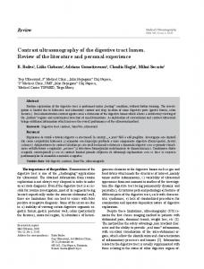
Figure 4 Sex and reproductive differences in ISCs and ECs. (A) Contributions of the intrinsic sex differentiation pathway and the mating-triggered rise in circulating JH to ISC and EC homeostasis in females. (B) Contributions of the intrinsic sex differentiation pathway to ISC and EC homeostasis in males. See section Sex differences and reproductive plasticity for details. This figure was inspired by Portman and Biteau (2016).
a-amylase in Digestive enzymes and their regulation), or the systemic signals produced by EC/EEs (reviewed in Systemic and EE signals). Sex differences and reproductive plasticity
The nature and significance of sex differences in the Drosophila intestine had remained unexplored until recently, despite circumstantial observations pointing to their existence. Indeed, lineage tracing had suggested faster turnover of the adult midgut epithelium in females [data not shown in Jiang et al. (2009)], and a method based on quantifying visual features of excreta had revealed modulation of physiological features such as pH and concentration of intestinal contents by sex and reproductive state (Cognigni et al. 2011). A more recent study explored sexual dimorphisms more comprehensively in the midgut of adult virgin flies, and revealed extensive sex differences in the expression of genes with putative roles in proliferation, redox, and carbohydrate metabolism (Hudry et al. 2016). Adult female ISCs divide more readily than their male counterparts, both in homeostasis and in response to epithelial damage. Increased proliferation in females results from a noncanonical sex differentiation pathway active inside their ISCs. This pathway involves the sex-specific actions of the Transformer (Tra)
366
I. Miguel-Aliaga, H. Jasper, and B. Lemaitre
RNA binding protein downstream of sex chromosome/autosome sensing mechanisms, but is independent of the canonical Tra partner Transformer-2 and the two Tra targets Doublesex and Fruitless (Hudry et al. 2016) (Figure 4). Why are female ISCs more proliferative? A clue was provided by close examination of what happens inside a female fly after mating. In addition to previously described behavioral changes, a single mating dramatically remodels the midgut of a female fly in only 3 days (Reiff et al. 2015). Stem cell proliferation, the number of differentiated ECs and the size of the midgut are all increased. Changes in the expression and activity of lipid metabolism regulators in ECs are also apparent. Preventing at least some of these mating-induced intestinal changes reduces egg production, indicating that the reproductive plasticity of the female intestine is important for reproductive success (Reiff et al. 2015). While at least one signal upstream of these reproductive changes is a postmating rise in JH (Figure 4), ISCs require their intrinsic female identity to respond to the mating signal(s) by increasing their proliferation (Hudry et al. 2016). Hence, the female sexual identity of adult ISCs allows organ resizing for reproductive purposes. The sex difference in midgut size (virgin female midguts are larger and longer than male counterparts, and this dimorphism is further enhanced by mating; Reiff et al.
2015; Hudry et al. 2016)), also provides an attractive experimental paradigm to explore regulation of organ size. Changes in organ size may not only involve changes in the rate and mode (asymmetric vs. symmetric) of stem cell division, but also in the turnover of their progeny; persistent masculinization of adult ISC/EBs in virgin females shrink their gut to a male-like size (Hudry et al. 2016). Because virgin male and female guts have comparable ISC density and division mode, the finding that females require a higher proliferation rate to maintain organ size suggests that their EC turnover may also be faster than that of males. Consistent with this idea, genetic manipulations that interfere with EC survival and adhesion in mated females can affect midgut size (Liang et al. 2017). Are there trade-offs to the enhanced plasticity of the female midgut? The female gut is more susceptible to tumorigenic insults, at least partly as a result of the female sexual identity of its intestinal progenitors (Hudry et al. 2016) (Figure 4). Another study has begun to shed mechanistic light on the molecular defects contributing to spontaneous, age-related neoplasia and their different prevalence in males and females. The authors also reported a positive correlation between neoplasia and ISC proliferation rate, and further showed that, in males, spontaneous neoplasia often arises through genomic deletions and large structural rearrangements leading to loss of heterozygosity of X-linked tumor suppressors (present in a single copy in males) (Siudeja et al. 2015). As well as neoplasia, other aspects of intestinal (patho)physiology may contribute to differences in life span between the sexes (Figure 4). It has long been known that genetically limiting intestinal proliferation extends the life span of females, but not that of males (Biteau et al. 2010). A more recent study reported that age-related dysplasia and intestinal barrier breakdown are both more pronounced in females than males (mated flies were used), and can be ameliorated by dietary restriction: an intervention known to extend life span (Regan et al. 2016). Not everything is bad news for female flies; in the same study, male flies were found to be more susceptible to acute intestinal infection and xenobiotic stress. Like the tumors, all these observations could at least partly be explained by the higher proliferation rate of female ISCs, which may help females regenerate their midgut faster after infections, but may also render it more vulnerable to age-related dysplasia. In light of these recently reported, but apparently extensive sex differences, we encourage the community to control for sex and reproductive state in any future studies.
Functions In addition to its obvious roles in nutrient extraction and utilization, the digestive tract responds to the food and bacteria in its lumen to adapt both its own physiology and that of remote organs. In the following sections, we review how the digestive tract senses, digests, and absorbs nutrients, how it
interacts with commensal microbes and opportunistic pathogens, and how its different cell populations adapt and signal to the rest of the fly. Digestion and absorption
Once food enters the digestive tract, its complex macromolecules are broken down by digestive enzymes before being absorbed by the intestinal epithelium. It is generally accepted that the midgut is the main site of digestion in Drosophila, despite evidence for extraoral digestion and enzymatic conversions in the foregut and/or crop of other insects (Lehane and Billingsley 1996). Work primarily in other insects has revealed that digestion can be further modulated by temperature, redox potential, pH, and intestinal transit (Lehane and Billingsley 1996; Douglas 2013). The amount and composition of food available for digestion may also be modulated by gut bacteria (Huang and Douglas 2015). This section describes how nutrients are broken down and absorbed in the adult midgut, as well as the (so far limited) evidence in Drosophila for roles of gut acidity and intestinal transit in the context of digestion. Digestive enzymes and their regulation: Drosophila feeds on various kinds of decaying plant and fungal material. The relatively complex composition of the material it ingests is paralleled by an impressive array of digestive enzymes dedicated to the handling of carbohydrates, proteins, and lipids; as many as 349, based on bioinformatics predictions, with the largest families corresponding to endo/exo peptidases as well as proteins with carbohydrate or lipase activity (Carlson and Hogness 1985; Ross et al. 2003; Horne and Haritos 2008; Horne et al. 2009; Tamaki et al. 2012; see https://lemaitrelab.epfl.ch/resources for a complete list). Flies may also be able to digest both bacteria and the microbial material found in rotting fruits. Indeed, the presence of 15 different lysozymes in the Drosophila genome with no known immune functions suggests that flies may use them to digest peptidoglycan: a major component of bacterial walls (Kylsten et al. 1992). Flies also appear to be equipped with chitinases and glucanases that may aid in the digestion of yeasts. Intriguingly, families of related digestive enzymes are often found as gene clusters in the genome. This applies to Jonah proteases, trypsins, a-esterases, mannosidases, and lipases (Buchon et al. 2013b). These gene clusters may have arisen by gene duplication to enhance digestive capacity, and/or to tailor digestive activities to specific portions of the digestive tract following gene duplication and subsequent divergence: a possibility suggested by evolutionary analysis of the a-amylase gene family (Da Lage et al. 2000; Zhang et al. 2003). Consistent with regional specialization of digestive functions, the expression of most digestive enzymes is confined to specific segments of the digestive tract (Abraham and Doane 1978; Buchon et al. 2013b; Dutta et al. 2015a). For example, the expression of some genes coding for enzymes involved in
The Drosophila melanogaster Intestine
367
the breakdown of sugars are enriched in anterior (R1/R3) portions of the adult midgut, whereas peptidase genes may be expressed more posteriorly (Dutta et al. 2015a). Regional expression of digestive enzymes is also striking in the larva (Harrop et al. 2014; Overend et al. 2016). However, it is important to consider that the ultimate site of enzymatic activity may not necessarily be equivalent to the site of transcript expression, as enzymes may diffuse in the gut lumen. They may also differ in their positioning and interactions with EC villi or the peritrophic matrix. Indeed, work in other insects has shown that enzymes involved in earlier steps of digestion of macromolecules (e.g. a-amylases, proteases) tend to localize to the lumen of the digestive tract, whereas those involved in later steps (maltases, di-peptidases) are more often found in the space between the epithelium and the peritrophic matrix, and are often associated with the surface of gut epithelial cells (Terra et al. 1979; Douglas 2013). The enzymatic activity of the intestine is a key factor determining availability of certain nutrients. It is therefore not surprising that the expression and/or activity of digestive enzymes are tightly regulated in many insects. Modulation by nutrient quality and quantity, neuronal activity, and endocrine signals has been described in insects such as mosquitoes, locusts, cockroaches, or crickets (Wigglesworth 1972; Clissold et al. 2010; Douglas 2013), but has not been extensively investigated in Drosophila. A substantial reduction of intestinal digestive enzyme activities including trypsin, chymotrypsin, aminopeptidase, and acetate esterase has been reported in flies lacking EE cells, in the absence of obvious effects on food intake (Amcheslavsky et al. 2014). The transcription of digestive enzymes with a putative function in breaking down carbohydrate (such as intestinal amylases) is induced by starvation in both larvae and adults (Zinke et al. 2002; Chatterjee et al. 2014). Their transcription is also sexually dimorphic in adult flies, with many of them showing upregulated expression in males (Hudry et al. 2016). It has also long been known that the end products of digestive processes can be repressed by expression of enzymes involved in their production; for example, sucrose and its products, glucose and fructose, have been shown to repress amylase gene expression, an effect known as glucose repression (Hickey and Benkel 1982; Benkel and Hickey 1986; Zinke et al. 2002; Chng et al. 2014). This reduction in digestive capacity may represent an adaptation to limit dietary sugar absorption during periods of nutritional abundance, given that Drosophila is poorly adapted to nutritional excess. Two recent studies have shed light on the repression mechanism. One such mechanism involves the TGF-b/Activin ligand Dawdle (Daw) which, upon refeeding with nutritious sugars (but not non-nutritious sugars) after a period of starvation, reduces the expression of carbohydrate digestive enzymes in the ECs of adult flies (Chng et al. 2014). Experiments using whole larvae have also revealed that activation of the intracellular sugar sensor complex MondoBigmax promotes the expression of both daw and the transcription factor sugarbabe (sug) (Mattila et al. 2015). sug is
368
I. Miguel-Aliaga, H. Jasper, and B. Lemaitre
both necessary and sufficient to repress the expression of amylases. Although further work will be required to clarify how Mondo-Bigmax and TGF-b/Activin signaling intersect, the current data are consistent with a model whereby ECs integrate information about sugar uptake (sensed intrinsically in the intestine by Mondo-Bigmax) and the carbohydrate status of the fat body (relayed by TGF-b/Activin signaling) to modulate expression of the carbohydrate digestive enzymes. Luminal bacteria can also affect the expression of digestive enzymes, which may, in turn, affect digestive capacity. Indeed, gut-associated bacteria have been shown to modulate the expression of enzymes such as amylases, proteases, and maltases (Erkosar et al. 2014). Importantly, the positive effect of microbiota on peptidase gene expression is at least partly responsible for their larval growth-promoting effects in nutrient-poor conditions (Storelli et al. 2011; Erkosar et al. 2015). Oral bacterial infection is often associated with reduced expression of a broad range of digestive enzymes (e.g. lipases, trypsins, amylases) (Buchon et al. 2009b; Chakrabarti et al. 2012; Erkosar et al. 2015; Loudhaief et al. 2017; Troha et al. 2018), and can lead to hypophagia and changes in excretion (Vallet-Gely et al. 2008; Ayres and Schneider 2009; Du et al. 2016). The causal links between gut infection, damage, reduced feeding, and expression of digestive enzymes remain to be fully elucidated. Digestive arrest upon infection may be a consequence of gut damage and/or reduced feeding, but might also involve bacteria-tohost signaling. Reduced digestion and some of these other intestinal/feeding changes may represent a host strategy to help limit bacterial ingestion. Alternatively, they may also be a strategy used by pathogenic bacteria to counteract peristalsis and persist in the gut. In contrast to the relatively abundant data illustrating their dynamic expression, the functions of digestive enzymes remain largely unexplored. Two notable exceptions concern the roles of amylases and a lipase. A functional role for amylases in breaking down complex sugars was revealed by mutants lacking both amylase p and d (Hickey et al. 1988). Unlike wild-type flies, these mutants die on a starch-only diet, but their lethality can be rescued by dietary supplementation with simple sugars: the end products of amylase digestion. Interestingly, mutant lethality can also be bypassed by cohousing the flies lacking amylases with wild-type flies, suggestive of extraoral digestion. Such extraoral digestion may be enabled by regurgitation and/or excretion of amylases. Another functional study concerned the intestinal triacylglyceride (TAG) lipase/cholesterol esterase Magro (Mag). In response to low cholesterol in the diet, expression of the Hr96 nuclear receptor (homologous to the vertebrate LXR receptor involved in regulated cholesterol homeostasis) is upregulated (Bujold et al. 2010). Hr96 binds cholesterol and promotes the expression of genes involved in cholesterol homeostasis and lipid breakdown including mag (Horner et al. 2009; Sieber and Thummel 2009; Bujold et al. 2010). Mutant and knockdown experiments are consistent with a
dual role for intestinal Mag in breaking down intestinal cholesterol esters to maintain cholesterol homeostasis, as well as enabling TAG breakdown, required for intestinal lipid absorption and peripheral fat accumulation (Sieber and Thummel 2009, 2012). Intestinal mag expression can also repressed by a sugar-rich diet in a foxo-dependent manner (Karpac et al. 2013). Interestingly, this adaptive mechanism becomes chronically active in the aging intestine as a result of JNK pathway activation, disrupting lipid homeostasis and contributing to the age-associated breakdown of metabolic homeostasis (Karpac et al. 2013). Absorption of carbohydrates: Following the breakdown of complex carbohydrates by digestive enzymes, a diverse array of transporters internalizes simple sugars into the ECs for further digestion and/or absorption [see Miguel-Aliaga (2012) for a comparative review]. The two major types of glucose transporters known to function in animals, the GLUT/Slc2 family of facilitative glucose transporters and the SGLT/Slc5 family of Na+-glucose symporters, have been shown to be expressed and/or active in the intestine of other insects [see, for example, Caccia et al. (2005, 2007), Price et al. (2007, 2010), Bifano et al. (2010)]. One such GLUT-like gene was described in Drosophila (Escher and RasmusonLestander 1999), and homologs of other glucose transporters can be found in the Drosophila genome. However, their expression and function remain to be investigated. The Drosophila genome also harbors a homolog of the more recently characterized SWEET family of sugar transporters (Artero et al. 1998; Baker et al. 2012). A disaccharide transporter similar to the transmembrane sugar transporters found in prokaryotes and fungi has also been described in flies [Slc45-1, referred to as Scrt in Meyer et al. (2011)]. Slc45-1 belongs to the relatively obscure Slc45 family of transporters, which also includes several human homologs. It is expressed in the embryonic and adult hindgut and can transport sucrose (Meyer et al. 2011; Vitavska and Wieczorek 2013). Disaccharide transport may also be achieved by two other trehalose transporters: Tret1-1 and Tret1-2, although the latter shows no trehalose uptake (Kanamori et al. 2010). The possible intestinal activity of these transporters deserves further investigation, not least because of their plastic expression. Indeed, genes with predicted functions in glucose transport are expressed at higher levels in male than female flies (Hudry et al. 2016) and, like digestive enzymes, are repressed by a high-glucose diet (Chng et al. 2014). It may also be of interest to explore whether putative sweet taste receptors recently shown to be expressed in ECs, enteric neurons, and EE cells (see Interorgan signaling for details) are functionally relevant in the context of sugar transport and/or absorption. Absorption of proteins: Proteins are broken down into products of a diverse chemical nature: di- and tri-peptides and a mixture of amino acids. This chemical diversity is paralleled by
a broad range of apical and basolateral transport systems, many of which are homologous to known mammalian transporter systems (Boudko 2012; Miguel-Aliaga 2012). These include Drosophila homologs of cationic amino acid transporters (Colombani et al. 2003), ion-dependent and independent amino acid transporters for neutral amino acids (Martin et al. 2000; Goberdhan et al. 2005; Miller et al. 2008; Reynolds et al. 2009) and oligopeptide transporters (Roman et al. 1998; Capo et al. 2017). Intestinal expression has been reported for the amino acid transporters Pathetic (Goberdhan et al. 2005), Minidiscs (Martin et al. 2000), NAT1 and other Slc6 family members (Thimgan et al. 2006; Miller et al. 2008), and the oligopeptide transporters Yin and CG2930, with enriched expression in proventriculus/hindgut and midgut, respectively (Roman et al. 1998; Capo et al. 2017). The nature, physiological modulation, and/or significance of many of these amino acid/oligopeptide transporters remains to be investigated. While many of these transport systems may handle dietary nutrients, some may be involved in detection and/or absorption of bacterially derived products. A possible contribution of three Drosophila transporters belonging to the Slc15 family of electrogenic (H+-coupled) oligopeptide transporters was tested in the context of the Drosophila immune response to microbially derived peptidoglycan. A previous study based on expression of ectopic Yin (one of the Drosophila Slc15 transporters) in mammalian cells had pointed to potential roles in NF-kB activation downstream of Nod receptors, involved in recognizing peptidoglycan (Charrière et al. 2010). However, in flies (which lack Nod-like receptors, and where NF-kB pathway activation by bacteria depends on recognition of peptidoglycan by Peptidoglycan recognition protein (PGRP) family members), neither endogenous mutation of Yin nor that of the two other Slc15 family members CG2930 and CG9444 was found to affect NF-kB activation, peptidoglycan internalization, or its transport from the gut lumen to the circulating hemolymph (Capo et al. 2017; Paik et al. 2017). One of these two studies found that CG8046, a member of the Slc46 H+-driven cotransporter, may instead be involved in the transport of peptidoglycan monomers into the cytosol (Paik et al. 2017). Finally, the broad neutral amino acid transporter NAT1 might also mediate absorption of bacterially derived metabolites (Boudko 2012). NAT1 is expressed in the larval posterior midgut, and is able to transport both L and D isomers of several amino acids (Miller et al. 2008). D isomers are particularly abundant in the cell walls of bacteria and can substitute essential L amino acids in the Drosophila diet (Geer 1966). Absorption of lipids and sterols: The products of lipid digestion include free fatty acids, glycerol, mono- and diacylglycerols, and phospholipid derivatives. These are absorbed, along with dietary sterols, by intestinal cells. Our knowledge of intestinal lipid transport in Drosophila is still rudimentary. Absorption may be at least partly achieved by diffusion of
The Drosophila melanogaster Intestine
369
some of these breakdown products across membranes, and may be facilitated by emulsification (Chapman 2013). In contrasts to vertebrates, which emulsify by covering lipids with bile salts, insects achieve emulsification by forming fatty acidamino acid and glycolipid complexes, as well as fatty acids and lysophospholipid micelles (Chapman 2013). In ECs, the products of lipid breakdown are used to resynthesize diacylglycerols and TAG. These are packaged together with cholesterol and fat body-derived carrier proteins to form lipoprotein particles, which are trafficked throughout the body (Palm et al. 2012). This process may help ensure that the products of lipid breakdown are kept at low concentrations inside the ECs, which may facilitate diffusion. Assessment of intestinal lipid accumulation in mutants in which lipoprotein secretion from the fat body is compromised has revealed both anterior and posterior midgut regions as sites of lipid efflux (Palm et al. 2012). In addition to passive diffusion mechanisms, membrane proteins may also contribute to the transport of specific lipid breakdown products. Members of the Cluster of Differentiation 36 (CD36)/Scavenger Receptor Class B type 1 family can mediate the transport of lipoproteins and fatty acids in mammals. Fourteen Drosophila homologs of these mammalian genes have been identified (Herboso et al. 2011). The intestinal expression of 12 of these Drosophila CD36-like genes (Herboso et al. 2011) points to their possible function in lipid uptake or handling. Niemann-Pick C1 (Npc1) proteins are 13-transmembrane proteins possessing a sterol-sensing domain that play a key role in intestinal absorption and intracellular trafficking of sterol in mammals (Jia et al. 2011). The absorption of sterols is crucial to insects because, unlike mammals, they are unable to synthesize sterols from acetate and thus require a dietary source of sterol for the synthesis of the steroid molting hormone ecdysone. The Drosophila genome encodes eight Npc2 and two Npc1 homologs. Npc1a and Npc2a are broadly required for intracellular sterol trafficking (Huang et al. 2007), whereas Npc1b is expressed in the midgut and is required for intestinal sterol absorption (Voght et al. 2007). Its mutation causes early larval lethality, possibly due to a defect in ecdysone synthesis resulting from sterol deficit. Further double-mutant analyses involving both Npc1 and Npc2 family members have, however, pointed to additional, Npc1-independent mechanisms of sterol absorption, possibly involving Npc2 family members (Huang et al. 2007; Voght et al. 2007). Interestingly, Npc genes are targets of the nuclear hormone receptor Hr96, the activity of which is enhanced upon cholesterol scarcity, providing a homeostatic link between dietary cholesterol and its transport machinery (Bujold et al. 2010). The amount of neutral lipid found in the intestine can differ depending on environmental conditions and/or internal state. For example, it accumulates in ECs following changes in the expression of p38 kinase or the Atf3 and Foxo transcription factors (Karpac et al. 2013; Chakrabarti et al. 2014). Neutral lipid is also increased following depletion of the EE hormone Tk (Song et al. 2014), or in sterile female flies after mating
370
I. Miguel-Aliaga, H. Jasper, and B. Lemaitre
(Reiff et al. 2015). While accumulation of neutral lipid may result from changes in lipid transport and/or absorption (Song et al. 2014; Reiff et al. 2015), it may also be reflective of increased intestinal lipogenesis; insect ECs can also make lipids de novo from absorbed sugars such as glucose or galactose (Chapman 2013). Consistent with de novo intestinal lipogenesis, upregulation/activation of the single Drosophila homolog of the mammalian family of sterol regulatory element-binding proteins (SREBPs) and/or some of its targets involved in fatty acid synthesis/activation have been reported in female flies after mating (Reiff et al. 2015), in response to skeletal muscle-specific foxo manipulations that affect Adipokinetic hormone (Akh) release (Zhao and Karpac 2017), or following Tk depletion (Song et al. 2014). Modulation of intestinal lipid handling appears to be physiologically significant and has been investigated in the context of dietary and reproductive challenges. In addition to the above described diet-dependent regulation of cholesterol levels and peripheral fat stores by HR96 via the TAG lipase/cholesterol esterase Mag (Sieber and Thummel 2009, 2012; Bujold et al. 2010), activation of intestinal lipogenesis is key to survival in diet-restricted flies. Indeed, nutrient scarcity induces expression of the sugar sensor transcription factor sug in the intestine which, in turn, promotes intestinal lipogenesis. Genetic interference with this response resulted in reduced survival in nutrient-poor conditions (Luis et al. 2016). Internal nutritional challenges may be equally dependent on deployment of these intestinal adaptations; for example, to maximize reproductive output in mated female flies (Reiff et al. 2015). The signaling mechanism in this case involves a postmating rise in circulating JH which, acting through bHLH-PAS domain proteins Methoprene-tolerant (Met) and Germ cell-expressed (Gce) in ECs, increased SREBP activity and upregulated expression of genes involved in fatty acid synthesis and activation. When the mating-triggered lipid remodeling of ECs was genetically prevented (by means of EC-specific SREBP or JH receptor downregulation), reproductive output was reduced (Reiff et al. 2015). Intestinal pH: It is common for many animals to generate localized regions of low pH inside the intestinal lumen. Low luminal pH facilitates protein breakdown, absorption of minerals and metals, and limits the survival of ingested microbes. While mammalian digestion takes place in acidic conditions, insect digestion occurs at neutral or basic pH. Drosophila digestion is no exception and large portions of its gut are indeed neutral or mildly alkaline. Luminal pH does, however, display consistent transitions along the length of the intestine and becomes strongly acidic (pH 2–4) in the copper cell region of both larvae and adults (Dubreuil et al. 1998; Shanbhag and Tripathi 2009; Overend et al. 2016). Posterior to this region, the midgut lumen becomes mildly alkaline again (pH 7–9), but is again acidified in the hindgut (pH 5), partly as a result of discharges from the Malpighian tubules, occurring at the junction between the midgut and hindgut. Final pH adjustments may take place in the rectal
ampulla, the acidity of which is strongly affected by diet (Cognigni et al. 2011). The cellular and molecular mechanisms involved in establishing and maintaining these pH transitions remain largely unexplored, with one notable exception: the acidic R3 midgut region, where copper cells reside. Copper cells are specialized ECs with a highly invaginated apical membrane, similar to the mammalian gastric parietal cells (Dubreuil 2004). Their role in acid production has been suggested by mutants that interfere with their differentiation, structure, or maintenance. Indeed, acidity is lost in larvae lacking the copper cell–specific homeobox transcription factor Lab, required for copper cell differentiation, and in a-spectrin mutants, in which the shape and pattern of copper cells is abnormal (Lee et al. 1993; Hoppler and Bienz 1994; Dubreuil et al. 1998). In adult flies, genetic interference with copper cell identity or their progressive loss during normal aging are also associated with loss of gut acidity (H. Li et al. 2016), with physiological consequences (see Intestinal plasticity during aging). Until recently, the molecular mechanisms of acid secretion had remained puzzling in the absence of an obvious homolog of the H+/K+-ATPase (the mammalian stomach’s proton pump) in the Drosophila genome. Recent experiments have, however, pointed to an involvement of the H+ V-ATPase complex. V (vacuolar-type)-ATPases are large multisubunit pumps that transport hydrogen ions in exchange for energy, in the form of ATP. Many of the H+ V-ATPase complex subunits are expressed in the intestine, often in a region-specific manner (Allan et al. 2005; Buchon et al. 2013b; Overend et al. 2016), suggesting that the composition, functionality, and/or modulation of the complex may differ in different gut regions. In particular, several complex subunits are enriched in the acidic region of their midgut (Buchon et al. 2013b; H. Li et al. 2016; Overend et al. 2016). Genetic knockdown of the Vha16-1 gene in adults, which encodes the V1 c subunit, or the Vha100-2 and Vha100-4 genes in larvae, coding for the Vo a subunit, have both revealed their requirement in maintaining the low pH of this region (Lin et al. 2015; Overend et al. 2016). RNA interference (RNAi) knockdown experiments have also explored the contribution of ion transporters enriched in the acidic region to pH maintenance, and have identified five ion transporters that sustain low pH (Overend et al. 2016). These include: the potassium/chloride symporter Kazachoc (Kcc), a member the Slc12 family of electroneutral cation-chloride transporters previously shown to be expressed in several organs including the intestine (Filippov et al. 2003; Sun et al. 2010); the Slowpoke poreforming subunit of a calcium-activated K+ channel, expressed in neurons, muscles, tracheal cells, and two types of midgut ECs in the copper and iron cell regions (Brenner and Atkinson 1997); the ligand-gated chloride channel pHCL-2 which, in addition to regulating fluid secretion in Malpighian tubules, is expressed in the copper cell, iron, and large flat cell regions of the midgut (Feingold et al. 2016; Remnant et al. 2016); the carbonic anhydrase CAH1; and the bicarbonate/chloride exchanger CG8177, belonging
to the Slc4a1-3 subfamily of anion exchangers expressed in a specific midgut pattern similar to that of pHCl-2 (Dubreuil et al. 2010). Of note, a CG8177 mutant generated in the latter study failed to revel a contribution of this transporter to gut acidity, in contrast to the RNAi knockdown in Overend et al. (2016). Differences in the pH indicator dyes used in the two studies may account for this discrepancy. Collectively, these findings suggest that the transport of H+, Cl2, K+, and HCO32 contributes to acid generation in the Drosophila midgut. Recent work is also beginning to shed light on the physiological significance of the luminal pH transitions. Somewhat surprisingly, lab mutants lacking copper cells (and, consequently, acid secretion in this region) develop normally, suggesting that maintaining a low pH is not essential for digestion, at least during larval development (Dubreuil et al. 2001). More recent studies have revealed links between gut acidity and luminal bacteria. Indeed, preventing acidification of the intestinal lumen in the copper cell region of larvae (achieved by interfering with the V-ATPase complex in copper cells or by downregulating lab) is associated with increased bacterial abundance (Overend et al. 2016). Preventing acidification by means of lab downregulation also increased bacterial abundance in both larvae (Storelli et al. 2018) and adults (H. Li et al. 2016), and further revealed changes in species composition and their regional localization, with more of them colonizing the posterior midgut (H. Li et al. 2016; Storelli et al. 2018). Similarly, and as mentioned in Intestinal plasticity during aging, the age-dependent decline in copper cell number may contribute to aging from the dysbiosis resulting from acidity loss. The significance of gut acidity during normal development and physiology remains to be elucidated. An intriguing study reported increased adiposity following global knockdown of V-ATPase (Lin et al. 2015); an effect that was, however, not apparent when knockdown was confirmed to the midgut in a subsequent study (Overend et al. 2016). Absorption of water and osmolytes: To maintain hydration and ionic balance, Drosophila flies need to extract water from their diet. This compensates for substantial water loss resulting from metabolic and physiological processes such as respiration. The Malpighian tubules associated with the insect intestine are key to this process, discharging into the junction between the midgut and the hindgut, but there is also a contribution from the intestine itself. In the insect gut, water absorption from the food occurs in the midgut and in the rectum (specifically, in the rectal pads) (Douglas 2013). The rectal pads in the hindgut are also the primary site for reabsorption of ions. The transport of water and ions also plays a key role in the maintenance of ion gradients that sustain active transport in the intestinal epithelium. This is an energetically costly process sustained by ATPases which, like the V-ATPase complex described in the previous section, generate electrochemical gradients that, in turn, drive ion transport through channels, cotransporters, and antiporters.
The Drosophila melanogaster Intestine
371
In insects, ions and water can cross the intestinal epithelium through or between cells (via transcellular and paracellular transport, respectively). Although the relative importance of both mechanisms has not been directly investigated in Drosophila, ultrastructural analysis of the larval gut argues against substantial paracellular transport (Shanbhag and Tripathi 2005). The scanning ion-selective electrode technique (SIET) provides a way to probe intestinal gradients for ions such as K+, Na+, H+, or Cl2 (Shanbhag and Tripathi 2009; Naikkhwah and O’Donnell 2012). SIET has revealed regional differences in ion concentrations in the larval gut, indicating that K+ and Na+ absorption occur primarily in the large flat cell and posterior regions of the midgut and, in the case of Na+, also in the anterior hindgut (Naikkhwah and O’Donnell 2012). Combined with dietary manipulations, SIET has also uncovered that the mechanisms of intestinal ion transport are plastic; for example, salt stress leads to reductions in K+ and Na+ absorption and concomitant increases in K+ and Na+ secretion (Naikkhwah and O’Donnell 2012). The presence or absence of microbiota also seems to have a strong effect on conductance, imparting asymmetry to the epithelium by activating apical membrane conductance (Shanbhag et al. 2017). A possible mechanistic link between dietary challenges and transepithelial transport has been provided by the observation that peptide hormones can also modulate trans-epithelial ion transport, and they appear to do so in a region-specific manner. Indeed, treatment of larval guts ex vivo with Allatostatin A increased K+ absorption across the anterior midgut, but reduced it across the copper cells and large flat cells of the middle midgut (Vanderveken and O’Donnell 2014). The molecular machinery involved in sustaining electrochemical gradients across the intestinal epithelium may be heterogeneous and region-specific, and is only beginning to be characterized. A recent study combining SIET with the use of pharmacological inhibitors has suggested that H+ V-ATPase drives H+ absorption in the larval caeca and midgut (D’Silva et al. 2017). Together with the genetic experiments described in the previous section (Lin et al. 2015; Overend et al. 2016), this finding lends further support to the idea that the H+ V-ATPase complex generates the low luminal pH of the copper cell region. However, this study also showed that there may be other energizing mechanisms involved in establishing intestinal ion gradients, and invoked a Na+/K+ ATPase that would promote K+ secretion in the anterior midgut and the large flat cell zone of the middle midgut: a hypothesis that remains to be genetically tested. The nature of the channels involved in the transport of ions and water is only beginning to be established. In addition to the Cl2, K+, and HCO32 channels contributing to the maintenance of acidity in the copper cell region (see previous section), cation/H+ antiporters such as the Nhe and Nha channels may, in turn, use the H+ electrochemical gradient to achieve trans-epithelial transport of other ions (Wieczorek et al. 1991; Azuma et al. 1995; Chintapalli et al. 2015). The activity of the two Drosophila Nha members has been ex-
372
I. Miguel-Aliaga, H. Jasper, and B. Lemaitre
plored in some detail. Both are broadly expressed in epithelia including the intestine, and their ubiquitous knockdown decrease survival, especially under salt (specifically Na+) stress (Chintapalli et al. 2007, 2015; Day et al. 2008; Buchon et al. 2013b). Salt stress is associated with increased transcription of both channels, intriguingly, in the crop (Chintapalli et al. 2015). Experiments in Xenopus oocytes indicate that, while Nha2 acts as a canonical Na+/H+ exchanger, Nha1 may function as a H+/Cl2 cotransporter (Chintapalli et al. 2015). In addition to kcc (described in the previous section in the context of gut acidity), four other genes encoding homologs of the cation-Cl2 Slc12 cotransporters are expressed in osmoregulatory organs (gut, anal pads, and Malpighian tubules) (Filippov et al. 2003; Sun et al. 2010). Single deletions of two of these genes (Ncc69 and CG10413) are viable, but absence of Ncc69 does compromise fluid homeostasis in both the Malpighian tubules and the brain (Leiserson et al. 2011; Rodan et al. 2012). The Drosophila genome also encodes seven water aquaporins: small, integral membrane proteins that transport water across cell membranes in response to osmotic gradients created by active solute transport (Verkman 2013). Several aquaporins appear to be expressed in specific intestinal regions (Kaufmann et al. 2005; Chintapalli et al. 2007; Buchon et al. 2013b), but their role in maintaining fluid balance remains to be established. As we acquire better understanding of the mechanisms involved in shuttling ions and water across intestinal membranes, it will also be important to gain deeper insight into the functional significance of these processes. In addition to their contributions to maintaining gut pH gradients and water/ion balance (described above), recent studies are also pointing to roles for ion gradients in metal absorption (see next section) and cold tolerance (Andersen et al. 2017; Overgaard and MacMillan 2017). Absorption of metal ions: Metal ions such as iron, copper, and zinc are essential micronutrients required for the correct folding and/or activity of a broad range of enzymes. The biological actions of metal ions such as iron and copper often rely on the fact that they can exist as ions of multiple valences. This redox activity can contribute to oxidative stress and therefore requires sophisticated mechanisms to avoid metal toxicity by controlling their availability. The contribution of the intestine to metal homeostasis has not been extensively investigated, but several studies have identified important (and, often, evolutionary conserved) molecular mediators of uptake, storage and export. Early studies pointed to two specific midgut regions, the so-called copper cell and iron regions, as the most likely sites of metal ion absorption. The copper cell region becomes bright luminescent orange upon copper ingestion due to the fixation of copper by metallothionein (Poulson and Bowen 1952; McNulty et al. 2001), and appears to be an important site of accumulation of ingested radioactively labeled copper (Poulson et al. 1952) [although see Lauverjat et al. (1989)]. Similarly, the iron cell region in R4a is stained by Prussian blue (a histological stain
commonly used to visualize the presence of iron) and also accumulates exogenously administered radioactive iron (Poulson and Bowen 1952; Missirlis et al. 2007). Subsequent studies have explored the molecular machinery involved in the intestinal uptake, intracellular trafficking, and efflux of metal ions. These studies, described in some detail below, have confirmed roles for the copper/ion regions, but have also revealed that the mediators of metal handling are more broadly distributed along the midgut than previously thought. Copper: The uptake of copper may be mediated by three Copper transporter 1 family importers: Ctr1A, B, and C. None of these transporters are uniquely expressed in the copper cell region (Chintapalli et al. 2007; Buchon et al. 2013b; Marianes and Spradling 2013; Overend et al. 2016). Ctr1C expression may be confined to the male gonad. Ctr1A is broadly expressed and leads to developmental arrest when mutated (Turski and Thiele 2007; Hua et al. 2010). Based on its expression in EC membranes (Selvaraj et al. 2005), Ctr1B may be devoted to intestinal copper transport. It is expressed during larval stages, and is up-regulated in response to low dietary copper (Zhou et al. 2003). It is also the only copper importer identified as a direct target of the metal-responsive enzyme MTF-1 (Southon et al. 2004; Selvaraj et al. 2005). Ctr1B mutants die as larvae when copper is scarce, while heterozygotes are viable but show pigmentation defects, reflecting the requirement for copper as a cofactor for enzymes involved in pigmentation such as the tyrosinase enzyme (Zhou et al. 2003). In addition to Ctr1 family members, another protein has been proposed to contribute to copper transport: Malvolio (Mvl), the Drosophila homolog of the divalent transporter ion transporter 1 (DMT1). Mvl appears to be primarily involved in the transport of dietary iron in the gut (see Iron section below), but its expression levels can modulate copper content in both S2 cells and in the gut (Southon et al. 2008). Flies lacking Mvl are viable, but are sensitive to dietary copper excess. Copper export from the intestine to the hemolymph is mediated by the P-type ATPase ATP7, which is localized at the basal membrane (Burke et al. 2008). ATP7 expression is both copper- and MTF-1-dependent (Burke et al. 2008; Mercer et al. 2017). Unlike its mammalian counterparts, however, it may not translocate from the Golgi to the plasma membrane in response to copper (Burke et al. 2008; Mercer et al. 2017). In addition to the transporters of copper into and out of cells, a third group of proteins known as copper chaperones is known to play a key role in copper homeostasis in mammalian cells (Palumaa 2013; Navarro and Schneuwly 2017). Copper chaperones ensure the safe handling and specific delivery of potentially harmful copper ions to a variety of essential copper proteins. However, their intestinal roles remain to be investigated. Iron: Iron functions as a cofactor in nearly 100 enzymes. Earlier findings defined the midgut iron cell region as the site of Prussian blue stain accumulation (Poulson and Bowen 1952), showed that iron was present in the form of ferritin
(Locke and Leung 1984), and established that excess dietary iron resulted in its active excretion from cells in the copper cell region into the intestinal lumen (Poulson and Bowen 1952). Since then, studies of iron regulation in flies have identified molecular mediators of iron import, export, and storage. These studies have also revealed that, in contrast to what we know so far about the transport of other metals, several of the key proteins involved in iron homeostasis in mammals, such as EPO or hepcidin, are not found in flies. Different anatomy and physiological requirements may account for these molecular differences; for example, the different way in which oxygen is distributed across the body and, probably as a consequence, the fact that Drosophila and other insects lack oxygen-carrying blood cells (Mandilaras et al. 2013). Iron uptake into ECs is, at least in part, mediated by Mvl, the Drosophila homolog of the DMT1 that mediates iron import in mammals. Mvl is expressed in anterior and posterior parts of the midgut as well as in Malpighian tubules, brain, and testis (Folwell et al. 2006), and its absence results in depleted iron stores and reduced intestinal iron accumulation (Bettedi et al. 2011). Dietary iron export from ECs into the hemolymph requires an intracellular iron transfer step mediated by iron transporter Zip99C (also known as Zip13): an ER/ Golgi-resident protein previously presumed to function as a zinc importer (Xiao et al. 2014). Zip99C knockdown results in whole-body (but not midgut) iron deficiency. Zip99C may be required for ferric iron loading into the ferritin storage complex, which titrates free iron and is highly expressed in the midgut, particularly in iron cells (Mehta et al. 2009; Xiao et al. 2014). Unlike mammals, Drosophila ferritin chains contain secretion signals and may be secreted into the hemolymph (Nichol et al. 2002). Consistent with a role for ferritin in iron export from ECs, midgut-specific ferritin knockdown results in intestinal iron accumulation and systemic iron deficiency (Tang and Zhou 2013). Multicopper oxidase (Mco) proteins may also play a role in dietary iron absorption. Drosophila has four Mco proteins that might function as ferroxidases (Lang et al. 2012; Wang et al. 2018) [although see Peng et al. (2015)]. Knockdown of Mco1, located on the basal surfaces of the intestine and Malpighian tubules, results in decreased iron accumulation in both midguts and whole insects (Lang et al. 2012). A role for Mco3 in intestinal iron absorption has also been suggested by the observation that Mco3 mutants accumulate iron in the iron region of the intestine, and restore the depleted iron stores of Mvl mutants (Bettedi et al. 2011). Its effects on overall iron homeostasis seem, however, milder than those of Mco1 mutants (Bettedi et al. 2011; Lang et al. 2012; Wang et al. 2018). The fate of absorbed iron once it has reached the hemolymph remains to be investigated, as does the mechanism involved in its trafficking. Transferrins serve as iron transport carriers between cells in mammals, and the Drosophila homologs may play similar roles in flies (Mandilaras et al. 2013). Alternatively (or additionally), transferrins may also play developmental roles in the intestine, such as that
The Drosophila melanogaster Intestine
373
described for the transferrin-2 septate junction assembly (Tiklová et al. 2010). Zinc: Zinc is a redox-neutral element that plays key structural, catalytic, or coactivator roles in enzymes, as well as acting as a cofactor in the zinc finger domain of hundreds of transcription factors. Several transporters have been identified that mediate zinc uptake and efflux. Generally, zinc uptake into cells is mediated by ZIP transporters, whereas its efflux from into the hemolymph is mediated by ZnT transporters. The Drosophila genome encodes 10 Zip genes (Richards and Burke 2016) and seven ZnT genes, although some of these proteins may mediate intracellular zinc transport (Lye et al. 2012; Richards and Burke 2016; Xiao and Zhou 2016). The zinc transport activity of several of these ZIP/ZnT proteins has often been indirectly inferred from homology to their mammalian counterparts, or from the effect of their overexpression/knockdown (alone or in combination) in the adult fly eye, where developmental defects in zinc handling can lead to a rough eye phenotype. A few studies have attempted to characterize the endogenous transporters mediating intestinal zinc transport in larvae. These studies are consistent with a partially redundant role for Zip42C.1 and Zip42C.2 (expressed on the apical side of a subset of ECs) in uptake from the lumen into the EC (Wang et al. 2009; Qin et al. 2013). Larvae lacking another Zip, Zip89B, are resistant to high dietary zinc and upregulate expression of Zip42C.1 and Zip42C.2, suggesting that Zip89B may also contribute to zinc uptake (Richards et al. 2015). Zip88E was also once thought to contribute to the dietary absorption of zinc (Dechen et al. 2015). However, the recent generation of a Zip88E-Gal4 reporter is suggestive of expression in subsets of neurons and EE cells, pointing to possible roles in metal sensing or transport these cell subsets (Richards et al. 2017). Exit of zinc from the ECs into the circulation appears to be primarily mediated by zinc exporters ZnT63C and ZnT77C (Wang et al. 2009; Qin et al. 2013), and may not require handling by intracellular organelles (Qin et al. 2013; Richards and Burke 2015). ZnT63C is expressed at the basal membrane of a subset of ECs in an unspecified midgut region, as well as in Malpighian tubules. Downregulation of ZnT63C results in developmental arrest when dietary zinc is scarce, while its overexpression causes hypersensitivity to zinc (Wang et al. 2009). Strikingly, the human homolog of ZnT63C (human ZnT1) can rescue the sensitivity of ZnT63C knockdown flies to dietary zinc restriction (Wang et al. 2009). The expression and contribution of ZnT77C are less well characterized, and a possible role in manganese rather than zinc transport has been suggested (Richards and Burke 2016). While these studies have begun to shed light on the relative importance of these transporters in the intestine, more work will be needed to confirm their metal specificity, establish the contribution of specific intestinal regions to zinc handling, and clarify where in the cells these proteins function. Functional analyses of zinc homeostasis at the cell and tissue levels will be
374
I. Miguel-Aliaga, H. Jasper, and B. Lemaitre
facilitated by methods to visualize and quantify zinc with cellular or subcellular resolution. Changes in intracellular zinc levels have been inferred from activation of the zincactivating reporter MtnB-eYFP (Lye et al. 2012), but recent approaches have also made use of synchrotron X-ray fluorescence microscopy (M. W. Jones et al. 2015) or zinc-sensitive dyes such as Fluozin-3 (Filipiak et al. 2012; Groth et al. 2013; Tejeda-Guzmán et al. 2018). Functional studies of zinc homeostasis may also benefit from the recent transcriptional characterization of zinc detoxification responses in Drosophila cells (Mohr et al. 2018). Of note, the white mutant strain commonly used as an experimental control strain lacks zinc storage granules (Tejeda-Guzmán et al. 2018). Investigations of zinc homeostasis or zinc-related phenotypes may consequently require the use of wild-type flies, or flies containing mini-White transgenes. Maintaining metal homeostasis: The expression of many genes involved in metal handling is dynamically regulated by dietary metal availability. This includes the above-mentioned Ctr1B and ZIP/ZnT transporters, but also the five known Drosophila metallothioneins (MtnA, MtnB, MtnC, MtnD, and MtnE) (Egli et al. 2006a; Atanesyan et al. 2011). The expression of Mtns (cysteine-rich proteins able to sequester metals) is sensitive to copper, cadmium, and zinc load and, in the intestine, both their endogenous expression and induction by metals are highly regionalized (Durliat et al. 1995; Egli et al. 2006a,b; Atanesyan et al. 2011). Consistent with a role for Mtns in metal detoxification, flies lacking all four of them are sensitive to copper, cadmium, and zinc load (Egli et al. 2006b). Similarly, the expression of the conserved copper-binding zinc finger transcription factor MTF-1 is also dynamically regulated. Strikingly, MTF-1 appears to be able to promote the expression of different target genes depending on whether a specific metal is scarce or too abundant (Selvaraj et al. 2005; Chen et al. 2008; Sims et al. 2012): the copper transporter Ctr1B when copper is scarce (Zhang et al. 2001), and the Mtn proteins in intestines exposed to high-copper diets (Egli et al. 2006b; Balamurugan et al. 2007; Norgate et al. 2007). Relatively little is known about how the iron absorption machinery is physiologically regulated. In mammalian cells, iron-response proteins (IRPs) repress the translation of proteins such as ferritin while increasing expression of DMT1 and Transferrin in response to low dietary iron (Mandilaras et al. 2013). Two IRP1 homologs have been described in flies (Rothenberger et al. 1990; Muckenthaler et al. 1998; Lind et al. 2006) that might play similar roles (Mandilaras et al. 2013). As well as regulating transport and sequestering metal excess in proteins such as Mtns, maintaining metal homeostasis may also require active regulation of metal storage. For example, larvae have been shown to accumulate copper following a period of copper scarcity (Balamurugan et al. 2007). The subcellular localization of the different components of the uptake/efflux machinery may also be tightly regulated. A recent study illustrated the importance of
localizing transporters correctly, and began to shed light on the mechanisms involved. In this study, midgut-specific knockdown of the vacuolar-type H+ ATPase subunit VhaPPA1-2 or the aquaporin homolog big brain resulted in mislocalization of apical copper and zinc uptake proteins, as well as whole-body pigmentation phenotypes consistent with copper deficiency (J. Wang et al. 2014). Possible interactions between the molecular machinery involved in handling different metals also deserve further investigation; for example, the finding that elevated levels of dietary zinc lead to increased ferritin protein in the posterior midgut points to possible links between intestinal zinc and ion homeostasis (Gutiérrez et al. 2010). Similarly, high levels of dietary copper have long been known to reduce midgut iron accumulation (Poulson and Bowen 1952) and whole-body iron stores (Bettedi et al. 2011). The mechanism involved remains to be established but might involve inhibition of iron-sulfur cluster biosynthesis by copper (Vallières et al. 2017; Marelja et al. 2018). Finally, behavioral adaptations (e.g. avoidance of high copper food) may also contribute to maintaining metal homeostasis (Balamurugan et al. 2007). Transit and excretion: Adjusting the repertoire of digestive enzymes and nutrient transporters is only one of several mechanisms by which digestion and absorption can be coupled to internal or environmental challenges. There is now evidence that both the transit of food along the alimentary canal as well as its subsequent excretion are also subject to active modulation, which may affect nutrient extraction and/ or utilization. In larvae, excretion appears to be stereotypical and cyclic; sequential contractions of the posterior hindgut and anal sphincter have been described, leading to opening of the anal slit to expel feces out of the lumen every 38 sec at 25° (Zhang et al. 2014). In the adult, pulse-chase experiments using food dyes have revealed that food can travel the entire length of the digestive tract in less than 1 hr (Wong et al. 2008). However, this process can be regulated at the levels of intestinal capacity, transit, and excretion by multiple external and internal factors. For example, the amount of food retained in the crop is much larger in starved then refed flies than in flies fed ad libitum (Edgecomb et al. 1994; Wong et al. 2008), and starvation also reduces defecation rate long before the gut is emptied (Cognigni et al. 2011). Chronic food deprivation in larval life has been shown to subsequently increase excretion in adult flies, even in nutritional conditions or genetic backgrounds in which no differences in food ingestion were observed (Urquhart-Cronish and Sokolowski 2014). One mechanism by which nutrient availability may be coupled with intestinal transit and excretion may involve the Diuretic hormone 44 (Dh44) brain-derived neuropeptide (described in Gut-innervating neurons). Several other peptides have also been shown to affect peristalsis of intestinal muscles (see Systemic and EE signals for details). However, the relevant source of these peptides (brain vs. EE cells) remain to be investigated, as does their physiological regulation.
Internal challenges such as reproduction or infection can also affect transit and excretion. For example, female flies actively engaged in reproduction reduce their defecation rate despite increasing their food intake (Cognigni et al. 2011). By contrast, ingestion of food-borne pathogens such as Erwinia carotovora increases defecation rate, promoting bacteria expulsion (Du et al. 2016). In the latter case, the mechanism involved requires the TrpA1 channel in EE cells. Bacteriaderived uracil leads to Duox-dependent production of highly reactive HOCl, which activates the TrpA1 channel in EE cells. Loss of TrpA1 results in increase bacterial persistence and can exacerbate the mortality rate of immunocompromised flies (Du et al. 2016). In addition to adapting transit time and excretion frequency, flies can also change the nature of their excreta. For example, a recent study has pointed to the existence of a diet-dependent factor in excreta, only produced following ingestion of nutritive sugars (Abu et al. 2018). This factor may convey the presence of food to other flies, causing them to aggregate around it. The pH of excreta is also physiologically regulated in response to both external (nutritional) and internal (reproductive) challenges. The hindgut may contribute to the pH adjustment, which may help offset the excess acid produced by these physiological stressors (Cognigni et al. 2011). The water content of excreta can also be actively modulated: excreta become more concentrated in response to metformin treatment (Slack et al. 2012) or, more physiologically, during reproduction. Indeed, as well as reducing defecation rate, mated females flies also retain fluid, yielding fewer, more concentrated excreta (Cognigni et al. 2011). Changes in intestinal fluid retention are likely to involve the distal part of the hindgut (namely, the rectum and/or rectal glands), given its known role in water reabsorption in other insects (Douglas 2013), and may help maximize absorption at a time of high nutritional demand. Mechanistically, the fluid retention observed in mated females does not result from passive allocation of fluid/nutrients to egg production, or from changes in renal function. Instead, it is at least partly mediated by the sex peptide transferred by males during copulation (Cognigni et al. 2011; Apger-McGlaughon and Wolfner 2013), which ultimately affects the HGN1 subset of hindgut-innervating neurons (Cognigni et al. 2011; see Gut-innervating neurons for details). In future, it may be important to clarify the connections between intestinal fluid retention, absorption, peristalsis, and excretion. The diverticulated crop may well prove to be a key organ in this regard, given that its differential peristalsis and engorgement can determine whether food is temporarily stored or released into the midgut for digestion and absorption (Stoffolano and Haselton 2013). Intriguingly, mutations in the drop dead gene affect the nervous system but are also associated with increased crop size, reduced transfer of ingested food from the crop to the midgut, and reduced defecation (Buchanan and Benzer 1993; Peller et al. 2009). A link between muscle peristalsis and epithelial absorption has been suggested by characterization of mutants lacking the
The Drosophila melanogaster Intestine
375
key regulator of energy homeostasis AMPK (Bland et al. 2010). AMPKa mutants are developmentally delayed but, strikingly, this phenotype can be rescued by visceral-muscle specific reintroduction of AMPKa which, by promoting muscle peristalsis, enhances nutrient intake and supports growth of the whole animal. It will also be important to consider possible effects of transit and excretion on microbiota, recently shown to modulate the energy of their host through their consumption of dietary nutrients (Huang and Douglas 2015). More generally, it will also be interesting to explore how differences in intestinal transit and excretion affect as yet unrelated aspects of intestinal homeostasis, such as stem cell renewal, immunity, and senescence. Gut microbiota and immunity
Intestinal microbiota: Drosophila and its microbial communities: Our understanding of the nature and impact of Drosophila microbial communities has increased dramatically in the past decade. Drosophila live and feed on an ephemeral ecological niche, rotting fruit, where it is constantly exposed to yeasts and bacteria (Broderick and Lemaitre 2012). Unlike mammals or social insects (Engel and Moran 2013), Drosophila do not harbor a core microbiota of defined bacterial species like mammals or social insects. However, Drosophilaassociated microbes still have a profound effect on its physiology. The associations between Drosophila and its microbiota can be qualified as “open,” as there is connection between the microbial communities inside the gut and in the external environment; external microbes colonize the host, and gut microbes are released in the external environment as part of excreta. Released bacteria can, in turn, modify the ecological niche in a way that favors colonization by bacteria that are beneficial to the fly (Winans et al. 2017; Storelli et al. 2018). Contrasting with the general view that the gut microbiota of Drosophila is unstable, two stably associated bacterial strains have been recently reported; such stability would favor their spread into the environment (Pais et al. 2018). Both the stability and subsequent dynamics of this association have been shown to differ between bacteria from the wild or from laboratory flies (Obadia et al. 2017). Studies comparing wild-caught and laboratory fly stocks have shown that the gut of D. melanogaster is an environment with low bacterial diversity (1–30 species), and that the most commonly found species are members of three major families: Lactobacillaceae (e.g. Lactobacillus, Leuconostoc), Acetobacteraceae (e.g. Acetobacter, Gluconobacter) and, sometimes, Enterobacteriaceae. Yeasts such as Hanseniaspora or Saccharomyces are also found (Chandler et al. 2011; Wong et al. 2011; Broderick and Lemaitre 2012). Interestingly, flies tend to favor microbiota diversity, and are attracted to specific bacteriayeast compositions able to provide specific metabolites, such as derivatives of ethanol and acetate catabolism (Fischer et al. 2017). Different bacterial strain differ in their spatial colonization of the gut, which may help maintain diversity (Obadia et al. 2017).
376
I. Miguel-Aliaga, H. Jasper, and B. Lemaitre
The Drosophila microbiota found in the laboratory is less diverse than that found in the wild, and can be shaped by food composition. For instance, a food medium enriched in sucrose should favor the predominance of Acetobacter that are efficient at processing it (Huang and Douglas 2015), and starvation decreases bacterial loads and affects interfly variation differentially depending on whether bacteria are wild or laboratory isolates (Obadia et al. 2017). The composition of the fly’s microbiota is further influenced by the host genotype (Wong et al. 2013; Dobson et al. 2015; Early et al. 2017). Thus, bacteria found in the gut are therefore a subset of those found in the food substratum. Larvae, which are continuous feeders, have a more constant and abundant microbiota (Storelli et al. 2011). In the laboratory, emerging adults have almost no bacteria in their gut, and frequent flipping of flies on sterile medium tends to reduce the microbiota load of adults (Blum et al. 2013; Wong et al. 2013; Broderick et al. 2014). Many studies have shown that microbial loads increase upon aging (see Intestinal plasticity during aging for details), with a shift in bacterial composition (Buchon et al. 2009b; Clark et al. 2015) The effect of microbiota on host traits: Germ-free flies can be easily cultivated in the laboratory, but the presence of live bacteria becomes crucial when flies are raised on a poor diet. The importance of bacteria as a food source is suggested by the large repertoire of fly enzymes that digest bacterial cell wall (see Digestive enzymes and their regulation). The microbiota also complements Drosophila metabolism by providing vitamins (notably, thiamine), cholesterol from yeast, and by enhancing survival on ethanol or high-glucose diets (Whon et al. 2017; Sannino et al. 2018). Microbiota is not just a food source: it needs to be alive to mediate its beneficial effects. Analysis of germ-free and gnotobiotic flies has unveiled key roles of bacteria in promoting larval growth and oogenesis (Shin et al. 2011; Storelli et al. 2011; Ridley et al. 2012; Elgart et al. 2016). Drosophila-associated bacteria can simulate digestion and generate metabolites that impact insulin or TOR growth pathways (Shin et al. 2011; Storelli et al. 2011). Many laboratory experiments, however, do not allow us to distinguish whether these effects result from the direct action of bacteria on the gut, or from their processing of the food medium the flies feed on, which could facilitate its digestion. The presence of indigenous bacteria also maintains a basal level of ISC activity by stimulating Nox and Duox reactive oxygen species (ROS) signaling in the gut (R. M. Jones et al. 2015). At the same time, it promotes low-level antibacterial immunity which, in turn, regulate microbiota loads together with ROS and acidity (Buchon et al. 2009a; Broderick et al. 2014). Interestingly, the Immune deficiency (Imd) immune pathway that mediates most antibacterial response in the gut also regulates the expression of a subset of microbiota-regulated digestive enzymes (Erkosar et al. 2014, 2015; Matos et al. 2017). There may be trade-offs to the microbiota’s broadly beneficial effects; the high levels of ROS and AMPs produced by old flies to control microbiota stress the epithelium. This increases epithelial cell turnover,
contributing to a shift from epithelium renewal to dysplasia, which eventually compromises gut function (Buchon et al. 2009a; Guo et al. 2014). As discussed above, one of the initial triggers for the age-dependent microbiota expansion may be the loss of acidity resulting from loss of copper cell identity (see Intestinal plasticity during aging for details). Many contradictory findings involving (or not) the microbiota may be accounted for by the specific diets used in different studies. Thus, the role of the microbiota can only be established as an interaction with the nutritional environment. For instance, Drosophila bacteria can promote the lifespan of flies raised on a poor diet by complementing the food (Yamada et al. 2015). By contrast, germ-free flies are generally longer-lived if raised on a rich diet. Microbiota composition can shape food choice and egg laying behaviors by buffering adult flies from the lack of dietary essential amino acids (Leitão-Gonçalves et al. 2017). Intriguingly, the Drosophila microbiota can influence mating preference (Sharon et al. 2010), although this finding was not reproduced by other laboratories (Leftwich et al. 2017). Changes in microbiota may also exert the long-term effects of early-life stress; indeed, transient exposure to low concentrations of oxidants was found to extend adult lifespan by selectively depleting microbiota of an Acetobacter species (Obata et al. 2018a). In conclusion, there is increasing evidence for strong effects of gut-associated microbes on a broad range of host traits. Such effects may result, at least partly, from the different ways in which microbes modulate Drosophila nutrition. More broadly, the Drosophila microbiota studies have provided a new perspective on host-microbe interactions by shedding light on the plasticity, flexibility and mutual benefits of such interactions, from the microbial as well as the host side. Mucosal immunity: The digestive tract of Drosophila is a major entry route for infectious agents such as viruses, bacteria, fungi, and parasites. It also contains transient bacteria such as plant pathogens that use the fruit fly as a vehicle for transmission. Some of these bacteria express virulence factors that promote gut colonization (Basset et al. 2003). Several constitutive or inducible layers of defense help the fly protect against ingested pathogens. To provide protection against abrasive food particles and enteric pathogens, the intestinal epithelium is entirely lined by a chitinous matrix (Hegedus et al. 2009). While the foregut and hindgut are lined by an impermeable cuticle, a more permeable structure — the peritrophic matrix — protects the midgut. The peritrophic matrix is composed of chitin fibrils and chitin-binding proteins that are assembled in the proventriculus, remodeled along the midgut and eventually degraded at the mid-hindgut junction (Kenmoku et al. 2016). The presence of a peritrophic matrix and epithelial tight septate junctions explains why only rare pathogens are capable of breaching the gut and penetrate the hemolymph compartment (Nehme et al. 2007; Kuraishi et al. 2011; Bonnay et al. 2013; Shibata et al. 2015). Most entomopathogenic bacteria, such as Bacillus thuringiensis, Pseudomonas entomophila, or Serratia marcescens, harm
their host while in the lumen by producing pore-forming toxins, which cross the peritrophic matrix and target the midgut epithelium (Opota et al. 2011; Lee et al. 2016). Some bacteria such as P. entomophila also secrete proteases that degrade the peritrophic matrix, thus facilitating the action of pore-forming toxins (Shibata et al. 2015). Peritrophic matrix components are cross-linked by enzymes such as transglutaminase. There is a trade-off between high cross-linking to allow resistance to the action of pore forming toxins and low cross-linking to increase permeability for nutrient absorption (Kuraishi et al. 2011; Shibata et al. 2015). Interestingly, intestinal epithelia exposed to hemolysin, a poreforming toxin secreted by Serratia marcescens, undergo an evolutionarily conserved process of thinning followed by recovery of their initial thickness within a few hours. In this process, ECs extrude most of their apical cytoplasm, including organelles, yet do not lyse. Epithelial thinning may allow fast and efficient recovery to intestinal infections, with poreforming toxins acting as alarm signals (Lee et al. 2016). Ingestion of pathogenic bacteria also induces a transient blockage of food uptake: a behavioral response that limits further microbial contamination (Chakrabarti et al. 2012; Keita et al. 2017). As described in Intestinal pH, the acidity of the copper cell region contributes to the elimination of ingested bacteria. Harsh conditions in the crop, protease activities, lysozymes, and peristalsis are all thought to contribute to host defense, but their precise roles have not yet been investigated. In addition to these constitutive host defense mechanisms, local production of antimicrobial peptides and ROS provides two complementary and inducible defense mechanisms in the gut. Ingestion of Gram-negative bacteria triggers the expression of several antimicrobial peptide genes in specific domains along the digestive tract (Buchon et al. 2009b). This response is regulated by the Imd pathway upon recognition of peptidoglycan by either a transmembrane recognition receptor PGRP-LC in the ectodermal parts of the gut or an intracellular receptor PGRP-LE in the midgut (Bosco-Drayon et al. 2012; Neyen et al. 2012). Intracellular sensing by PGRP-LE likely involves the translocation of peptidoglycan monomers from the gut lumen into ECs. The gut antibacterial response is kept in check by several negative regulators of the Imd pathway, notably enzymatic PGRPs that scavenge peptidoglycan (Lhocine et al. 2008; Paredes et al. 2011; Charroux et al. 2018). Excessive and deleterious immune activation is observed in flies lacking these negative regulators (Paredes et al. 2011). Peptidoglycan fragments can also cross the gut and remotely induce the production of antimicrobial peptides by the fat body (Neyen et al. 2012; Charroux et al. 2018), and can interfere with neuronal octopamine release to reduce egg laying (Kurz et al. 2017). Together with JNK signaling, the Imd pathway also promotes EC delamination; cell shedding is likely to contribute to bacterial elimination (Loudhaief et al. 2017; Zhai et al. 2018). Both Toll and melanization pathways are functional in the foregut and hindgut but not in the midgut (Buchon et al. 2013b). The Toll pathway contributes to resistance to oral infection by Drosophila C virus, but the
The Drosophila melanogaster Intestine
377
protection mechanism, whether physiological or immune, is not yet known (Ferreira et al. 2014). Transcriptionally, transcription factors such as Nub coordinate the response to bacterial infection (Lindberg et al. 2018). The second inducible host defense mechanism is the production of ROS by Duox or the NADPH oxidase Nox (Ha et al. 2005; Jones et al. 2013). Duox is activated upon the sensing of uracil released by pathogenic bacteria (Lee et al. 2013). ROS are not only bactericidal, but increase defecation of food-borne pathogens by activating the HOCl receptor TrpA1 in EE cells (Du et al. 2016). A side effect of ROS production is damage to the intestinal barrier. Efficient and rapid recovery from a bacterial infection is possible only when the immune response is coordinated with epithelial renewal to repair the damage caused by infection (Amcheslavsky et al. 2009; Buchon et al. 2009a; Jiang and Edgar 2009). While the regulation of Duox has been extensively studied (Lee et al. 2018), the precise contribution of both Duox and Nox in immunity and epithelium renewal remains to be elucidated. High epithelial turnover is triggered in response to infection by the release of secreted ligands of the Upd family, which activate the Jak/ Stat pathway in progenitors. The Jak/Stat pathway further regulates a subset of putative antifungal peptides with homology to Drosomycin (Osman et al. 2012). Epithelial renewal has also been shown to function against oral viral infection (Sansone et al. 2015). While the midgut has been the focus of most studies, the crop and the proventriculus are both expected to play a role in the defense against pathogens. Systemic infections have an effect on the gut, which results in increased epithelial renewal since circulating hemocytes can remotely activate ISC proliferation by providing a source of Upd (Takeishi et al. 2013; Chakrabarti et al. 2016; Guillou et al. 2016). Upd3 expression is induced in hemocytes by JNK signaling after septic injury, and hemocyte-derived Upd2 and Upd3 can activate ISC proliferation (Chakrabarti et al. 2016). The gut thus turns out to be a critical organ to survival by systemic infection (Takeishi et al. 2013; Chtarbanova et al. 2014; Chakrabarti et al. 2016). Upon oral infection, hemocytes are further recruited to the midgut and release Dpp, which can trigger ISC proliferation in the early phase of the injury response but, as a consequence of changes in Dpp receptor composition, also restrain continuous proliferation in the recovery phase (Ayyaz et al. 2015). The sources and regulation of both Upds and Dpp in these responses are complex, with ligands being produced by hemocytes, epithelial cells, and even the visceral muscle in a dynamic fashion during the regenerative response (Osman et al. 2012; Guo et al. 2013; Tian and Jiang 2014, 2017; Ayyaz et al. 2015; Chakrabarti et al. 2016; Jiang et al. 2016; Houtz et al. 2017; Tian et al. 2017). Recent work has started to explore the role of specific signaling pathways and transcription factors in the regulation of Upd3 expression in the gut, and has found key roles for the transcription factors Scalloped (Sd), Mothers against dpp (Mad), and D-Fos. Accordingly, the Hippo, TGF-b/Dpp, Src, and p38-dependent MAPK pathways were found to regulate Upd3 in ECs. It can be anticipated that such studies will pro-
378
I. Miguel-Aliaga, H. Jasper, and B. Lemaitre
vide the basis for a more detailed exploration of the dynamics of Upd expression, both during regeneration and in normal homeostasis (Houtz et al. 2017; Zhai et al. 2018). The role of hemocytes in oral infection models remains to be further investigated as a recent study failed to observe a defect in ISC proliferation following Ecc15 oral infection in flies in which hemocytes were ablated through the expression of BAX (Chakrabarti et al. 2016). While further work is thus needed to elucidate the relative and temporal contribution of specific signals and cell types to the infection-triggered increase in epithelial turnover, it is increasingly apparent that intestinal epithelial renewal is sensitive to insults originating both from the lumen and the hemocoel. Interorgan signaling
Throughout this review, we have encountered multiple examples of coordination between different intestinal functions such as digestion, absorption, transit, and excretion. We have also summarized evidence for coupling between intestinal physiology and the functions of other organs; for example, the effects that several intestinal manipulations can have on food intake. Such coordination and coupling rely on both intercellular and interorgan communication. Enteric neurons and endocrine signals are emerging as important mediators of these processes. Gut-innervating neurons: Anatomy of enteric innervation: The adult digestive tract receives innervation from three distinct sources: the stomatogastric nervous system (Hartenstein et al. 1994; Gonzalez-Gaitan and Jackle 1995; Pankratz and Hoch 1995; Spiess et al. 2008); the corpora cardiaca, neurosecretory structures which, in adult flies, are fused with one of the stomatogastric ganglia (the hypocerebral ganglion) (Lee and Park 2004); and neurons located in the central nervous system (CNS), which extend their axons toward three different portions of the digestive tract (MiguelAliaga and Thor 2004; Miguel-Aliaga et al. 2008; Cognigni et al. 2011; Talsma et al. 2012; Schoofs et al. 2014b) (Figure 5, A and B). The Ret receptor tyrosine kinase, crucial for the development of the mammalian enteric nervous system, has recently been shown to contribute to the development of stomatogastric ganglia in flies (Myers et al. 2018). Ret remains expressed in most, if not all, gut-innervating neurons in adults, including stomatogastric as well as CNS neurons (Perea et al. 2017). The anatomy of different stomatogastric ganglia and their interconnectivity are summarized in Figure 5B for both larvae and adults. In contrast to mammalian gastrointestinal tracts, profusely innervated throughout their entire length, innervation of the fly’s digestive tract is confined to three distinct portions: its anterior-most portion comprising the pharynx, esophagus, crop and anterior midgut; the midgut/hindgut junction; and the posterior hindgut (Hartenstein et al. 1994; Spiess et al. 2008; Cognigni et al. 2011; Schoofs et al. 2014b) (Figure 5, A and B). Muscle valves are present in all three regions, consistent with the idea that peristaltic regulation and
Figure 5 Innervation of the adult intestine. (A) Enteric innervation of the adult digestive tract. Neurons located in the brain and enteric ganglia contribute to the innervation of anterior portions (see B and Gut-innervating neurons section for details). Central neurons with cell bodies in the posterior segments of the abdominal ganglion of the ventral nerve cord (VNC) send axons in the hindgut nerves toward the pylorus, which extend anterior along the posterior portion of the midgut. The hindgut nerves also branch to innervate the rectal ampulla and rectum. (B) Stomatogastric nervous system ganglia and nerves contribute to enteric innervation of the larval esophagus and anterior midgut (left) and adult esophagus, crop, and anterior midgut (right). In adults, the larval proventricular and hypocerebral ganglia have fused. The fused hypocerebral ganglion is adjacent to the corpus allatum and corpora cardiaca, which have moved posteriorly from their larval head position to the main body of the adult fly.
intestinal transit are important functions of gut-innervating neurons. Most neurites terminate on the visceral muscles, but some appear to reach the underlying epithelium, particularly in the esophagus, proventriculus, pyloric valve, and rectal ampulla (all of which are ectodermally derived epithelia) (Cognigni et al. 2011; Kenmoku et al. 2016). This is suggestive of neuronal regulation of epithelial properties such as secretion or absorption (see next section for two examples). Not all innervation is efferent; taste neuron afferents from the pharynx are known to send their axons to the suboesophageal zone (the taste center of the fly brain), where they target a distinct domain adjacent to the projections of other (leg/ labellum) gustatory receptor neurons (Stocker 1994; Wang et al. 2004; Marella et al. 2006; Ito et al. 2014). Posterior to the pharynx, sensory innervation is relatively scarce. Dendrites emanating from peripheral sensory neurons are apparent in the anterior and posterior-most regions of the digestive tract (Cognigni et al. 2011), and appear most abundant in the esophagus and anterior midgut (P. Cognigni and I. MiguelAliaga, unpublished data). Few peripheral cell bodies are apparent on, or close to the intestine (Cognigni et al. 2011; P. Cognigni and I. Miguel-Aliaga, unpublished data), and many of them are likely to be efferent neurons residing in
the stomatogastric ganglia. An anal sensory neuron controlling defecation in larvae has been investigated in some detail (Zhang et al. 2014; see below for functional relevance). Enteric innervation is chemically diverse. In the anterior portion of the larval and/or adult midgut, neurites positive for serotonin and various neuropeptides have been described, including Akh, Dh44, Myosuppressin, and possibly Allatostatin C and FMRFamide (or an FMRFamide-like peptide such as the NPY-like neuropeptide short neuropeptide F [sNPF]) (Budnik et al. 1989; McCormick and Nichols 1993; Nichols et al. 2002; Lee and Park 2004; Schoofs et al. 2014b; Dus et al. 2015). On the larval and/or hindgut, neurites positive for Pdf, Ion transport peptide, and Proctolin are found (Anderson et al. 1988; Nässel et al. 1993; Miguel-Aliaga and Thor 2004; Dircksen et al. 2008; Dircksen 2009). Notably, all three innervated regions receive insulinergic innervation from the CNS; the pars intercerebralis insulin-producing cells extend axons beyond the ring gland that innervate the anterior midgut and crop in adult flies, and the insulin-like peptide 7 (Ilp7)-producing neurons of the abdominal ganglion innervate the midgut/hindgut junction and the rectal ampulla (Cao and Brown 2001; Miguel-Aliaga et al. 2008). Intriguingly, putative dendritic termini of both kinds of
The Drosophila melanogaster Intestine
379
insulin-producing neurons are found in very close proximity in the CNS, suggesting that the release of different insulins to the different portions of the digestive tract may be coregulated centrally (Cognigni et al. 2011; see following section). Intestinal and nonintestinal roles of enteric neurons: Functional studies of insect innervation have predominantly concerned the control of peristalsis and peptide hormone secretion. The neuronal mechanisms that generate and propagate the peristaltic waves that allow transit and digestion are not well understood, but recent studies have suggested that the central pattern generators that generate feeding rhythms in larvae are located in the suboesophageal zone (Hückesfeld et al. 2015). Clusters of serotonergic neurons located in this area innervate the anterior portions of the digestive tract including stomatogastric ganglia, and play an important role in modulating the frequency of food ingestion and esophageal peristalsis (Schoofs et al. 2014b, 2018). Once initiated, peristaltic waves may propagate by myogenic transmission, given the lack of innervation of some portions of the digestive tract, including a significant portion of the midgut (Cognigni et al. 2011). There is precedent for myogenic transmission of electrical activity in the absence of innervation across the gastroduodenal junction of cats, dogs, and primates (Bortoff and Davis 1968). Studies of peristaltic regulation in Drosophila have primarily concerned the effects of neuropeptides such as Allatostatins, Myosuppressin, or Drosulfakinins on ex vivo intestinal preparations (Kaminski et al. 2002; Price et al. 2002; Palmer et al. 2007; Vanderveken and O’Donnell 2014). They have also ascribed distinct roles for these peptides in the modulation of crop or anterior midgut contractions in adults. Two studies have combined these classical contraction assays with more modern genetic and imaging techniques to demonstrate both intestinal and nonintestinal roles in the regulation of muscle peristalsis for a set of hindgut-innervating neurons located in the abdominal ganglion of the CNS: the Pdf-expressing neurons (Talsma et al. 2012; Zhang et al. 2014). It was found that this neural source of Pdf (a neuropeptide related to mammalian vasoactive intestinal polypeptides, better known for its roles in the central circadian clock) promotes peristalsis of hindgut muscles and sustains the defecation cycle in larvae (Zhang et al. 2014). Pdf can also promote contractions of the muscles of the ureters, the proximal part of the Malpighian tubules (Talsma et al. 2012). Hence, some enteric neurons may use the digestive tract as a docking site to exert their functions on other internal organs at some distance. Less is known about the physiological modulation of peristalsis and the roles of neurons in this context. A link between nutrient availability and peristalsis has been provided by the finding that six neurosecretory cells in the pars intercerebralis of the adult brain secrete Dh44 in response to nutritious sugars (Dus et al. 2015). At least some of these neurons innervate the anterior midgut and crop, and gain- and loss-of-function experiments targeting Dh44 or its receptor(s) revealed a role for Dh44 in promoting intestinal motility and excretion (Dus et al. 2015). Both Dh44 neurons and the gut-innervating in-
380
I. Miguel-Aliaga, H. Jasper, and B. Lemaitre
sulin-producing neurons of the pars intercerebralis are innervated by Hugin-producing neurons that suppress food intake and induce locomotion, providing a possible link between food-related behaviors and intestinal physiology (Schoofs et al. 2014a). Beyond peristalsis, recent studies have revealed epithelial roles for gut-innervating neurons. Their role in the control of fluid balance have been revealed by a method based on the semiautomated analysis of defecation behavior in adult flies, providing quantitative readouts for food intake, fluid/ ion balance, and intestinal transit (Cognigni et al. 2011; Wayland et al. 2014). The HGN1 neurons, a subset of 2-5 CNS neurons located in the posterior segments of the abdominal ganglion, innervate the hindgut and the rectum, with a subset of their neurites projecting through the visceral muscles to reach the underlying epithelium (Cognigni et al. 2011). Silencing of HGN1 neurons resulted in increased defecation rate, and further revealed that, perhaps as a result of their epithelial innervation, HGN1 neurons are required for the postmating changes in intestinal fluid retention (described in Transit and excretion). A more recent study of larval innervation has confirmed a contribution of both HGN1 neurons and the Pdf hindgut-innervating neurons to the defecation cycle. It has further revealed that these two neuronal populations are activated sequentially, and has established their direct action on the hindgut and/or anal sphincter muscles (Zhang et al. 2014). Intriguingly, this study has further revealed that these neurons receive presynaptic input from anal sensory neurons that may sense anal opening via the NompC TRP channel. It will be interesting to explore whether the anal sensory neurons are part of the neuronal circuit that relays the sex-peptide driven changes that ultimately affect the function of HGN1 neurons, possibly providing a neuronal link between intestinal fluid retention and the reduced defecation observed in mated females. A role for gut-innervating neurons in the maintenance of epithelial turnover has also been suggested by the finding of anatomical proximity between enteric neurites in the posterior midgut and adult somatic intestinal progenitors, and the reduced ISC to EC differentiation resulting from downregulating Hedgehog (Hh) signaling (albeit pan-neuronally) (Han et al. 2015). The more anterior innervation of the proventriculus (the gut portion where peritrophic matrix is made) may also play a role in maintaining gut permeability. This was inferred from the finding that inactivation of a relatively broad subset of neurons, including a subset of anterior midgut-innervating neurons results in an abnormal proventricular structure, increased permeability of the epithelial barrier, and increased susceptibility to oral bacterial infection: all suggestive of defects in the production of peritrophic matrix (Kenmoku et al. 2016). Finally, functional analyses of the two different sets of gutinnervating, insulin-producing neurons has revealed that, in addition to the intestinal muscles and epithelium, intestinal trachea are an important cellular target of their actions (Linneweber et al. 2014). During larval life, the branching
of the tracheal terminal cells is plastic and highly dependent on dietary yeast. Such plasticity is controlled by different insulin- and/or Pdf-expressing gut-innervating neurons, which act via insulin and Pdf receptors in tracheal cells. In vivo calcium imaging of larval Ilp7 neurons has suggested that these neuropeptides are released in response to nutrients or hypoxia to promote tracheal branching, providing a neuronal link between nutrients and tracheal plasticity. Such plasticity during larval life is important in the context of adaptive responses to malnutrition in later life; flies with reduced gut tracheation readily mobilize their lipid stores and survive better when faced with nutrient scarcity. The mechanism by which reduced gut tracheal branching results in these adaptations remains to be established. In adult flies, genetic inactivation of insulin-producing neurons resulted in opposing effects on the hyperphagic response triggered by nutrient scarcity; Kir2.1-mediated silencing of the insulin-producing cells of the brain pars intercerebralis that innervate the anterior midgut reduced this response, whereas silencing of the hindgut-innervating Ilp7 neurons increased it, and also resulted in higher circulating glucose (Cognigni et al. 2011; Olds and Xu 2014). It remains to be established, however, whether these effects result from the effects of the insulins on trachea, their systemic actions, or their effects on other intestinal cell populations. Little is known about the significance of the sparse sensory innervation of the intestine. One notable exception at its anterior-most end are the pharyngeal taste neurons. poxneuro (poxn) mutant flies lacking taste sensory function in the legs and labial palps retain expression of sweet taste receptors in their pharynx and a preference for sweet compounds, suggesting a pharyngeal contribution to sugar detection (Galindo and Smith 2001; LeDue et al. 2015). Further insight into the taste circuit relaying this pharyngeal sensory signal was provided by the identification of a subset of interneurons (the so-called IN1 neurons) receiving input from the pharyngeal sensory neurons. The activity of IN1 neurons is exquisitely dependent on the amount and duration of feeding (Yapici et al. 2016). Posterior to the pharynx, in the gastrointestinal tract, the contribution of sensory innervation to nutritional homeostasis remains to be investigated. Postingestive sensory feedback from the gut has been assumed to inhibit feeding based on work in other insects; for example, severing the recurrent nerve or the medial abdominal nerve, which transmit information from the gut to the brain, results in overconsumption in blowflies (Dethier and Gelperin 1967). Recent work in flies lends support to this idea; whereas severing the medial abdominal nerve did not affect food consumption, severing the recurrent nerve elevated consumption of sucrose but not water or bitter solutions (Pool et al. 2014). The existence of neuronal stretch receptors on the gut that monitor the volume of ingested food is, to some degree, supported by both neurophysiological and anatomical data in several other insects (Dethier and Gelperin 1967; Belzer 1978; Chapman 2013; Stoffolano and Haselton 2013). However, the existence and molecular
nature of these receptors in Drosophila remains to be established. Intriguingly, six peripheral neurons on the proventriculus have been shown to express the gustatory receptor Gr43a, which is also expressed by some pharyngeal neurons and can function as a fructose receptor in central neurons (Miyamoto et al. 2012; Mishra et al. 2013; Miyamoto and Amrein 2014). These proventricular neurons extend dendritic processes into the foregut lumen, and a subset of their axons innervate the midgut, whereas another subset extend along the esophagus, forming a nerve bundle with axons of gustatory receptor neurons projecting toward the suboesophageal ganglion. Hence, they may relay nutritional information back to central/more anterior neurons or act locally on the gut. Establishing their roles will require genetic tools able to target the enteric subset without affecting the central or peripheral Gr43a-expressing neurons. Systemic and enteroendocrine signals
As well as using neurons, the Drosophila intestine can also communicate with other organs through systemic signals. Intestinal physiology is modulated by both extrinsic hormonal signals (emanating from endocrine glands, neuroendocrine structures, or organs such as the fat body) as well as by its own peptide hormones, produced by EE cells. In turn, gut-derived signals such as EE cell-derived peptide hormones can have long-range effects on other internal organs. Enteroendocrine hormones: EE cells are relatively abundant in flies, accounting for 5–10% of midgut epithelial cells compared to 0.4–0.6% in the mammalian small intestine (Cheng and Leblond 1974; Micchelli and Perrimon 2006; BeehlerEvans and Micchelli 2015). At least 95% of them express peptide hormones, often more than one and with regional stereotypy (Veenstra et al. 2008; Veenstra 2009; Reiher et al. 2011; Beehler-Evans and Micchelli 2015). The developmental program of EE cells shares similarities with that of neurons, probably reflecting a common phylogenetic origin (Hartenstein 2006; Hartenstein et al. 2010, 2017). Consistent with this idea, all known EE peptide hormones (possibly with the exception of one of the insect CCHamides; S. Li et al. 2013) are also produced by the brain. Acting through these hormones, EE cells may play “neural-like” roles in regulating intestinal physiology and/or conveying intestinal/nutritional state to other cell types or organs—roles that may be particularly prominent in the midgut given the relatively sparse innervation of this gut region. Flies lacking all EE cells (scute mutants) are viable, relatively normal in terms of food intake and fertility, but are shorter-lived and, as described below, display abnormal intestinal homeostasis (Amcheslavsky et al. 2014). There is some evidence for local paracrine actions of EE peptides. A role for EE cells on muscle peristalsis was suggested by the finding that ablation of Diuretic hormone 31 (Dh31)-expressing EE cells or Dh31 downregulation both reduced muscle peristalsis of muscles in the larval anterior midgut, which may function as a valve to minimize mixing of acidified and nonacidified food in the acidic region of the
The Drosophila melanogaster Intestine
381
midgut (LaJeunesse et al. 2010). Two links between EE cells and ISC proliferation have also been described, both involving a visceral muscle relay (Amcheslavsky et al. 2014; Scopelliti et al. 2014). Adult EE cells produce Bursicon which, by signaling through the Bursicon/DLGR2 receptor in visceral muscle, represses the production of the visceral muscle-derived mitogen Vein and, consequently, ISC proliferation. The net effect of EE cells on epithelial turnover may, however, be the opposite: an independent study found that EE cell depletion in scute mutants compromised the nutrient-dependent midgut growth that occurs posteclosion (Amcheslavsky et al. 2014). This effect was partly accounted for by the lack of EE cell-derived Tk, which normally promotes expression of the visceral muscle-derived Ilp3 insulin-like peptide shown to sustain ISC proliferation and nutrient-dependent midgut growth (O’Brien et al. 2011; Amcheslavsky et al. 2014). Somewhat paradoxically, another study found that Tk production in EE cells was increased during starvation, and that EE cell-derived Tk acts on its receptors in ECs to suppress SREBP-mediated intestinal lipogenesis, contributing to the loss of systemic lipid storage (Song et al. 2014). Finally, a recent comparative fly–mouse– human study has pointed to neurotensin-like signaling from EE cells to ECs in flies, with effects on lipid metabolism and AMPK activation. Indeed, expression of mouse neurotensin from Drosophila EE cells (and possibly also peripheral sensory neurons) promoted lipid accumulation in both standard and high-fat diets in the midgut, fat body, and oenocytes, and also decreased gut AMPK activation (J. Li et al. 2016). The effect was dependent on expression of the Pyrokinin 1 receptor in ECs, but did not seem to be mediated by EE cell-derived Pyrokinin 1, pointing to an involvement of a different ligand (J. Li et al. 2016). Recent work has also provided evidence for systemic roles for EE-derived peptide hormones. A high-sugar diet leads to increased midgut EE cell number and enhanced production of EE-derived Activin ligand (in this case, Activin-b rather than Daw) (Song et al. 2017). Mirroring the activin-mediated fatto-gut signaling involved in sucrose repression (described in Digestive enzymes and their regulation), midgut-derived Activinb binds to the TGF-b receptor Baboon in fat cells which, in turn, leads to enhancement of Akh signaling it the fat body and consequent hyperglycemia (Song et al. 2017). There may also be a contribution of EE CCHamides to feeding, growth rate, sensory perception, and olfactory behavior (Farhan et al. 2013; Ren et al. 2015; Sano et al. 2015). CCHamides are recently discovered insect hormones (Roller et al. 2008; Hansen et al. 2011). Their expression is promoted by nutrient availability and sites of expression include the gut EE cells, a subset of central neurons and, possibly, the fat body (S. Li et al. 2013; Ren et al. 2015; Sano et al. 2015). Their receptors are expressed in the nervous system including the insulinproducing neurons, and are absent from the gut (S. Li et al. 2013; Sano et al. 2015). Although this expression is suggestive of gut to brain (or peripheral nervous system) signaling, definitive proof that EE cell-derived hormones can reach
382
I. Miguel-Aliaga, H. Jasper, and B. Lemaitre
central neurons awaits further investigation. Although not strictly gut-derived, a new peptide hormone produced not by EE cells, but by an adjacent secretory gland may have provided the most compelling example to date of gut-to-brain communication. Indeed, Limostatin (Lst) peptide is produced by the corpus cardiacum: the Akh-producing gland which, in the adult, is found adjacent to the hypocerebral ganglion on the gastrointestinal tract, at the junction between the esophagus and anterior midgut. Lst is released in response to nutrient restriction and suppresses insulin production by the insulin-producing cells of the brain pars intercerebralis. Lst mutant flies accumulate excess fat and display phenotypes associated with insulin excess (Alfa et al. 2015). Relatively little is known about the physiological modulation of EE peptide hormone release. Experiments using the cytoplasmic calcium reporter CaLexA—normally used to visualize neuronal activation—have suggested that a subset of Dh31- and Tk-expressing EE cells are activated by dietary protein and amino acids (Park et al. 2016). At least 12 gustatory receptors have been recently reported to be expressed in subsets of EE cells (Park and Kwon 2011). Together with the dietary regulation of EE peptides illustrated by some examples above, these findings suggest that, like in mammals, EE hormone release may be modulated in response to nutrient quality or quantity. In animals with a vascular system, peptides secreted from EE cells can enter the bloodstream and reach tissues at a considerable distance, ranging from other cells in the digestive tract to brain centers regulating appetite (Clemmensen et al. 2017). While the above-described studies have provided functional evidence for actions of Drosophila EE-derived peptides on receptors in remote organs, implying systemic release into the open circulation of flies, practically nothing is known about how EE peptide hormones are secreted through the visceral muscles and basement membranes into the hemolymph. As well as modulating EE peptide release, nutrient availability can also affect the number of EE cells; signaling through the nuclear hormone receptor Hr96, dietary lipids control EE differentiation during the first few days of adult life, providing another way to couple nutrient availability with tissue architecture and physiology (Obniski et al. 2018). Interorgan signaling involving other intestinal cell types: The Drosophila gut expresses a battery of receptors for neurotransmitters or peptides not produced by gut-innervating neurons or EE cells (Veenstra et al. 2008; Veenstra 2009), suggesting significant modulation of intestinal physiology by systemic signals. Examples of striking systemic effects include the above described control of epithelial turnover by insulinlike peptides or JH, and the coupling of dietary availability of sugars with EC digestive enzyme production via the fat bodyderived Activin ligand Daw. Another example is provided by the actions of the diuretic peptide Leucokinin (Lk), secreted into the circulation from CNS-derived nerves that terminate at the abdominal wall (O’Donnell et al. 1996; Radford et al. 2002; Cognigni et al. 2011). Downregulation of either this peptide or its receptor leads to abnormal excreta and extreme
fluid retention that can rupture the abdominal wall (Cognigni et al. 2011), although a possible contribution of the intestinal Lk receptor remains to be established. Finally, a link between energy balance, intestinal permeability, and immunity has been suggested by the finding that sNPF is a target of the Crtc/CREB energy sensing pathway, and functions to maintain epithelial barrier integrity acting through its receptor in ECs (Shen et al. 2016). Although the precise source of sNPF remains to be established, sNPF does not appear to be expressed in the gut (Lee et al. 2004), and tissue-specific genetic and expression data points to roles in neurosecretory cells (Shen et al. 2016), consistent with roles as a neuroendocrine hormone or in gut-innervating neurons. Conversely, the gut can also produce long-range signals to affect the physiology of other organs, and can do so using signals other than neurons or EE peptides. A case in point is the production of the signaling protein Hh by larval EC (Rodenfels et al. 2014). Circulating Hh regulates developmental timing by controlling ecdysteroid production in the prothoracic gland, and is required for mobilization of fat body TAG stores during starvation (Rodenfels et al. 2014). Finally, localized activation of AMPK and/or the autophagy-specific protein kinase Atg1 in the intestinal epithelium or in neurons can remotely affect autophagy, life span, and/or insulin production, but further work will be required to establish the signals involved as well as their physiological significance (Ulgherait et al. 2014).
Conclusions and Outlook More than a century of using Drosophila to study how an animal is made have equipped us with an arsenal of genetic tools that we are now using to explore how Drosophila functions. The excitement that the study of the adult fly intestine has sparked is an excellent illustration of how, in only a decade, we have repurposed the powerful genetics of Drosophila to make significant discoveries in areas of physiology as diverse as immunity, neurobiology, stem cell biology, and aging. Some of the intestinal mechanisms that we first found in flies have recently been shown to be active in mammals, and may therefore become relevant in the context of human pathologies such as gastrointestinal cancers, aging, or obesity. In common with many other disciplines (and often biased by historical accidents, laboratory pedigrees, research fashions, etc.), our community has focused its efforts on a few topics, such as stem cell biology and aging, and on specific gut portions, cell types, and developmental stages—typically, the intestinal epithelium of two adult midgut regions. Consequently, functions such as digestion and transport, or organs such as the crop and the proventiculus, remain poorly characterized. We may need to delve deeper into the biology of these, not only to shed light on the logic of gut organization and function, but also to investigate what may be their key role in human or insect (patho)physiology. Looking ahead, what has been our biggest asset may also become our most significant challenge. Indeed, the knowl-
edge that we have so far acquired is effectively a compendium of necessities and sufficiencies: phenotypes arising from genetic screens and gain- and loss-of-function experiments. Although these approaches have proved to be extremely powerful in identifying the effects that certain gene products can have in a particular process, or in revealing how the gut can change and adapt, it is perhaps time to think about how we can best explore physiological drives: what the gut does do and when. What is the relative importance of all the genetic mechanisms that we have found to modulate epithelial turnover? When and how are they deployed? What are the key physiological triggers for specific adaptations or in age-related pathology? Which are the key intestinal sensors? More “holistic” and quantitative approaches may be required to answer these questions, and we may need to integrate spatial and temporal information about genetic events more comprehensively, so that cause and effect can be uncoupled in a physiological context.
Acknowledgments We thank Angela E. Douglas, Julian Dow, Fanis Missirlis, and Zongzhao Zhai for advice, and Dafni Hadjieconomou and George King for assistance with preparation of the figures. I.M.-A. is funded by an European Research Council Advanced Grant (ERC-2017-AdG-787470), a Biotechnology and Biological Sciences Research Council project grant (BB/N000528/1), and Medical Research Council intramural funding. B.L. was supported by a Swiss National Science Foundation grant (3100A0 12079/1). H.J. was funded by National Institutes of Health grants AG047497, AG050104, GM117412, and AG055864.
Literature Cited Abraham, I., and W. W. Doane, 1978 Genetic regulation of tissuespecific expression of amylase structural genes in Drosophila melanogaster. Proc. Natl. Acad. Sci. USA 75: 4446–4450. https:// doi.org/10.1073/pnas.75.9.4446 Abu, F., J. G. Wang, Y. Oh, J. Deng, T. A. Neubert et al., 2018 Communicating the nutritional value of sugar in Drosophila. Proc. Natl. Acad. Sci. USA 115: E2829–E2838. https://doi.org/ 10.1073/pnas.1719827115 Alfa, R. W., S. Park, K. R. Skelly, G. Poffenberger, N. Jain et al., 2015 Suppression of insulin production and secretion by a decretin hormone. Cell Metab. 21: 323–334 (corrigenda: Cell Metab. 27: 479 (2018)). https://doi.org/10.1016/j.cmet.2015.01.006 Allan, A. K., J. Du, S. A. Davies, and J. A. Dow, 2005 Genomewide survey of V-ATPase genes in Drosophila reveals a conserved renal phenotype for lethal alleles. Physiol. Genomics 22: 128–138. https://doi.org/10.1152/physiolgenomics.00233.2004 Amcheslavsky, A., J. Jiang, and Y. T. Ip, 2009 Tissue damage-induced intestinal stem cell division in Drosophila. Cell Stem Cell 4: 49–61. https://doi.org/10.1016/j.stem.2008.10.016 Amcheslavsky, A., N. Ito, J. Jiang, and Y. T. Ip, 2011 Tuberous sclerosis complex and Myc coordinate the growth and division of Drosophila intestinal stem cells. J. Cell Biol. 193: 695–710. https://doi.org/10.1083/jcb.201103018
The Drosophila melanogaster Intestine
383
Amcheslavsky, A., W. Song, Q. Li, Y. Nie, I. Bragatto et al., 2014 Enteroendocrine cells support intestinal stem-cell-mediated homeostasis in Drosophila. Cell Rep. 9: 32–39. https://doi.org/ 10.1016/j.celrep.2014.08.052 Andersen, M. K., H. A. MacMillan, A. Donini, and J. Overgaard, 2017 Cold tolerance of Drosophila species is tightly linked to the epithelial K(+) transport capacity of the Malpighian tubules and rectal pads. J. Exp. Biol. 220: 4261–4269. https://doi.org/ 10.1242/jeb.168518 Anderson, M. S., M. E. Halpern, and H. Keshishian, 1988 Identification of the neuropeptide transmitter proctolin in Drosophila larvae: characterization of muscle fiber-specific neuromuscular endings. J. Neurosci. 8: 242–255. https://doi.org/10.1523/JNEUROSCI.0801-00242.1988 Antonello et al. 2015 EMBO J PMID: 26077448. Apger-McGlaughon, J., and M. F. Wolfner, 2013 Post-mating change in excretion by mated Drosophila melanogaster females is a long-term response that depends on sex peptide and sperm. J. Insect Physiol. 59: 1024–1030. https://doi.org/10.1016/j.jinsphys.2013.07.001 Artero, R. D., J. Terol-Alcayde, N. Paricio, J. Ring, M. Bargues et al., 1998 saliva, a new Drosophila gene expressed in the embryonic salivary glands with homologues in plants and vertebrates. Mech. Dev. 75: 159–162. https://doi.org/10.1016/S09254773(98)00087-2 Atanesyan, L., V. Gunther, S. E. Celniker, O. Georgiev, and W. Schaffner, 2011 Characterization of MtnE, the fifth metallothionein member in Drosophila. J. Biol. Inorg. Chem. 16: 1047–1056. https://doi.org/10.1007/s00775-011-0825-4 Ayres, J. S., and D. S. Schneider, 2009 The role of anorexia in resistance and tolerance to infections in Drosophila. PLoS Biol. 7: e1000150. https://doi.org/10.1371/journal.pbio.1000150 Ayyaz, A., and H. Jasper, 2013 Intestinal inflammation and stem cell homeostasis in aging Drosophila melanogaster. Front. Cell. Infect. Microbiol. 3: 98. https://doi.org/10.3389/fcimb.2013. 00098 Ayyaz, A., H. Li, and H. Jasper, 2015 Haemocytes control stem cell activity in the Drosophila intestine. Nat. Cell Biol. 17: 736–748. https://doi.org/10.1038/ncb3174 Azuma, M., W. R. Harvey, and H. Wieczorek, 1995 Stoichiometry of K+/H+ antiport helps to explain extracellular pH 11 in a model epithelium. FEBS Lett. 361: 153–156. https://doi.org/ 10.1016/0014-5793(95)00146-Z Baechler, B. L., C. McKnight, P. C. Pruchnicki, N. A. Biro, and B. H. Reed, 2015 Hindsight/RREB-1 functions in both the specification and differentiation of stem cells in the adult midgut of Drosophila. Biol. Open 5: 1–10. https://doi.org/10.1242/ bio.015636 Baker, R. F., K. A. Leach, and D. M. Braun, 2012 SWEET as sugar: new sucrose effluxers in plants. Mol. Plant 5: 766–768. https:// doi.org/10.1093/mp/SSS054 Balamurugan, K., D. Egli, H. Hua, R. Rajaram, G. Seisenbacher et al., 2007 Copper homeostasis in Drosophila by complex interplay of import, storage and behavioral avoidance. EMBO J. 26: 1035–1044. https://doi.org/10.1038/sj.emboj.7601543 Bardin, A. J., C. N. Perdigoto, T. D. Southall, A. H. Brand, and F. Schweisguth, 2010 Transcriptional control of stem cell maintenance in the Drosophila intestine. Development 137: 705– 714. https://doi.org/10.1242/dev.039404 Basset, A., P. Tzou, B. Lemaitre, and F. Boccard, 2003 A single gene that promotes interactions of a phytopathogenic bacterium with its insect vector, Drosophila melanogaster. Embo R 4: 205– 209. https://doi.org/10.1038/sj.embor.embor730 Beebe, K., W. C. Lee, and C. A. Micchelli, 2010 JAK/STAT signaling coordinates stem cell proliferation and multilineage differentiation in the Drosophila intestinal stem cell lineage. Dev. Biol. 338: 28–37. https://doi.org/10.1016/j.ydbio.2009.10.045
384
I. Miguel-Aliaga, H. Jasper, and B. Lemaitre
Beehler-Evans, R., and C. A. Micchelli, 2015 Generation of enteroendocrine cell diversity in midgut stem cell lineages. Development 142: 654–664. https://doi.org/10.1242/dev.114959 Belzer, W. R., 1978 Recurrent nerve inhibition of protein feeding in the blowfly Phormia regina. Physiol. Entomol. 3: 259–263. https://doi.org/10.1111/j.1365-3032.1978.tb00157.x Benkel, B. F., and D. A. Hickey, 1986 Glucose repression of amylase gene expression in DROSOPHILA MELANOGASTER. Genetics 114: 137–144. Bettedi, L., M. F. Aslam, J. Szular, K. Mandilaras, and F. Missirlis, 2011 Iron depletion in the intestines of Malvolio mutant flies does not occur in the absence of a multicopper oxidase. J. Exp. Biol. 214: 971–978. https://doi.org/10.1242/jeb.051664 Bienz, M., 1997 Endoderm induction in Drosophila: the nuclear targets of the inducing signals. Curr. Opin. Genet. Dev. 7: 683– 688. https://doi.org/10.1016/S0959-437X(97)80017-2 Bifano, T. D., T. G. Alegria, and W. R. Terra, 2010 Transporters involved in glucose and water absorption in the Dysdercus peruvianus (Hemiptera: Pyrrhocoridae) anterior midgut. Comp. Biochem. Physiol. B Biochem. Mol. Biol. 157: 1–9. https://doi. org/10.1016/j.cbpb.2010.05.014 Biteau, B., and H. Jasper, 2011 EGF signaling regulates the proliferation of intestinal stem cells in Drosophila. Development 138: 1045–1055. https://doi.org/10.1242/dev.056671 Biteau, B., and H. Jasper, 2014 Slit/Robo signaling regulates cell fate decisions in the intestinal stem cell lineage of Drosophila. Cell Rep. 7: 1867–1875. https://doi.org/10.1016/j.celrep.2014. 05.024 Biteau, B., C. E. Hochmuth, and H. Jasper, 2008 JNK activity in somatic stem cells causes loss of tissue homeostasis in the aging Drosophila gut. Cell Stem Cell 3: 442–455. https://doi.org/ 10.1016/j.stem.2008.07.024 Biteau, B., J. Karpac, S. Supoyo, M. Degennaro, R. Lehmann et al., 2010 Lifespan extension by preserving proliferative homeostasis in Drosophila. PLoS Genet. 6: e1001159. https://doi.org/ 10.1371/journal.pgen.1001159 Biteau, B., C. E. Hochmuth, and H. Jasper, 2011 Maintaining tissue homeostasis: dynamic control of somatic stem cell activity. Cell Stem Cell 9: 402–411. https://doi.org/10.1016/j.stem.2011.10.004 Bland, M. L., R. J. Lee, J. M. Magallanes, J. K. Foskett, and M. J. Birnbaum, 2010 AMPK supports growth in Drosophila by regulating muscle activity and nutrient uptake in the gut. Dev. Biol. 344: 293–303. https://doi.org/10.1016/j.ydbio.2010.05.010 Blum, J. E., C. N. Fischer, J. Miles, and J. Handelsman, 2013 Frequent replenishment sustains the beneficial microbiome of Drosophila melanogaster. MBio 4: e00860–e00813. https://doi.org/10.1128/mBio.00860-13 Bonnay, F., E. Cohen-Berros, M. Hoffmann, S. Y. Kim, G. L. Boulianne et al., 2013 big bang gene modulates gut immune tolerance in Drosophila. Proc. Natl. Acad. Sci. USA 110: 2957–2962. https:// doi.org/10.1073/pnas.1221910110 Bortoff, A., and R. S. Davis, 1968 Myogenic transmission of antral slow waves across the gastroduodenal junction in situ. Am. J. Physiol. 215: 889–897. Bosco-Drayon, V., M. Poidevin, I. G. Boneca, K. Narbonne-Reveau, J. Royet et al., 2012 Peptidoglycan sensing by the receptor PGRP-LE in the Drosophila gut induces immune responses to infectious bacteria and tolerance to microbiota. Cell Host Microbe 12: 153–165. https://doi.org/10.1016/j.chom.2012.06.002 Boudko, D. Y., 2012 Molecular basis of essential amino acid transport from studies of insect nutrient amino acid transporters of the SLC6 family (NAT-SLC6). J. Insect Physiol. 58: 433–449. https://doi.org/10.1016/j.jinsphys.2011.12.018 Brenner, R., and N. S. Atkinson, 1997 Calcium-activated potassium channel gene expression in the midgut of Drosophila. Comp. Biochem. Physiol. B Biochem. Mol. Biol. 118: 411–420. https://doi.org/10.1016/S0305-0491(97)00167-3
Broderick, N. A., and B. Lemaitre, 2012 Gut-associated microbes of Drosophila melanogaster. Gut Microbes 3: 307–321. https:// doi.org/10.4161/gmic.19896 Broderick, N. A., N. Buchon, and B. Lemaitre, 2014 Microbiotainduced changes in drosophila melanogaster host gene expression and gut morphology. MBio 5: e01117–e01114. https://doi. org/10.1128/mBio.01117-14 Buchanan, R. L., and S. Benzer, 1993 Defective glia in the Drosophila brain degeneration mutant drop-dead. Neuron 10: 839– 850. https://doi.org/10.1016/0896-6273(93)90200-B Buchon, N., and D. Osman, 2015 All for one and one for all: regionalization of the Drosophila intestine. Insect Biochem. Mol. Biol. 67: 2–8. https://doi.org/10.1016/j.ibmb.2015.05.015 Buchon, N., N. A. Broderick, S. Chakrabarti, and B. Lemaitre, 2009a Invasive and indigenous microbiota impact intestinal stem cell activity through multiple pathways in Drosophila. Genes Dev. 23: 2333–2344. https://doi.org/10.1101/gad.1827009 Buchon, N., N. A. Broderick, M. Poidevin, S. Pradervand, and B. Lemaitre, 2009b Drosophila intestinal response to bacterial infection: activation of host defense and stem cell proliferation. Cell Host Microbe 5: 200–211. https://doi.org/10.1016/j.chom. 2009.01.003 Buchon, N., N. A. Broderick, T. Kuraishi, and B. Lemaitre, 2010 Drosophila EGFR pathway coordinates stem cell proliferation and gut remodeling following infection. BMC Biol. 8: 152. https://doi.org/10.1186/1741-7007-8-152 Buchon, N., N. A. Broderick, and B. Lemaitre, 2013a Gut homeostasis in a microbial world: insights from Drosophila melanogaster. Nat. Rev. Microbiol. 11: 615–626. https://doi.org/ 10.1038/nrmicro3074 Buchon, N., D. Osman, F. P. David, H. Y. Fang, J. P. Boquete et al., 2013b Morphological and molecular characterization of adult midgut compartmentalization in Drosophila. Cell Rep. 3: 1725– 1738 (erratum: Cell Rep. 3: 1755). https://doi.org/10.1016/j. celrep.2013.04.001 Budnik, V., C. F. Wu, and K. White, 1989 Altered branching of serotonin-containing neurons in Drosophila mutants unable to synthesize serotonin and dopamine. J. Neurosci. 9: 2866–2877. https://doi.org/10.1523/JNEUROSCI.09-08-02866.1989 Bujold, M., A. Gopalakrishnan, E. Nally, and K. King-Jones, 2010 Nuclear receptor DHR96 acts as a sentinel for low cholesterol concentrations in Drosophila melanogaster. Mol. Cell. Biol. 30: 793–805. https://doi.org/10.1128/MCB.01327-09 Burke, R., E. Commons, and J. Camakaris, 2008 Expression and localisation of the essential copper transporter DmATP7 in Drosophila neuronal and intestinal tissues. Int. J. Biochem. Cell Biol. 40: 1850–1860. https://doi.org/10.1016/j.biocel.2008.01.021 Caccia, S., M. G. Leonardi, M. Casartelli, A. Grimaldi, M. de Eguileor et al., 2005 Nutrient absorption by Aphidius ervi larvae. J. Insect Physiol. 51: 1183–1192. https://doi.org/10.1016/j.jinsphys.2005. 06.010 Caccia, S., M. Casartelli, A. Grimaldi, E. Losa, M. de Eguileor et al., 2007 Unexpected similarity of intestinal sugar absorption by SGLT1 and apical GLUT2 in an insect (Aphidius ervi, Hymenoptera) and mammals. Am. J. Physiol. Regul. Integr. Comp. Physiol. 292: R2284–R2291. https://doi.org/10.1152/ajpregu. 00847.2006 Cao, C., and M. R. Brown, 2001 Localization of an insulin-like peptide in brains of two flies. Cell Tissue Res. 304: 317–321. https://doi.org/10.1007/s004410100367 Capo, F., D. Chaduli, A. Viallat-Lieutaud, B. Charroux, and J. Royet, 2017 Oligopeptide transporters of the SLC15 family are dispensable for peptidoglycan sensing and transport in Drosophila. J. Innate Immun. 9: 483–492. https://doi.org/10.1159/000475771 Carlson, J. R., and D. S. Hogness, 1985 The Jonah genes: a new multigene family in Drosophila melanogaster. Dev. Biol. 108: 341–354. https://doi.org/10.1016/0012-1606(85)90038-7
Casas-Tinto, S., M. Gomez-Velazquez, B. Granadino, and P. FernandezFunez, 2008 FoxK mediates TGF-beta signalling during midgut differentiation in flies. J. Cell Biol. 183: 1049–1060. https://doi. org/10.1083/jcb.200808149 Chakrabarti, S., P. Liehl, N. Buchon, and B. Lemaitre, 2012 Infection-induced host translational blockage inhibits immune responses and epithelial renewal in the Drosophila gut. Cell Host Microbe 12: 60–70. https://doi.org/10.1016/j. chom.2012.06.001 Chakrabarti, S., M. Poidevin, and B. Lemaitre, 2014 The Drosophila MAPK p38c regulates oxidative stress and lipid homeostasis in the intestine. PLoS Genet. 10: e1004659. https://doi.org/ 10.1371/journal.pgen.1004659 Chakrabarti, S., J. P. Dudzic, X. Li, E. J. Collas, J. P. Boquete et al., 2016 Remote control of intestinal stem cell activity by haemocytes in Drosophila. PLoS Genet. 12: e1006089. https://doi. org/10.1371/journal.pgen.1006089 Chandler, J. A., J. M. Lang, S. Bhatnagar, J. A. Eisen, and A. Kopp, 2011 Bacterial communities of diverse Drosophila species: ecological context of a host-microbe model system. PLoS Genet. 7: e1002272. https://doi.org/10.1371/journal.pgen.1002272 Chapman, R. F., 2013 The Insects. Structure and Function. Cambridge University Press, New York. Charrière, G. M., W. E. Ip, S. Dejardin, L. Boyer, A. Sokolovska et al., 2010 Identification of Drosophila Yin and PEPT2 as evolutionarily conserved phagosome-associated muramyl dipeptide transporters. J. Biol. Chem. 285: 20147–20154. https://doi.org/ 10.1074/jbc.M110.115584 Charroux, B., F. Capo, C. L. Kurz, S. Peslier, D. Chaduli et al., 2018 Cytosolic and secreted peptidoglycan-degrading enzymes in Drosophila respectively control local and systemic immune responses to microbiota. Cell Host Microbe 23: 215–228.e4. https://doi.org/10.1016/j.chom.2017.12.007 Chatterjee, D., S. D. Katewa, Y. Qi, S. A. Jackson, P. Kapahi et al., 2014 Control of metabolic adaptation to fasting by dILP6induced insulin signaling in Drosophila oenocytes. Proc. Natl. Acad. Sci. USA 111: 17959–17964. https://doi.org/10.1073/ pnas.1409241111 Chen, C. H., A. Luhur, and N. Sokol, 2015 Lin-28 promotes symmetric stem cell division and drives adaptive growth in the adult Drosophila intestine. Development 142: 3478–3487. https:// doi.org/10.1242/dev.127951 Chen, H., X. Zheng, and Y. Zheng, 2014 Age-associated loss of lamin-B leads to systemic inflammation and gut hyperplasia. Cell 159: 829–843 (erratum: Cell 163: 256–257). https://doi. org/10.1016/j.cell.2014.10.028 Chen, J., N. Xu, H. Huang, T. Cai, and R. Xi, 2016 A feedback amplification loop between stem cells and their progeny promotes tissue regeneration and tumorigenesis. eLife 5: e14330. https://doi.org/10.7554/eLife.14330 Chen, J., A.-C. Sayadian, N. Lowe, H. E. Lovegrove, and D. St Johnston, 2018a An alternative mode of epithelial polarity in the Drosophila midgut. bioRxiv. https://doi.org/10.1101/ 307579 Chen, J., N. Xu, C. Wang, P. Huang, H. Huang et al., 2018b Transient Scute activation via a self-stimulatory loop directs enteroendocrine cell pair specification from selfrenewing intestinal stem cells. Nat. Cell Biol. 20: 152–161 (erratum: Nat. Cell Biol. 20: 991). https://doi.org/10.1038/ s41556-017-0020-0 Chen, X., H. Hua, K. Balamurugan, X. Kong, L. Zhang et al., 2008 Copper sensing function of Drosophila metal-responsive transcription factor-1 is mediated by a tetranuclear Cu(I) cluster. Nucleic Acids Res. 36: 3128–3138. https://doi.org/10. 1093/nar/gkn103 Cheng, H., and C. P. Leblond, 1974 Origin, differentiation and renewal of the four main epithelial cell types in the mouse small
The Drosophila melanogaster Intestine
385
intestine. III. Entero-endocrine cells. Am. J. Anat. 141: 503–519. https://doi.org/10.1002/aja.1001410405 Chintapalli, V. R., J. Wang, and J. A. Dow, 2007 Using FlyAtlas to identify better Drosophila melanogaster models of human disease. Nat. Genet. 39: 715–720. https://doi.org/10.1038/ ng2049 Chintapalli, V. R., A. Kato, L. Henderson, T. Hirata, D. J. Woods et al., 2015 Transport proteins NHA1 and NHA2 are essential for survival, but have distinct transport modalities. Proc. Natl. Acad. Sci. USA 112: 11720–11725. https://doi.org/10.1073/ pnas.1508031112 Chng, W. B., M. S. Bou Sleiman, F. Schupfer, and B. Lemaitre, 2014 Transforming growth factor beta/activin signaling functions as a sugar-sensing feedback loop to regulate digestive enzyme expression. Cell Rep. 9: 336–348. https://doi.org/10.1016/ j.celrep.2014.08.064 Choi, N. H., J. G. Kim, D. J. Yang, Y. S. Kim, and M. A. Yoo, 2008 Age-related changes in Drosophila midgut are associated with PVF2, a PDGF/VEGF-like growth factor. Aging Cell 7: 318– 334. https://doi.org/10.1111/j.1474-9726.2008.00380.x Choi, N. H., E. Lucchetta, and B. Ohlstein, 2011 Nonautonomous regulation of Drosophila midgut stem cell proliferation by the insulin-signaling pathway. Proc. Natl. Acad. Sci. USA 108: 18702–18707. https://doi.org/10.1073/pnas.1109348108 Chtarbanova, S., O. Lamiable, K. Z. Lee, D. Galiana, L. Troxler et al., 2014 Drosophila C virus systemic infection leads to intestinal obstruction. J. Virol. 88: 14057–14069. https://doi.org/10.1128/ JVI.02320-14 Clark, R. I., A. Salazar, R. Yamada, S. Fitz-Gibbon, M. Morselli et al., 2015 Distinct shifts in microbiota composition during Drosophila aging impair intestinal function and drive mortality. Cell Rep. 12: 1656–1667. https://doi.org/10.1016/j.celrep.2015.08.004 Clemmensen, C., T. D. Muller, S. C. Woods, H. R. Berthoud, R. J. Seeley et al., 2017 Gut-brain cross-talk in metabolic control. Cell 168: 758–774. https://doi.org/10.1016/j.cell.2017.01.025 Clissold, F. J., B. J. Tedder, A. D. Conigrave, and S. J. Simpson, 2010 The gastrointestinal tract as a nutrient-balancing organ. Proc. Biol. Sci. 277: 1751–1759. https://doi.org/10.1098/rspb. 2009.2045 Cognigni, P., A. P. Bailey, and I. Miguel-Aliaga, 2011 Enteric neurons and systemic signals couple nutritional and reproductive status with intestinal homeostasis. Cell Metab. 13: 92–104. https://doi.org/10.1016/j.cmet.2010.12.010 Colombani, J., S. Raisin, S. Pantalacci, T. Radimerski, J. Montagne et al., 2003 A nutrient sensor mechanism controls Drosophila growth. Cell 114: 739–749. https://doi.org/10.1016/S00928674(03)00713-X Cordero, J. B., R. K. Stefanatos, A. Scopelliti, M. Vidal, and O. J. Sansom, 2012 Inducible progenitor-derived Wingless regulates adult midgut regeneration in Drosophila. EMBO J. 31: 3901–3917. https://doi.org/10.1038/emboj.2012.248 Da Lage, J. L., F. Maczkowiak, and M. L. Cariou, 2000 Molecular characterization and evolution of the amylase multigene family of Drosophila ananassae. J. Mol. Evol. 51: 391–403. https://doi. org/10.1007/s002390010102 Day, J. P., S. Wan, A. K. Allan, L. Kean, S. A. Davies et al., 2008 Identification of two partners from the bacterial Kef exchanger family for the apical plasma membrane V-ATPase of Metazoa. J. Cell Sci. 121: 2612–2619. https://doi.org/10.1242/ jcs.033084 Dechen, K., C. D. Richards, J. C. Lye, J. E. Hwang, and R. Burke, 2015 Compartmentalized zinc deficiency and toxicities caused by ZnT and Zip gene over expression result in specific phenotypes in Drosophila. Int. J. Biochem. Cell Biol. 60: 23–33. https://doi.org/10.1016/j.biocel.2014.12.017 Demerec, M. (Editor), 1950 Biology of Drosophila. John Wiley & Sons, Inc, New York.
386
I. Miguel-Aliaga, H. Jasper, and B. Lemaitre
de Navascués, J., C. N. Perdigoto, Y. Bian, M. H. Schneider, A. J. Bardin et al., 2012 Drosophila midgut homeostasis involves neutral competition between symmetrically dividing intestinal stem cells. EMBO J. 31: 2473–2485. https://doi.org/10.1038/ emboj.2012.106 Deng, H., A. A. Gerencser, and H. Jasper, 2015 Signal integration by Ca(2+) regulates intestinal stem-cell activity. Nature 528: 212–217. https://doi.org/10.1038/nature16170 Denton, D., B. Shravage, R. Simin, K. Mills, D. L. Berry et al., 2009 Autophagy, not apoptosis, is essential for midgut cell death in Drosophila. Curr. Biol. 19: 1741–1746. https://doi. org/10.1016/j.cub.2009.08.042 Denton, D., T. K. Chang, S. Nicolson, B. Shravage, R. Simin et al., 2012 Relationship between growth arrest and autophagy in midgut programmed cell death in Drosophila. Cell Death Differ. 19: 1299–1307. https://doi.org/10.1038/cdd.2012.43 Denton, D., T. Xu, S. Dayan, S. Nicolson, and S. Kumar, 2018 Dpp regulates autophagy-dependent midgut removal and signals to block ecdysone production. Cell Death Differ. DOI: 10.1038/ s41418-018-0154-z. https://doi.org/10.1038/s41418-018-0154-z Dethier, V. G., and A. Gelperin, 1967 Hyperphagia in the blowfly. J. Exp. Biol. 47: 191–200. Dircksen, H., 2009 Insect ion transport peptides are derived from alternatively spliced genes and differentially expressed in the central and peripheral nervous system. J. Exp. Biol. 212: 401– 412. https://doi.org/10.1242/jeb.026112 Dircksen, H., L. K. Tesfai, C. Albus, and D. R. Nassel, 2008 Ion transport peptide splice forms in central and peripheral neurons throughout postembryogenesis of Drosophila melanogaster. J. Comp. Neurol. 509: 23–41. https://doi.org/10.1002/cne.21715 Dobson, A. J., J. M. Chaston, P. D. Newell, L. Donahue, S. L. Hermann et al., 2015 Host genetic determinants of microbiota-dependent nutrition revealed by genome-wide analysis of Drosophila melanogaster. Nat. Commun. 6: 6312 (erratum: Nat. Commun. 6: 7296). https://doi.org/10.1038/ncomms7312 Douglas, A. E., 2013 The alimentary canal, pp. 46–80 in The Insects: Structure and Function, edited by R. F. Chapman, S. J. Simpson, and A. E. Douglas. Cambridge University Press, Cambridge. Driver, I., and B. Ohlstein, 2014 Specification of regional intestinal stem cell identity during Drosophila metamorphosis. Development 141: 1848–1856. https://doi.org/10.1242/dev.104018 D’Silva, N. M., A. Donini, and M. J. O’Donnell, 2017 The roles of V-type H(+)-ATPase and Na(+)/K(+)-ATPase in energizing K (+) and H(+) transport in larval Drosophila gut epithelia. J. Insect Physiol. 98: 284–290. https://doi.org/10.1016/j.jinsphys.2017.01.019 Du, E. J., T. J. Ahn, I. Kwon, J. H. Lee, J. H. Park et al., 2016 TrpA1 regulates defecation of food-borne pathogens under the control of the duox pathway. PLoS Genet. 12: e1005773. https://doi.org/10.1371/journal.pgen.1005773 Dubreuil, R. R., 2004 Copper cells and stomach acid secretion in the Drosophila midgut. Int. J. Biochem. Cell Biol. 36: 745–752. https://doi.org/10.1016/j.biocel.2003.07.004 Dubreuil, R. R., J. Frankel, P. Wang, J. Howrylak, M. Kappil et al., 1998 Mutations of alpha spectrin and labial block cuprophilic cell differentiation and acid secretion in the middle midgut of Drosophila larvae. Dev. Biol. 194: 1–11. https://doi.org/10. 1006/dbio.1997.8821 Dubreuil, R. R., T. Grushko, and O. Baumann, 2001 Differential effects of a labial mutation on the development, structure, and function of stomach acid-secreting cells in Drosophila melanogaster larvae and adults. Cell Tissue Res. 306: 167–178. https://doi.org/10.1007/s004410100422 Dubreuil, R. R., A. Das, C. Base, and G. H. Mazock, 2010 The Drosophila anion exchanger (DAE) lacks a detectable interaction with the spectrin cytoskeleton. J. Negat. Results Biomed. 9: 5. https://doi.org/10.1186/1477-5751-9-5
Durliat, M., F. Bonneton, E. Boissonneau, M. Andre, and M. Wegnez, 1995 Expression of metallothionein genes during the postembryonic development of Drosophila melanogaster. Biometals 8: 339–351. https://doi.org/10.1007/BF00141608 Dus, M., J. S. Lai, K. M. Gunapala, S. Min, T. D. Tayler et al., 2015 Nutrient sensor in the brain directs the action of the brain-gut axis in Drosophila. Neuron 87: 139–151. https:// doi.org/10.1016/j.neuron.2015.05.032 Dutta, D., N. Buchon, J. Xiang, and B. A. Edgar, 2015a Regional cell specific RNA expression profiling of FACS isolated Drosophila intestinal cell populations. Curr. Protoc Stem Cell Biol. 34: 2F.2.1– 2F.2.14. https://doi.org/10.1002/9780470151808.sc02f02s34 Dutta, D., A. J. Dobson, P. L. Houtz, C. Glasser, J. Revah et al., 2015b Regional cell-specific transcriptome mapping reveals regulatory complexity in the adult Drosophila midgut. Cell Rep. 12: 346–358. https://doi.org/10.1016/j.celrep.2015.06.009 Early, A. M., N. Shanmugarajah, N. Buchon, and A. G. Clark, 2017 Drosophila genotype influences commensal bacterial levels. PLoS One 12: e0170332. https://doi.org/10.1371/journal. pone.0170332 Edgecomb, R. S., C. E. Harth, and A. M. Schneiderman, 1994 Regulation of feeding behavior in adult Drosophila melanogaster varies with feeding regime and nutritional state. J. Exp. Biol. 197: 215–235. Egli, D., J. Domenech, A. Selvaraj, K. Balamurugan, H. Hua et al., 2006a The four members of the Drosophila metallothionein family exhibit distinct yet overlapping roles in heavy metal homeostasis and detoxification. Genes Cells 11: 647–658. https:// doi.org/10.1111/j.1365-2443.2006.00971.x Egli, D., H. Yepiskoposyan, A. Selvaraj, K. Balamurugan, R. Rajaram et al., 2006b A family knockout of all four Drosophila metallothioneins reveals a central role in copper homeostasis and detoxification. Mol. Cell. Biol. 26: 2286–2296. https://doi. org/10.1128/MCB.26.6.2286-2296.2006 Elgart, M., S. Stern, O. Salton, Y. Gnainsky, Y. Heifetz et al., 2016 Impact of gut microbiota on the fly’s germ line. Nat. Commun. 7: 11280. https://doi.org/10.1038/ncomms11280 Engel, P., and N. A. Moran, 2013 Functional and evolutionary insights into the simple yet specific gut microbiota of the honey bee from metagenomic analysis. Gut Microbes 4: 60–65. https:// doi.org/10.4161/gmic.22517 Erkosar, B., A. Defaye, N. Bozonnet, D. Puthier, J. Royet et al., 2014 Drosophila microbiota modulates host metabolic gene expression via IMD/NF-kappaB signaling. PLoS One 9: e94729 (erratum: PLoS One 9: e104120). https://doi.org/10.1371/ journal.pone.0094729 Erkosar, B., G. Storelli, M. Mitchell, L. Bozonnet, N. Bozonnet et al., 2015 Pathogen virulence impedes mutualist-mediated enhancement of host juvenile growth via inhibition of protein digestion. Cell Host Microbe 18: 445–455. https://doi.org/10. 1016/j.chom.2015.09.001 Escher, S. A., and A. Rasmuson-Lestander, 1999 The Drosophila glucose transporter gene: cDNA sequence, phylogenetic comparisons, analysis of functional sites and secondary structures. Hereditas 130: 95–103. https://doi.org/10.1111/j.1601-5223. 1999.00095.x Farhan, A., J. Gulati, E. Grobetae-Wilde, H. Vogel, B. S. Hansson et al., 2013 The CCHamide 1 receptor modulates sensory perception and olfactory behavior in starved Drosophila. Sci. Rep. 3: 2765. https://doi.org/10.1038/srep02765 Feingold, D., T. Starc, M. J. O’Donnell, L. Nilson, and J. A. Dent, 2016 The orphan pentameric ligand-gated ion channel pHCl-2 is gated by pH and regulates fluid secretion in Drosophila Malpighian tubules. J. Exp. Biol. 219: 2629–2638. https://doi.org/ 10.1242/jeb.141069 Ferreira, A. G., H. Naylor, S. S. Esteves, I. S. Pais, N. E. Martins et al., 2014 The Toll-dorsal pathway is required for resistance
to viral oral infection in Drosophila. PLoS Pathog. 10: e1004507. https://doi.org/10.1371/journal.ppat.1004507 Filer, D., M. A. Thompson, V. Takhaveev, A. J. Dobson, I. Kotronaki et al., 2017 RNA polymerase III limits longevity downstream of TORC1. Nature 552: 263–267. Filipiak, M., G. Tylko, and E. Pyza, 2012 Zinc induces caspasedependent mitochondrial pathway of the programmed cell death in haemocytes of Drosophila melanogaster. Biometals 25: 507–516. https://doi.org/10.1007/s10534-012-9530-1 Filippov, V., K. Aimanova, and S. S. Gill, 2003 Expression of an Aedes aegypti cation-chloride cotransporter and its Drosophila homologues. Insect Mol. Biol. 12: 319–331. https://doi.org/10. 1046/j.1365-2583.2003.00415.x Fischer, C. N., E. P. Trautman, J. M. Crawford, E. V. Stabb, J. Handelsman et al., 2017 Metabolite exchange between microbiome members produces compounds that influence Drosophila behavior. eLife 6: e18855. https://doi.org/10.7554/ eLife.18855 Folwell, J. L., C. H. Barton, and D. Shepherd, 2006 Immunolocalisation of the D. melanogaster Nramp homologue Malvolio to gut and Malpighian tubules provides evidence that Malvolio and Nramp2 are orthologous. J. Exp. Biol. 209: 1988–1995. https://doi.org/ 10.1242/jeb.02193 Fox, D. T., and A. C. Spradling, 2009 The Drosophila hindgut lacks constitutively active adult stem cells but proliferates in response to tissue damage. Cell Stem Cell 5: 290–297. https:// doi.org/10.1016/j.stem.2009.06.003 Galindo, K., and D. P. Smith, 2001 A large family of divergent Drosophila odorant-binding proteins expressed in gustatory and olfactory sensilla. Genetics 159: 1059–1072. Geer, B. W., 1966 Utilization of D-amino acids for growth by Drosophila melanogaster larvae. J. Nutr. 90: 31–39. https:// doi.org/10.1093/jn/90.1.31 Gervais, L., and A. J. Bardin, 2017 Tissue homeostasis and aging: new insight from the fly intestine. Curr. Opin. Cell Biol. 48: 97– 105. https://doi.org/10.1016/j.ceb.2017.06.005 Goberdhan, D. C., D. Meredith, C. A. Boyd, and C. Wilson, 2005 PAT-related amino acid transporters regulate growth via a novel mechanism that does not require bulk transport of amino acids. Development 132: 2365–2375. https://doi.org/ 10.1242/dev.01821 Gonzalez-Gaitan, M., and H. Jackle, 1995 Invagination centers within the Drosophila stomatogastric nervous system anlage are positioned by Notch-mediated signaling which is spatially controlled through wingless. Development 121: 2313–2325. Groth, C., T. Sasamura, M. R. Khanna, M. Whitley, and M. E. Fortini, 2013 Protein trafficking abnormalities in Drosophila tissues with impaired activity of the ZIP7 zinc transporter Catsup. Development 140: 3018–3027. https://doi.org/10.1242/dev.088336 Guillou, A., K. Troha, H. Wang, N. C. Franc, and N. Buchon, 2016 The Drosophila CD36 homologue croquemort is required to maintain immune and gut homeostasis during development and aging. PLoS Pathog. 12: e1005961. https://doi.org/ 10.1371/journal.ppat.1005961 Guo, L., J. Karpac, S. L. Tran, and H. Jasper, 2014 PGRP-SC2 promotes gut immune homeostasis to limit commensal dysbiosis and extend lifespan. Cell 156: 109–122. https://doi.org/ 10.1016/j.cell.2013.12.018 Guo, Z., and B. Ohlstein, 2015 Stem cell regulation. Bidirectional Notch signaling regulates Drosophila intestinal stem cell multipotency. Science 350: aab0988. https://doi.org/10.1126/science.aab0988 Guo, Z., I. Driver, and B. Ohlstein, 2013 Injury-induced BMP signaling negatively regulates Drosophila midgut homeostasis. J. Cell Biol. 201: 945–961. https://doi.org/10.1083/jcb.201302049 Guo, Z., E. Lucchetta, N. Rafel, and B. Ohlstein, 2016 Maintenance of the adult Drosophila intestine: all roads lead to homeostasis.
The Drosophila melanogaster Intestine
387
Curr. Opin. Genet. Dev. 40: 81–86. https://doi.org/10.1016/j. gde.2016.06.009 Gutiérrez, L., N. Sabaratnam, R. Aktar, L. Bettedi, K. Mandilaras et al., 2010 Zinc accumulation in heterozygous mutants of fumble, the pantothenate kinase homologue of Drosophila. FEBS Lett. 584: 2942–2946. https://doi.org/10.1016/j.febslet.2010.05.029 Ha, E. M., C. T. Oh, Y. S. Bae, and W. J. Lee, 2005 A direct role for dual oxidase in Drosophila gut immunity. Science 310: 847–850. https://doi.org/10.1126/science.1117311 Han, H., C. Pan, C. Liu, X. Lv, X. Yang et al., 2015 Gut-neuron interaction via Hh signaling regulates intestinal progenitor cell differentiation in Drosophila. Cell Discov. 1: 15006. https://doi. org/10.1038/celldisc.2015.6 Hansen, K. K., F. Hauser, M. Williamson, S. B. Weber, and C. J. Grimmelikhuijzen, 2011 The Drosophila genes CG14593 and CG30106 code for G-protein-coupled receptors specifically activated by the neuropeptides CCHamide-1 and CCHamide-2. Biochem. Biophys. Res. Commun. 404: 184–189. https://doi.org/ 10.1016/j.bbrc.2010.11.089 Harrop, T. W., S. L. Pearce, P. J. Daborn, and P. Batterham, 2014 Whole-genome expression analysis in the third instar larval midgut of Drosophila melanogaster. G3 (Bethesda) 4: 2197–2205. https://doi.org/10.1534/g3.114.013870 Hartenstein, V., 2006 The neuroendocrine system of invertebrates: a developmental and evolutionary perspective. J. Endocrinol. 190: 555–570. https://doi.org/10.1677/joe.1.06964 Hartenstein, V., U. Tepass, and E. Gruszynski-Defeo, 1994 Embryonic development of the stomatogastric nervous system in Drosophila. J. Comp. Neurol. 350: 367–381. https:// doi.org/10.1002/cne.903500304 Hartenstein, V., S. Takashima, and K. L. Adams, 2010 Conserved genetic pathways controlling the development of the diffuse endocrine system in vertebrates and Drosophila. Gen. Comp. Endocrinol. 166: 462–469. https://doi.org/10.1016/j.ygcen.2009.12.002 Hartenstein, V., S. Takashima, P. Hartenstein, S. Asanad, and K. Asanad, 2017 bHLH proneural genes as cell fate determinants of entero-endocrine cells, an evolutionarily conserved lineage sharing a common root with sensory neurons. Dev. Biol. 431: 36–47. https://doi.org/10.1016/j.ydbio.2017.07.013 He, L., G. Si, J. Huang, A. D. T. Samuel, and N. Perrimon, 2018 Mechanical regulation of stem-cell differentiation by the stretch-activated Piezo channel. Nature 555: 103–106. https:// doi.org/10.1038/nature25744 Hegedus, D., M. Erlandson, C. Gillott, and U. Toprak, 2009 New insights into peritrophic matrix synthesis, architecture, and function. Annu. Rev. Entomol. 54: 285–302. https://doi.org/ 10.1146/annurev.ento.54.110807.090559 Herboso, L., A. Talamillo, C. Perez, and R. Barrio, 2011 Expression of the scavenger receptor class B type I (SR-BI) family in Drosophila melanogaster. Int. J. Dev. Biol. 55: 603–611. https://doi.org/10.1387/ijdb.103254lh Hickey, D. A., and B. Benkel, 1982 Regulation of amylase activity in drosophila melanogaster: effects of dietary carbohydrate. Biochem. Genet. 20: 1117–1129. https://doi.org/10.1007/ BF00498936 Hickey, D. A., B. F. Benkel, S. Abukashawa, and S. Haus, 1988 DNA rearrangement causes multiple changes in gene expression at the amylase locus in Drosophila melanogaster. Biochem. Genet. 26: 757–768. https://doi.org/10.1007/BF00553874 Hochmuth, C. E., B. Biteau, D. Bohmann, and H. Jasper, 2011 Redox regulation by Keap1 and Nrf2 controls intestinal stem cell proliferation in Drosophila. Cell Stem Cell 8: 188–199. https://doi.org/10.1016/j.stem.2010.12.006 Hoppler, S., and M. Bienz, 1994 Specification of a single cell type by a Drosophila homeotic gene. Cell 76: 689–702. https://doi. org/10.1016/0092-8674(94)90508-8
388
I. Miguel-Aliaga, H. Jasper, and B. Lemaitre
Horne, I., and V. S. Haritos, 2008 Multiple tandem gene duplications in a neutral lipase gene cluster in Drosophila. Gene 411: 27–37. https://doi.org/10.1016/j.gene.2008.01.002 Horne, I., V. S. Haritos, and J. G. Oakeshott, 2009 Comparative and functional genomics of lipases in holometabolous insects. Insect Biochem. Mol. Biol. 39: 547–567. https://doi.org/ 10.1016/j.ibmb.2009.06.002 Horner, M. A., K. Pardee, S. Liu, K. King-Jones, G. Lajoie et al., 2009 The Drosophila DHR96 nuclear receptor binds cholesterol and regulates cholesterol homeostasis. Genes Dev. 23: 2711–2716. https://doi.org/10.1101/gad.1833609 Houtz, P., A. Bonfini, X. Liu, J. Revah, A. Guillou et al., 2017 Hippo, TGF-beta, and Src-MAPK pathways regulate transcription of the upd3 cytokine in Drosophila enterocytes upon bacterial infection. PLoS Genet. 13: e1007091. https://doi.org/ 10.1371/journal.pgen.1007091 Hua, H., O. Georgiev, W. Schaffner, and D. Steiger, 2010 Human copper transporter Ctr1 is functional in Drosophila, revealing a high degree of conservation between mammals and insects. J. Biol. Inorg. Chem. 15: 107–113. https://doi.org/10.1007/s00775-009-0599-0 Huang, J. H., and A. E. Douglas, 2015 Consumption of dietary sugar by gut bacteria determines Drosophila lipid content. Biol. Lett. 11: 20150469. https://doi.org/10.1098/rsbl.2015.0469 Huang, X., J. T. Warren, J. Buchanan, L. I. Gilbert, and M. P. Scott, 2007 Drosophila Niemann-Pick type C-2 genes control sterol homeostasis and steroid biosynthesis: a model of human neurodegenerative disease. Development 134: 3733–3742. https:// doi.org/10.1242/dev.004572 Hückesfeld, S., A. Schoofs, P. Schlegel, A. Miroschnikow, and M. J. Pankratz, 2015 Localization of motor neurons and central pattern generators for motor patterns underlying feeding behavior in Drosophila Larvae. PLoS One 10: e0135011. https://doi.org/ 10.1371/journal.pone.0135011 Hudry, B., S. Khadayate, and I. Miguel-Aliaga, 2016 The sexual identity of adult intestinal stem cells controls organ size and plasticity. Nature 530: 344–348. https://doi.org/10.1038/nature16953 Immerglück, K., P. A. Lawrence, and M. Bienz, 1990 Induction across germ layers in Drosophila mediated by a genetic cascade. Cell 62: 261–268. https://doi.org/10.1016/0092-8674(90)90364-K Ito, K., K. Shinomiya, M. Ito, J. D. Armstrong, G. Boyan et al., 2014 A systematic nomenclature for the insect brain. Neuron 81: 755–765. https://doi.org/10.1016/j.neuron.2013.12.017 Jia, L., J. L. Betters, and L. Yu, 2011 Niemann-pick C1-like 1 (NPC1L1) protein in intestinal and hepatic cholesterol transport. Annu. Rev. Physiol. 73: 239–259. https://doi.org/ 10.1146/annurev-physiol-012110-142233 Jiang, H., and B. A. Edgar, 2009 EGFR signaling regulates the proliferation of Drosophila adult midgut progenitors. Development 136: 483–493. https://doi.org/10.1242/dev.026955 Jiang, H., and B. A. Edgar, 2011 Intestinal stem cells in the adult Drosophila midgut. Exp. Cell Res. 317: 2780–2788. https://doi. org/10.1016/j.yexcr.2011.07.020 Jiang, H., P. H. Patel, A. Kohlmaier, M. O. Grenley, D. G. McEwen et al., 2009 Cytokine/Jak/Stat signaling mediates regeneration and homeostasis in the Drosophila midgut. Cell 137: 1343–1355. https://doi.org/10.1016/j.cell.2009.05.014 Jiang, H., M. O. Grenley, M. J. Bravo, R. Z. Blumhagen, and B. A. Edgar, 2011 EGFR/Ras/MAPK signaling mediates adult midgut epithelial homeostasis and regeneration in Drosophila. Cell Stem Cell 8: 84–95. https://doi.org/10.1016/j.stem.2010.11.026 Jiang, H., A. Tian, and J. Jiang, 2016 Intestinal stem cell response to injury: lessons from Drosophila. Cell. Mol. Life Sci. 73: 3337– 3349. https://doi.org/10.1007/s00018-016-2235-9 Jin, Y., J. Xu, M. X. Yin, Y. Lu, L. Hu et al., 2013 Brahma is essential for Drosophila intestinal stem cell proliferation and regulated by Hippo signaling. eLife 2: e00999. https://doi. org/10.7554/eLife.00999
Jin, Y., P. H. Patel, A. Kohlmaier, B. Pavlovic, C. Zhang et al., 2017 Intestinal stem cell pool regulation in Drosophila. Stem Cell Reports 8: 1479–1487. https://doi.org/10.1016/j.stemcr. 2017.04.002 Jones, M. W., M. D. de Jonge, S. A. James, and R. Burke, 2015 Elemental mapping of the entire intact Drosophila gastrointestinal tract. J. Biol. Inorg. Chem. 20: 979–987. https:// doi.org/10.1007/s00775-015-1281-3 Jones, R. M., L. Luo, C. S. Ardita, A. N. Richardson, Y. M. Kwon et al., 2013 Symbiotic lactobacilli stimulate gut epithelial proliferation via Nox-mediated generation of reactive oxygen species. EMBO J. 32: 3017–3028. https://doi.org/10.1038/emboj.2013.224 Jones, R. M., C. Desai, T. M. Darby, L. Luo, A. A. Wolfarth et al., 2015 Lactobacilli modulate epithelial cytoprotection through the Nrf2 pathway. Cell Rep. 12: 1217–1225. https://doi.org/ 10.1016/j.celrep.2015.07.042 Kaminski, S., E. Orlowski, K. Berry, and R. Nichols, 2002 The effects of three Drosophila melanogaster myotropins on the frequency of foregut contractions differ. J. Neurogenet. 16: 125– 134. https://doi.org/10.1080/01677060213156 Kanamori, Y., A. Saito, Y. Hagiwara-Komoda, D. Tanaka, K. Mitsumasu et al., 2010 The trehalose transporter 1 gene sequence is conserved in insects and encodes proteins with different kinetic properties involved in trehalose import into peripheral tissues. Insect Biochem. Mol. Biol. 40: 30–37. https://doi.org/10.1016/j.ibmb.2009.12.006 Kapuria, S., J. Karpac, B. Biteau, D. Hwangbo, and H. Jasper, 2012 Notch-mediated suppression of TSC2 expression regulates cell differentiation in the Drosophila intestinal stem cell lineage. PLoS Genet. 8: e1003045. https://doi.org/10.1371/ journal.pgen.1003045 Karasov, W. H., C. Martinez del Rio, and E. Caviedes-Vidal, 2011 Ecological physiology of diet and digestive systems. Annu. Rev. Physiol. 73: 69–93. https://doi.org/10.1146/annurevphysiol-012110-142152 Karpac, J., B. Biteau, and H. Jasper, 2013 Misregulation of an adaptive metabolic response contributes to the age-related disruption of lipid homeostasis in Drosophila. Cell Rep. 4: 1250– 1261. https://doi.org/10.1016/j.celrep.2013.08.004 Karpowicz, P., J. Perez, and N. Perrimon, 2010 The Hippo tumor suppressor pathway regulates intestinal stem cell regeneration. Development 137: 4135–4145. https://doi.org/10.1242/dev.060483 Kaufmann, N., J. C. Mathai, W. G. Hill, J. A. Dow, M. L. Zeidel et al., 2005 Developmental expression and biophysical characterization of a Drosophila melanogaster aquaporin. Am. J. Physiol. Cell Physiol. 289: C397–C407. https://doi.org/10.1152/ajpcell.00612.2004 Keita, S., A. Masuzzo, J. Royet, and C. L. Kurz, 2017 Drosophila larvae food intake cessation following exposure to Erwinia contaminated media requires odor perception, Trpa1 channel and evf virulence factor. J. Insect Physiol. 99: 25–32. https://doi. org/10.1016/j.jinsphys.2017.02.004 Kenmoku, H., H. Ishikawa, M. Ote, T. Kuraishi, and S. Kurata, 2016 A subset of neurons controls the permeability of the peritrophic matrix and midgut structure in Drosophila adults. J. Exp. Biol. 219: 2331–2339. https://doi.org/10.1242/jeb.122960 King, D., 1988 Cellular organization and peritrophic membrane formation in the Cardia (proventriculus) of Drosophila melanogaster. J. Morphol. 196: 253–282. https://doi.org/10.1002/ jmor.1051960302 Kolahgar, G., S. J. Suijkerbuijk, I. Kucinski, E. Z. Poirier, S. Mansour et al., 2015 Cell competition modifies adult stem cell and tissue population dynamics in a JAK-STAT-dependent manner. Dev. Cell 34: 297–309. https://doi.org/10.1016/j.devcel.2015.06.010 Korzelius, J., S. K. Naumann, M. A. Loza-Coll, J. S. Chan, D. Dutta et al., 2014 Escargot maintains stemness and suppresses differentiation in Drosophila intestinal stem cells. EMBO J. 33: 2967–2982. https://doi.org/10.15252/embj.201489072
Kuraishi, T., O. Binggeli, O. Opota, N. Buchon, and B. Lemaitre, 2011 Genetic evidence for a protective role of the peritrophic matrix against intestinal bacterial infection in Drosophila melanogaster. Proc. Natl. Acad. Sci. USA 108: 15966–15971. https:// doi.org/10.1073/pnas.1105994108 Kurz, C. L., B. Charroux, D. Chaduli, A. Viallat-Lieutaud, and J. Royet, 2017 Peptidoglycan sensing by octopaminergic neurons modulates Drosophila oviposition. eLife 6: e21937. https://doi. org/10.7554/eLife.21937 Kylsten, P., D. A. Kimbrell, S. Daffre, C. Samakovlis, and D. Hultmark, 1992 The lysozyme locus in Drosophila melanogaster: different genes are expressed in midgut and salivary glands. Mol. Gen. Genet. 232: 335–343. https://doi.org/10.1007/BF00266235 LaJeunesse, D. R., B. Johnson, J. S. Presnell, K. K. Catignas, and G. Zapotoczny, 2010 Peristalsis in the junction region of the Drosophila larval midgut is modulated by DH31 expressing enteroendocrine cells. BMC Physiol. 10: 14. https://doi.org/10.1186/ 1472-6793-10-14 Lang, M., C. L. Braun, M. R. Kanost, and M. J. Gorman, 2012 Multicopper oxidase-1 is a ferroxidase essential for iron homeostasis in Drosophila melanogaster. Proc. Natl. Acad. Sci. USA 109: 13337–13342. https://doi.org/10.1073/pnas.1208703109 Lauverjat, S., C. Ballan-Dufrancais, and M. Wegnez, 1989 Detoxification of cadmium. Ultrastructural study and electron-probe microanalysis of the midgut in a cadmium-resistant strain of Drosophila melanogaster. Biol. Met. 2: 97–107. https:// doi.org/10.1007/BF01129208 LeDue, E. E., Y. C. Chen, A. Y. Jung, A. Dahanukar, and M. D. Gordon, 2015 Pharyngeal sense organs drive robust sugar consumption in Drosophila. Nat. Commun. 6: 6667. https://doi. org/10.1038/ncomms7667 Lee, G., and J. H. Park, 2004 Hemolymph sugar homeostasis and starvation-induced hyperactivity affected by genetic manipulations of the adipokinetic hormone-encoding gene in Drosophila melanogaster. Genetics 167: 311–323. https://doi.org/10.1534/ genetics.167.1.311 Lee, J. K., R. S. Coyne, R. R. Dubreuil, L. S. Goldstein, and D. Branton, 1993 Cell shape and interaction defects in alphaspectrin mutants of Drosophila melanogaster. J. Cell Biol. 123: 1797–1809. https://doi.org/10.1083/jcb.123.6.1797 Lee, K. A., S. H. Kim, E. K. Kim, E. M. Ha, H. You et al., 2013 Bacterial-derived uracil as a modulator of mucosal immunity and gut-microbe homeostasis in Drosophila. Cell 153: 797–811. https://doi.org/10.1016/j.cell.2013.04.009 Lee, K. A., K. C. Cho, B. Kim, I. H. Jang, K. Nam et al., 2018 Inflammation-modulated metabolic reprogramming is required for DUOX-dependent gut immunity in Drosophila. Cell Host Microbe 23: 338–352.e5. https://doi.org/10.1016/j. chom.2018.01.011 Lee, K. S., K. H. You, J. K. Choo, Y. M. Han, and K. Yu, 2004 Drosophila short neuropeptide F regulates food intake and body size. J. Biol. Chem. 279: 50781–50789. https://doi. org/10.1074/jbc.M407842200 Lee, K. Z., M. Lestradet, C. Socha, S. Schirmeier, A. Schmitz et al., 2016 Enterocyte purge and rapid recovery is a resilience reaction of the gut epithelium to pore-forming toxin attack. Cell Host Microbe 20: 716–730. https://doi.org/10.1016/j.chom. 2016.10.010 Lee, W. C., K. Beebe, L. Sudmeier, and C. A. Micchelli, 2009 Adenomatous polyposis coli regulates Drosophila intestinal stem cell proliferation. Development 136: 2255–2264. https://doi.org/10.1242/dev.035196 Leftwich, P. T., N. V. E. Clarke, M. I. Hutchings, and T. Chapman, 2017 Gut microbiomes and reproductive isolation in Drosophila. Proc. Natl. Acad. Sci. USA 114: 12767–12772 (corrigenda: Proc. Natl. Acad. Sci. USA 115: E2487 (2018)). https://doi.org/ 10.1073/pnas.1708345114
The Drosophila melanogaster Intestine
389
Lehane, M. J., and P. F. Billingsley, 1996 Biology of the Insect Midgut. Chapman & Hall, London. https://doi.org/10.1007/ 978-94-009-1519-0 Leiserson, W. M., B. Forbush, and H. Keshishian, 2011 Drosophila glia use a conserved cotransporter mechanism to regulate extracellular volume. Glia 59: 320–332. https://doi.org/10.1002/ glia.21103 Leitão-Gonçalves, R., Z. Carvalho-Santos, A. P. Francisco, G. T. Fioreze, M. Anjos et al., 2017 Commensal bacteria and essential amino acids control food choice behavior and reproduction. PLoS Biol. 15: e2000862. https://doi.org/10.1371/journal. pbio.2000862 Lemaitre, B., and I. Miguel-Aliaga, 2013 The digestive tract of Drosophila melanogaster. Annu. Rev. Genet. 47: 377–404. https://doi.org/10.1146/annurev-genet-111212-133343 Lhocine, N., P. S. Ribeiro, N. Buchon, A. Wepf, R. Wilson et al., 2008 PIMS modulates immune tolerance by negatively regulating Drosophila innate immune signaling. Cell Host Microbe 4: 147–158. https://doi.org/10.1016/j.chom.2008.07.004 Li, H., Y. Qi, and H. Jasper, 2013 Dpp signaling determines regional stem cell identity in the regenerating adult Drosophila gastrointestinal tract. Cell Rep. 4: 10–18. https://doi.org/ 10.1016/j.celrep.2013.05.040 Li, H., Y. Qi, and H. Jasper, 2016 Preventing age-related decline of gut compartmentalization limits microbiota dysbiosis and extends lifespan. Cell Host Microbe 19: 240–253. https://doi. org/10.1016/j.chom.2016.01.008 Li, J., J. Song, Y. Y. Zaytseva, Y. Liu, P. Rychahou et al., 2016 An obligatory role for neurotensin in high-fat-diet-induced obesity. Nature 533: 411–415. https://doi.org/10.1038/nature17662 Li, S., T. Torre-Muruzabal, K. C. Sogaard, G. R. Ren, F. Hauser et al., 2013 Expression patterns of the Drosophila neuropeptide CCHamide-2 and its receptor may suggest hormonal signaling from the gut to the brain. PLoS One 8: e76131. https://doi.org/ 10.1371/journal.pone.0076131 Li, Y., Z. Pang, H. Huang, C. Wang, T. Cai et al., 2017 Transcription factor antagonism controls enteroendocrine cell specification from intestinal stem cells. Sci. Rep. 7: 988. https://doi.org/10.1038/s41598-017-01138-z Li, Z., Y. Zhang, L. Han, L. Shi, and X. Lin, 2013 Trachea-derived dpp controls adult midgut homeostasis in Drosophila. Dev. Cell 24: 133–143. https://doi.org/10.1016/j.devcel.2012.12.010 Liang, J., S. Balachandra, S. Ngo, and L. E. O’Brien, 2017 Feedback regulation of steady-state epithelial turnover and organ size. Nature 548: 588–591. https://doi.org/10.1038/ nature23678 Lin, G., N. Xu, and R. Xi, 2008 Paracrine Wingless signalling controls self-renewal of Drosophila intestinal stem cells. Nature 455: 1119–1123. https://doi.org/10.1038/nature07329 Lin, G., N. Xu, and R. Xi, 2010 Paracrine unpaired signaling through the JAK/STAT pathway controls self-renewal and lineage differentiation of drosophila intestinal stem cells. J. Mol. Cell Biol. 2: 37–49. https://doi.org/10.1093/jmcb/mjp028 Lin, W. S., C. W. Huang, Y. S. Song, J. H. Yen, P. C. Kuo et al., 2015 Reduced gut acidity induces an obese-like phenotype in Drosophila melanogaster and in mice. PLoS One 10: e0139722. https://doi.org/10.1371/journal.pone.0139722 Lind, M. I., F. Missirlis, O. Melefors, H. Uhrigshardt, K. Kirby et al., 2006 Of two cytosolic aconitases expressed in Drosophila, only one functions as an iron-regulatory protein. J. Biol. Chem. 281: 18707–18714. https://doi.org/10.1074/jbc.M603354200 Lindberg, B. G., X. Tang, W. Dantoft, P. Gohel, S. Seyedoleslami Esfahani et al., 2018 Nubbin isoform antagonism governs Drosophila intestinal immune homeostasis. PLoS Pathog. 14: e1006936. https://doi.org/10.1371/journal.ppat.1006936 Linneweber, G. A., J. Jacobson, K. E. Busch, B. Hudry, C. P. Christov et al., 2014 Neuronal control of metabolism through nutrient-
390
I. Miguel-Aliaga, H. Jasper, and B. Lemaitre
dependent modulation of tracheal branching. Cell 156: 69–83. https://doi.org/10.1016/j.cell.2013.12.008 Locke, M., and H. Leung, 1984 The induction and distribution of an insect ferritin–a new function for the endoplasmic reticulum. Tissue Cell 16: 739–766. https://doi.org/10.1016/0040-8166(84)90007-7 Loudhaief, R., A. Brun-Barale, O. Benguettat, M. P. Nawrot-Esposito, D. Pauron et al., 2017 Apoptosis restores cellular density by eliminating a physiologically or genetically induced excess of enterocytes in the Drosophila midgut. Development 144: 808– 819. https://doi.org/10.1242/dev.142539 Loza-Coll, M. A., T. D. Southall, S. L. Sandall, A. H. Brand, and D. L. Jones, 2014 Regulation of Drosophila intestinal stem cell maintenance and differentiation by the transcription factor Escargot. EMBO J. 33: 2983–2996. https://doi.org/10.15252/ embj.201489050 Lucchetta, E. M., and B. Ohlstein, 2017 Amitosis of polyploid cells regenerates functional stem cells in the Drosophila intestine. Cell Stem Cell 20: 609–620.e6. https://doi.org/10.1016/j. stem.2017.02.012 Luhur, A., K. Buddika, I. S. Ariyapala, S. Chen, and N. S. Sokol, 2017 Opposing post-transcriptional control of InR by FMRP and LIN-28 adjusts stem cell-based tissue growth. Cell Rep. 21: 2671–2677. https://doi.org/10.1016/j.celrep.2017.11.039 Luis, N. M., L. Wang, M. Ortega, H. Deng, S. D. Katewa et al., 2016 Intestinal IRE1 is required for increased triglyceride metabolism and longer lifespan under dietary restriction. Cell Rep. 17: 1207–1216. https://doi.org/10.1016/j.celrep.2016.10.003 Lye, J. C., C. D. Richards, K. Dechen, D. Paterson, M. D. de Jonge et al., 2012 Systematic functional characterization of putative zinc transport genes and identification of zinc toxicosis phenotypes in Drosophila melanogaster. J. Exp. Biol. 215: 3254–3265. https://doi.org/10.1242/jeb.069260 Mandilaras, K., T. Pathmanathan, and F. Missirlis, 2013 Iron absorption in Drosophila melanogaster. Nutrients 5: 1622–1647. https://doi.org/10.3390/nu5051622 Marelja, Z., S. Leimkuhler, and F. Missirlis, 2018 Iron sulfur and molybdenum cofactor enzymes regulate the Drosophila life cycle by controlling cell metabolism. Front. Physiol. 9: 50. https:// doi.org/10.3389/fphys.2018.00050 Marella, S., W. Fischler, P. Kong, S. Asgarian, E. Rueckert et al., 2006 Imaging taste responses in the fly brain reveals a functional map of taste category and behavior. Neuron 49: 285–295. https://doi.org/10.1016/j.neuron.2005.11.037 Marianes, A., and A. C. Spradling, 2013 Physiological and stem cell compartmentalization within the Drosophila midgut. eLife 2: e00886. https://doi.org/10.7554/eLife.00886 Martin, J. F., E. Hersperger, A. Simcox, and A. Shearn, 2000 Minidiscs encodes a putative amino acid transporter subunit required nonautonomously for imaginal cell proliferation. Mech. Dev. 92: 155– 167. https://doi.org/10.1016/S0925-4773(99)00338-X Mathies, L. D., S. Kerridge, and M. P. Scott, 1994 Role of the teashirt gene in Drosophila midgut morphogenesis: secreted proteins mediate the action of homeotic genes. Development 120: 2799–2809. Mathur, D., A. Bost, I. Driver, and B. Ohlstein, 2010 A transient niche regulates the specification of Drosophila intestinal stem cells. Science 327: 210–213. https://doi.org/10.1126/science. 1181958 Matos, R. C., M. Schwarzer, H. Gervais, P. Courtin, P. Joncour et al., 2017 D-Alanylation of teichoic acids contributes to Lactobacillus plantarum-mediated Drosophila growth during chronic undernutrition. Nat. Microbiol. 2: 1635–1647. https://doi.org/ 10.1038/s41564-017-0038-x Mattila, J., E. Havula, E. Suominen, M. Teesalu, I. Surakka et al., 2015 Mondo-Mlx mediates organismal sugar sensing through the Gli-similar transcription factor sugarbabe. Cell Rep. 13: 350–364. https://doi.org/10.1016/j.celrep.2015.08.081
McCormick, J., and R. Nichols, 1993 Spatial and temporal expression identify dromyosuppressin as a brain-gut peptide in Drosophila melanogaster. J. Comp. Neurol. 338: 278–288. https:// doi.org/10.1002/cne.903380210 McLeod, C. J., L. Wang, C. Wong, and D. L. Jones, 2010 Stem cell dynamics in response to nutrient availability. Curr. Biol. 20: 2100–2105. https://doi.org/10.1016/j.cub.2010.10.038 McNulty, M., M. Puljung, G. Jefford, and R. R. Dubreuil, 2001 Evidence that a copper-metallothionein complex is responsible for fluorescence in acid-secreting cells of the Drosophila stomach. Cell Tissue Res. 304: 383–389. https://doi.org/ 10.1007/s004410100371 Mehta, A., A. Deshpande, L. Bettedi, and F. Missirlis, 2009 Ferritin accumulation under iron scarcity in Drosophila iron cells. Biochimie 91: 1331–1334. https://doi.org/10.1016/j. biochi.2009.05.003 Mercer, S. W., J. Wang, and R. Burke, 2017 In vivo modeling of the pathogenic effect of copper transporter mutations that cause Menkes and Wilson diseases, motor neuropathy, and susceptibility to Alzheimer’s disease. J. Biol. Chem. 292: 4113–4122. https://doi.org/10.1074/jbc.M116.756163 Meyer, H., O. Vitavska, and H. Wieczorek, 2011 Identification of an animal sucrose transporter. J. Cell Sci. 124: 1984–1991. https://doi.org/10.1242/jcs.082024 Micchelli, C. A., and N. Perrimon, 2006 Evidence that stem cells reside in the adult Drosophila midgut epithelium. Nature 439: 475–479. https://doi.org/10.1038/nature04371 Miguel-Aliaga, I., 2012 Nerveless and gutsy: intestinal nutrient sensing from invertebrates to humans. Semin. Cell Dev. Biol. 23: 614–620. https://doi.org/10.1016/j.semcdb.2012.01.002 Miguel-Aliaga, I., and S. Thor, 2004 Segment-specific prevention of pioneer neuron apoptosis by cell-autonomous, postmitotic Hox gene activity. Development 131: 6093–6105. https://doi. org/10.1242/dev.01521 Miguel-Aliaga, I., S. Thor, and A. P. Gould, 2008 Postmitotic specification of Drosophila insulinergic neurons from pioneer neurons. PLoS Biol. 6: e58. https://doi.org/10.1371/journal.pbio.0060058 Miller, D. F., S. L. Holtzman, A. Kalkbrenner, and T. C. Kaufman, 2001a Homeotic Complex (Hox) gene regulation and homeosis in the mesoderm of the Drosophila melanogaster embryo: the roles of signal transduction and cell autonomous regulation. Mech. Dev. 102: 17–32. https://doi.org/10.1016/S0925-4773(01)00300-8 Miller, D. F., B. T. Rogers, A. Kalkbrenner, B. Hamilton, S. L. Holtzman et al., 2001b Cross-regulation of Hox genes in the Drosophila melanogaster embryo. Mech. Dev. 102: 3–16. https://doi.org/ 10.1016/S0925-4773(01)00301-X Miller, M. M., L. B. Popova, E. A. Meleshkevitch, P. V. Tran, and D. Y. Boudko, 2008 The invertebrate B(0) system transporter, D. melanogaster NAT1, has unique d-amino acid affinity and mediates gut and brain functions. Insect Biochem. Mol. Biol. 38: 923–931. https://doi.org/10.1016/j.ibmb.2008.07.005 Mishra, D., T. Miyamoto, Y. H. Rezenom, A. Broussard, A. Yavuz et al., 2013 The molecular basis of sugar sensing in Drosophila larvae. Curr. Biol. 23: 1466–1471. https://doi.org/10.1016/j. cub.2013.06.028 Missirlis, F., S. Kosmidis, T. Brody, M. Mavrakis, S. Holmberg et al., 2007 Homeostatic mechanisms for iron storage revealed by genetic manipulations and live imaging of Drosophila ferritin. Genetics 177: 89–100. https://doi.org/10.1534/genetics.107.075150 Miyamoto, T., and H. Amrein, 2014 Diverse roles for the Drosophila fructose sensor Gr43a. Fly (Austin) 8: 19–25. https://doi. org/10.4161/fly.27241 Miyamoto, T., J. Slone, X. Song, and H. Amrein, 2012 A fructose receptor functions as a nutrient sensor in the Drosophila brain. Cell 151: 1113–1125. https://doi.org/10.1016/j.cell.2012.10.024 Mohr, S. E., K. Rudd, Y. Hu, W. R. Song, Q. Gilly et al., 2018 Zinc detoxification: a functional genomics and transcriptomics anal-
ysis in Drosophila melanogaster cultured cells. G3 (Bethesda) 8: 631–641. https://doi.org/10.1534/g3.117.300447 Muckenthaler, M., N. Gunkel, D. Frishman, A. Cyrklaff, P. Tomancak et al., 1998 Iron-regulatory protein-1 (IRP-1) is highly conserved in two invertebrate species–characterization of IRP-1 homologues in Drosophila melanogaster and Caenorhabditis elegans. Eur. J. Biochem. 254: 230–237. https://doi.org/10.1046/j.14321327.1998.2540230.x Murakami, R., A. Shigenaga, A. Matsumoto, I. Yamaoka, and T. Tanimura, 1994 Novel tissue units of regional differentiation in the gut epithelium of Drosopbila, as revealed by P-elementmediated detection of enhancer. Rouxs Arch. Dev. Biol. 203: 243–249. https://doi.org/10.1007/BF00360519 Myers, L., H. Perera, M. G. Alvarado, and T. Kidd, 2018 The Drosophila Ret gene functions in the stomatogastric nervous system with the Maverick TGFb ligand and the Gfrl co-receptor. Development 145: dev157446. https://doi.org/10.1242/dev.157446 Nagy, P., Z. Szatmari, G. O. Sandor, M. Lippai, K. Hegedus et al., 2017 Drosophila Atg16 promotes enteroendocrine cell differentiation via regulation of intestinal Slit/Robo signaling. Development 144: 3990–4001. https://doi.org/10.1242/dev.147033 Naikkhwah, W., and M. J. O’Donnell, 2012 Phenotypic plasticity in response to dietary salt stress: Na+ and K+ transport by the gut of Drosophila melanogaster larvae. J. Exp. Biol. 215: 461– 470. https://doi.org/10.1242/jeb.064048 Nakagoshi, H., 2005 Functional specification in the Drosophila endoderm. Dev. Growth Differ. 47: 383–392. https://doi.org/ 10.1111/j.1440-169X.2005.00811.x Nakagoshi, H., M. Hoshi, Y. Nabeshima, and F. Matsuzaki, 1998 A novel homeobox gene mediates the Dpp signal to establish functional specificity within target cells. Genes Dev. 12: 2724–2734. https://doi.org/10.1101/gad.12.17.2724 Nässel, D. R., S. Shiga, C. J. Mohrherr, and K. R. Rao, 1993 Pigment-dispersing hormone-like peptide in the nervous system of the flies Phormia and Drosophila: immunocytochemistry and partial characterization. J. Comp. Neurol. 331: 183– 198. https://doi.org/10.1002/cne.903310204 Navarro, J. A., and S. Schneuwly, 2017 Copper and zinc homeostasis: lessons from Drosophila melanogaster. Front. Genet. 8: 223. https://doi.org/10.3389/fgene.2017.00223 Nehme, N. T., S. Liegeois, B. Kele, P. Giammarinaro, E. Pradel et al., 2007 A model of bacterial intestinal infections in Drosophila melanogaster. PLoS Pathog. 3: e173. https://doi.org/10.1371/ journal.ppat.0030173 Neyen, C., M. Poidevin, A. Roussel, and B. Lemaitre, 2012 Tissueand ligand-specific sensing of gram-negative infection in drosophila by PGRP-LC isoforms and PGRP-LE. J. Immunol. 189: 1886–1897. https://doi.org/10.4049/jimmunol.1201022 Nichol, H., J. H. Law, and J. J. Winzerling, 2002 Iron metabolism in insects. Annu. Rev. Entomol. 47: 535–559. https://doi.org/ 10.1146/annurev.ento.47.091201.145237 Nichols, R., W. G. Bendena, and S. S. Tobe, 2002 Myotropic peptides in Drosophila melanogaster and the genes that encode them. J. Neurogenet. 16: 1–28. https://doi.org/10.1080/ 01677060290024592 Norgate, M., A. Southon, S. Zou, M. Zhan, Y. Sun et al., 2007 Copper homeostasis gene discovery in Drosophila melanogaster. Biometals 20: 683–697. https://doi.org/10.1007/ s10534-006-9075-2 Obadia, B., Z. T. Guvener, V. Zhang, J. A. Ceja-Navarro, E. L. Brodie et al., 2017 Probabilistic invasion underlies natural gut microbiome stability. Curr. Biol. 27: 1999–2006.e8. https://doi.org/ 10.1016/j.cub.2017.05.034 Obata, F., C. O. Fons, and A. P. Gould, 2018a Early-life exposure to low-dose oxidants can increase longevity via microbiome remodelling in Drosophila. Nat. Commun. 9: 975. https://doi. org/10.1038/s41467-018-03070-w
The Drosophila melanogaster Intestine
391
Obata, F., K. Tsuda-Sakurai, T. Yamazaki, R. Nishio, K. Nishimura et al., 2018b Nutritional control of stem cell division through S-adenosylmethionine in Drosophila intestine. Dev. Cell 44: 741–751.e3. https://doi.org/10.1016/j.devcel.2018.02.017 Obniski, R., M. Sieber, and A. C. Spradling, 2018 Dietary lipids modulate Notch signaling and influence adult intestinal development and metabolism in Drosophila. bioRxiv. https://www. biorxiv.org/content/early/2018/02/28/273813 O’Brien, L. E., S. S. Soliman, X. Li, and D. Bilder, 2011 Altered modes of stem cell division drive adaptive intestinal growth. Cell 147: 603–614. https://doi.org/10.1016/j.cell.2011.08.048 O’Donnell, M. J., J. A. Dow, G. R. Huesmann, N. J. Tublitz, and S. H. Maddrell, 1996 Separate control of anion and cation transport in malpighian tubules of Drosophila Melanogaster. J. Exp. Biol. 199: 1163–1175. Ohlstein, B., and A. Spradling, 2006 The adult Drosophila posterior midgut is maintained by pluripotent stem cells. Nature 439: 470–474. https://doi.org/10.1038/nature04333 Ohlstein, B., and A. Spradling, 2007 Multipotent Drosophila intestinal stem cells specify daughter cell fates by differential notch signaling. Science 315: 988–992. https://doi.org/10.1126/science.1136606 Olds, W. H., and T. Xu, 2014 Regulation of food intake by mechanosensory ion channels in enteric neurons. eLife 3: e04402. https://doi.org/10.7554/eLife.04402 Opota, O., I. Vallet-Gély, V. Vincentelli, C. Kellenberger, I. Iacovache et al., 2011 Monalysin, a novel b-pore-forming toxin from the Drosophila pathogen Pseudomonas entomophila, contributes to host intestinal damage and lethality. PLoS Pathog. 7: e1002259. https://doi.org/10.1371/journal.ppat.1002259 Osman, D., N. Buchon, S. Chakrabarti, Y. T. Huang, W. C. Su et al., 2012 Autocrine and paracrine unpaired signaling regulate intestinal stem cell maintenance and division. J. Cell Sci. 125: 5944–5949. https://doi.org/10.1242/jcs.113100 Overend, G., Y. Luo, L. Henderson, A. E. Douglas, S. A. Davies et al., 2016 Molecular mechanism and functional significance of acid generation in the Drosophila midgut. Sci. Rep. 6: 27242. https://doi.org/10.1038/srep27242 Overgaard, J., and H. A. MacMillan, 2017 The integrative physiology of insect chill tolerance. Annu. Rev. Physiol. 79: 187–208. https://doi.org/10.1146/annurev-physiol-022516-034142 Paik, D., A. Monahan, D. R. Caffrey, R. Elling, W. E. Goldman et al., 2017 SLC46 family transporters facilitate cytosolic innate immune recognition of monomeric peptidoglycans. J. Immunol. 199: 263–270. https://doi.org/10.4049/jimmunol.1600409 Pais, I. S., R. S. Valente, M. Sporniak, and L. Teixieira, 2018 Drosophila melanogaster establishes a species-specific mutualistic interaction with stable gut-colonizing bacteria. PLoS Biology. https://doi.org/10.1371/journal.pbio.2005710 Palm, W., J. L. Sampaio, M. Brankatschk, M. Carvalho, A. Mahmoud et al., 2012 Lipoproteins in Drosophila melanogaster–assembly, function, and influence on tissue lipid composition. PLoS Genet. 8: e1002828. https://doi.org/10.1371/journal.pgen.1002828 Palmer, G. C., T. Tran, A. Duttlinger, and R. Nichols, 2007 The drosulfakinin 0 (DSK 0) peptide encoded in the conserved Dsk gene affects adult Drosophila melanogaster crop contractions. J. Insect Physiol. 53: 1125–1133. https://doi.org/10.1016/j.jinsphys.2007.06.001 Palumaa, P., 2013 Copper chaperones. The concept of conformational control in the metabolism of copper. FEBS Lett. 587: 1902–1910. https://doi.org/10.1016/j.febslet.2013.05.019 Pankratz, M. J., and M. Hoch, 1995 Control of epithelial morphogenesis by cell signaling and integrin molecules in the Drosophila foregut. Development 121: 1885–1898. Paredes, J. C., D. P. Welchman, M. Poidevin, and B. Lemaitre, 2011 Negative regulation by amidase PGRPs shapes the Drosophila antibacterial response and protects the fly from innocuous infection. Immunity 35: 770–779. https://doi.org/10.1016/ j.immuni.2011.09.018
392
I. Miguel-Aliaga, H. Jasper, and B. Lemaitre
Park, J. H., and J. Y. Kwon, 2011 Heterogeneous expression of Drosophila gustatory receptors in enteroendocrine cells. PLoS One 6: e29022. https://doi.org/10.1371/journal.pone.0029022 Park, J. H., J. Chen, S. Jang, T. J. Ahn, K. Kang et al., 2016 A subset of enteroendocrine cells is activated by amino acids in the Drosophila midgut. FEBS Lett. 590: 493–500. https://doi.org/ 10.1002/1873-3468.12073 Patel, P. H., D. Dutta, and B. A. Edgar, 2015 Niche appropriation by Drosophila intestinal stem cell tumours. Nat. Cell Biol. 17: 1182–1192. https://doi.org/10.1038/ncb3214 Peller, C. R., E. M. Bacon, J. A. Bucheger, and E. M. Blumenthal, 2009 Defective gut function in drop-dead mutant Drosophila. J. Insect Physiol. 55: 834–839. https://doi.org/10.1016/j.jinsphys.2009.05.011 Peng, Z., N. T. Dittmer, M. Lang, L. M. Brummett, C. L. Braun et al., 2015 Multicopper oxidase-1 orthologs from diverse insect species have ascorbate oxidase activity. Insect Biochem. Mol. Biol. 59: 58–71. https://doi.org/10.1016/j.ibmb.2015.02.005 Perea, D., J. Guiu, B. Hudry, C. Konstantinidou, A. Milona et al., 2017 Ret receptor tyrosine kinase sustains proliferation and tissue maturation in intestinal epithelia. EMBO J. 36: 3029– 3045. https://doi.org/10.15252/embj.201696247 Petkau, K., M. Ferguson, S. Guntermann, and E. Foley, 2017 Constitutive immune activity promotes tumorigenesis in Drosophila intestinal progenitor cells. Cell Rep. 20: 1784–1793. https://doi.org/ 10.1016/j.celrep.2017.07.078 Pool, A. H., P. Kvello, K. Mann, S. K. Cheung, M. D. Gordon et al., 2014 Four GABAergic interneurons impose feeding restraint in Drosophila. Neuron 83: 164–177. https://doi.org/10.1016/j. neuron.2014.05.006 Portman, D. S., and B. Biteau, 2016 Neuter no more: fly stem cells know their sex. Cell Stem Cell 18: 429–430. https://doi.org/ 10.1016/j.stem.2016.03.009 Poulson, D. F., and L. E. Bowen, 1952 Organization and function of the inorganic constituents of nuclei. Exp. Cell Res. 2: 161–180. Poulson, D. F., V. T. Bowen, R. M. Hilse, and A. C. Rubinson, 1952 The copper metabolism of Drosophila. Proc. Natl. Acad. Sci. USA 38: 912–921. https://doi.org/10.1073/pnas.38.10.912 Price, D. R., H. S. Wilkinson, and J. A. Gatehouse, 2007 Functional expression and characterisation of a gut facilitative glucose transporter, NlHT1, from the phloem-feeding insect Nilaparvata lugens (rice brown planthopper). Insect Biochem. Mol. Biol. 37: 1138– 1148. https://doi.org/10.1016/j.ibmb.2007.07.001 Price, D. R., K. Tibbles, S. Shigenobu, A. Smertenko, C. W. Russell et al., 2010 Sugar transporters of the major facilitator superfamily in aphids; from gene prediction to functional characterization. Insect Mol. Biol. 19: 97–112. https://doi.org/10.1111/ j.1365-2583.2009.00918.x Price, M. D., J. Merte, R. Nichols, P. M. Koladich, S. S. Tobe et al., 2002 Drosophila melanogaster flatline encodes a myotropin orthologue to Manduca sexta allatostatin. Peptides 23: 787– 794. https://doi.org/10.1016/S0196-9781(01)00649-0 Qin, Q., X. Wang, and B. Zhou, 2013 Functional studies of Drosophila zinc transporters reveal the mechanism for dietary zinc absorption and regulation. BMC Biol. 11: 101. https://doi.org/ 10.1186/1741-7007-11-101 Quan, Z., P. Sun, G. Lin, and R. Xi, 2013 TSC1/2 regulates intestinal stem cell maintenance and lineage differentiation through Rheb-TORC1–S6K but independently of nutritional status or Notch regulation. J. Cell Sci. 126: 3884–3892. https:// doi.org/10.1242/jcs.125294 Radford, J. C., S. A. Davies, and J. A. Dow, 2002 Systematic G-protein-coupled receptor analysis in Drosophila melanogaster identifies a leucokinin receptor with novel roles. J. Biol. Chem. 277: 38810–38817. https://doi.org/10.1074/jbc.M203694200 Rahman, M. M., X. Franch-Marro, J. L. Maestro, D. Martin, and A. Casali, 2017 Local juvenile hormone activity regulates gut
homeostasis and tumor growth in adult Drosophila. Sci. Rep. 7: 11677. https://doi.org/10.1038/s41598-017-11199-9 Regan, J. C., M. Khericha, A. J. Dobson, E. Bolukbasi, N. Rattanavirotkul et al., 2016 Sex difference in pathology of the ageing gut mediates the greater response of female lifespan to dietary restriction. eLife 5: e10956. https://doi.org/10.7554/eLife.10956 Reiff, T., J. Jacobson, P. Cognigni, Z. Antonello, E. Ballesta et al., 2015 Endocrine remodelling of the adult intestine sustains reproduction in Drosophila. eLife 4: e06930. https://doi.org/ 10.7554/eLife.06930 Reiher, W., C. Shirras, J. Kahnt, S. Baumeister, R. E. Isaac et al., 2011 Peptidomics and peptide hormone processing in the Drosophila midgut. J. Proteome Res. 10: 1881–1892. https://doi. org/10.1021/pr101116g Remnant, E. J., A. Williams, C. Lumb, Y. T. Yang, J. Chan et al., 2016 Evolution, expression, and function of nonneuronal ligand-gated chloride channels in Drosophila melanogaster. G3 (Bethesda) 6: 2003–2012. https://doi.org/10.1534/g3.116.029546 Ren, F., B. Wang, T. Yue, E. Y. Yun, Y. T. Ip et al., 2010 Hippo signaling regulates Drosophila intestine stem cell proliferation through multiple pathways. Proc. Natl. Acad. Sci. USA 107: 21064–21069. https://doi.org/10.1073/pnas.1012759107 Ren, G. R., F. Hauser, K. F. Rewitz, S. Kondo, A. F. Engelbrecht et al., 2015 CCHamide-2 is an orexigenic brain-gut peptide in Drosophila. PLoS One 10: e0133017. https://doi.org/10.1371/ journal.pone.0133017 Rera, M., S. Bahadorani, J. Cho, C. L. Koehler, M. Ulgherait et al., 2011 Modulation of longevity and tissue homeostasis by the Drosophila PGC-1 homolog. Cell Metab. 14: 623–634. https:// doi.org/10.1016/j.cmet.2011.09.013 Rera, M., R. I. Clark, and D. W. Walker, 2012 Intestinal barrier dysfunction links metabolic and inflammatory markers of aging to death in Drosophila. Proc. Natl. Acad. Sci. USA 109: 21528– 21533. https://doi.org/10.1073/pnas.1215849110 Reuter, R., and M. P. Scott, 1990 Expression and function of the homoeotic genes Antennapedia and Sex combs reduced in the embryonic midgut of Drosophila. Development 109: 289–303. Reynolds, B., P. Roversi, R. Laynes, S. Kazi, C. A. Boyd et al., 2009 Drosophila expresses a CD98 transporter with an evolutionarily conserved structure and amino acid-transport properties. Biochem. J. 420: 363–372. https://doi.org/10.1042/BJ20082198 Richards, C. D., and R. Burke, 2015 Local and systemic effects of targeted zinc redistribution in Drosophila neuronal and gastrointestinal tissues. Biometals 28: 967–974. https://doi.org/10.1007/ s10534-015-9881-5 Richards, C. D., and R. Burke, 2016 A fly’s eye view of zinc homeostasis: novel insights into the genetic control of zinc metabolism from Drosophila. Arch. Biochem. Biophys. 611: 142–149. https://doi.org/10.1016/j.abb.2016.07.015 Richards, C. D., C. G. Warr, and R. Burke, 2015 A role for dZIP89B in Drosophila dietary zinc uptake reveals additional complexity in the zinc absorption process. Int. J. Biochem. Cell Biol. 69: 11– 19. https://doi.org/10.1016/j.biocel.2015.10.004 Richards, C. D., C. G. Warr, and R. Burke, 2017 A role for the Drosophila zinc transporter Zip88E in protecting against dietary zinc toxicity. PLoS One 12: e0181237. https://doi.org/10.1371/ journal.pone.0181237 Ridley, E. V., A. C. Wong, S. Westmiller, and A. E. Douglas, 2012 Impact of the resident microbiota on the nutritional phenotype of Drosophila melanogaster. PLoS One 7: e36765. https://doi.org/10.1371/journal.pone.0036765 Rodan, A. R., M. Baum, and C. L. Huang, 2012 The Drosophila NKCC Ncc69 is required for normal renal tubule function. Am. J. Physiol. Cell Physiol. 303: C883–C894. https://doi.org/ 10.1152/ajpcell.00201.2012 Rodenfels, J., O. Lavrynenko, S. Ayciriex, J. L. Sampaio, M. Carvalho et al., 2014 Production of systemically circulating Hedgehog by the
intestine couples nutrition to growth and development. Genes Dev. 28: 2636–2651. https://doi.org/10.1101/gad.249763.114 Rogers, R. P., and B. Rogina, 2014 Increased mitochondrial biogenesis preserves intestinal stem cell homeostasis and contributes to longevity in Indy mutant flies. Aging (Albany N.Y.) 6: 335–350. Roller, L., N. Yamanaka, K. Watanabe, I. Daubnerova, D. Zitnan et al., 2008 The unique evolution of neuropeptide genes in the silkworm Bombyx mori. Insect Biochem. Mol. Biol. 38: 1147–1157. https://doi.org/10.1016/j.ibmb.2008.04.009 Roman, G., V. Meller, K. H. Wu, and R. L. Davis, 1998 The opt1 gene of Drosophila melanogaster encodes a proton-dependent dipeptide transporter. Am. J. Physiol. 275: C857–C869. https:// doi.org/10.1152/ajpcell.1998.275.3.C857 Ross, J., H. Jiang, M. R. Kanost, and Y. Wang, 2003 Serine proteases and their homologs in the Drosophila melanogaster genome: an initial analysis of sequence conservation and phylogenetic relationships. Gene 304: 117–131. https://doi.org/10.1016/S03781119(02)01187-3 Rothenberger, S., E. W. Mullner, and L. C. Kuhn, 1990 The mRNAbinding protein which controls ferritin and transferrin receptor expression is conserved during evolution. Nucleic Acids Res. 18: 1175–1179. https://doi.org/10.1093/nar/18.5.1175 Sallé, J., L. Gervais, B. Boumard, M. Stefanutti, K. Siudeja et al., 2017 Intrinsic regulation of enteroendocrine fate by Numb. EMBO J. 36: 1928–1945. https://doi.org/10.15252/embj.201695622 Sandborn, E. B., S. Duclos, P. E. Messier, and J. J. Roberge, 1967 Atypical intestinal striated muscle in Drosophila melanogaster. J. Ultrastruct. Res. 18: 695–702. https://doi.org/ 10.1016/S0022-5320(67)80214-4 Sannino, D. R., A. J. Dobson, K. Edwards, E. R. Angert, and N. Buchon, 2018 The Drosophila melanogaster gut microbiota provisions thiamine to its host. MBio 9: e00155-18. https:// doi.org/10.1128/mBio.00155-18 Sano, H., A. Nakamura, M. J. Texada, J. W. Truman, H. Ishimoto et al., 2015 The nutrient-responsive hormone CCHamide-2 controls growth by regulating insulin-like peptides in the brain of Drosophila melanogaster. PLoS Genet. 11: e1005209 (erratum: PLoS Genet. 11: e1005481). https://doi.org/10.1371/ journal.pgen.1005209 Sansone, C. L., J. Cohen, A. Yasunaga, J. Xu, G. Osborn et al., 2015 Microbiota-dependent priming of antiviral intestinal immunity in Drosophila. Cell Host Microbe 18: 571–581. https:// doi.org/10.1016/j.chom.2015.10.010 Schell, J. C., D. R. Wisidagama, C. Bensard, H. Zhao, P. Wei et al., 2017 Control of intestinal stem cell function and proliferation by mitochondrial pyruvate metabolism. Nat. Cell Biol. 19: 1027–1036. https://doi.org/10.1038/ncb3593 Schoofs, A., S. Huckesfeld, P. Schlegel, A. Miroschnikow, M. Peters et al., 2014a Selection of motor programs for suppressing food intake and inducing locomotion in the Drosophila brain. PLoS Biol. 12: e1001893 (erratum: PLoS Biol. 12: e1002016). https://doi.org/10.1371/journal.pbio.1001893 Schoofs, A., S. Huckesfeld, S. Surendran, and M. J. Pankratz, 2014b Serotonergic pathways in the Drosophila larval enteric nervous system. J. Insect Physiol. 69: 118–125. https://doi.org/ 10.1016/j.jinsphys.2014.05.022 Schoofs, A., S. Huckesfeld, and M. J. Pankratz, 2018 Serotonergic network in the subesophageal zone modulates the motor pattern for food intake in Drosophila. J. Insect Physiol. 106: 36–46. https://doi.org/10.1016/j.jinsphys.2017.07.007 Scopelliti, A., J. B. Cordero, F. Diao, K. Strathdee, B. H. White et al., 2014 Local control of intestinal stem cell homeostasis by enteroendocrine cells in the adult Drosophila midgut. Curr. Biol. 24: 1199–1211. https://doi.org/10.1016/j.cub.2014.04.007 Selvaraj, A., K. Balamurugan, H. Yepiskoposyan, H. Zhou, D. Egli et al., 2005 Metal-responsive transcription factor (MTF-1) handles both extremes, copper load and copper starvation, by
The Drosophila melanogaster Intestine
393
activating different genes. Genes Dev. 19: 891–896. https://doi. org/10.1101/gad.1301805 Shanbhag, S., and S. Tripathi, 2005 Electrogenic H+ transport and pH gradients generated by a V-H+ -ATPase in the isolated perfused larval Drosophila midgut. J. Membr. Biol. 206: 61–72. https://doi.org/10.1007/s00232-005-0774-1 Shanbhag, S., and S. Tripathi, 2009 Epithelial ultrastructure and cellular mechanisms of acid and base transport in the Drosophila midgut. J. Exp. Biol. 212: 1731–1744. https://doi.org/ 10.1242/jeb.029306 Shanbhag, S. R., A. T. Vazhappilly, A. Sane, N. M. D’Silva, and S. Tripathi, 2017 Electrolyte transport pathways induced in the midgut epithelium of Drosophila melanogaster larvae by commensal gut microbiota and pathogens. J. Physiol. 595: 523–539. https://doi.org/10.1113/JP272617 Sharon, G., D. Segal, J. M. Ringo, A. Hefetz, I. Zilber-Rosenberg et al., 2010 Commensal bacteria play a role in mating preference of Drosophila melanogaster. Proc. Natl. Acad. Sci. USA 107: 20051–20056 (corrigenda: Proc. Natl. Acad. Sci. USA 110: 4853 (2013)). https://doi.org/10.1073/pnas.1009906107 Shen, R., B. Wang, M. G. Giribaldi, J. Ayres, J. B. Thomas et al., 2016 Neuronal energy-sensing pathway promotes energy balance by modulating disease tolerance. Proc. Natl. Acad. Sci. USA 113: E3307–E3314. https://doi.org/10.1073/pnas.1606106113 Shibata, T., K. Maki, J. Hadano, T. Fujikawa, K. Kitazaki et al., 2015 Crosslinking of a peritrophic matrix protein protects gut epithelia from bacterial exotoxins. PLoS Pathog. 11: e1005244 (corrigenda: PLoS Pathog. 12: e1005670 (2016)). https://doi.org/10.1371/journal.ppat.1005244 Shin, S. C., S. H. Kim, H. You, B. Kim, A. C. Kim et al., 2011 Drosophila microbiome modulates host developmental and metabolic homeostasis via insulin signaling. Science 334: 670–674. https://doi.org/10.1126/science.1212782 Sieber, M. H., and C. S. Thummel, 2009 The DHR96 nuclear receptor controls triacylglycerol homeostasis in Drosophila. Cell Metab. 10: 481–490. https://doi.org/10.1016/j.cmet.2009.10.010 Sieber, M. H., and C. S. Thummel, 2012 Coordination of triacylglycerol and cholesterol homeostasis by DHR96 and the Drosophila LipA homolog magro. Cell Metab. 15: 122–127. https://doi.org/10.1016/j.cmet.2011.11.011 Sims, H. I., G. W. Chirn, and M. T. Marr II. 2012 Single nucleotide in the MTF-1 binding site can determine metal-specific transcription activation. Proc. Natl. Acad. Sci. USA 109: 16516– 16521. https://doi.org/10.1073/pnas.1207737109 Singh, S. R., X. Zeng, Z. Zheng, and S. X. Hou, 2011 The adult Drosophila gastric and stomach organs are maintained by a multipotent stem cell pool at the foregut/midgut junction in the cardia (proventriculus). Cell Cycle 10: 1109–1120. https://doi.org/10.4161/cc.10.7.14830 Singh, S. R., X. Zeng, J. Zhao, Y. Liu, G. Hou et al., 2016 The lipolysis pathway sustains normal and transformed stem cells in adult Drosophila. Nature 538: 109–113. https://doi.org/10. 1038/nature19788 Siudeja, K., S. Nassari, L. Gervais, P. Skorski, S. Lameiras et al., 2015 Frequent somatic mutation in adult intestinal stem cells drives neoplasia and genetic mosaicism during aging. Cell Stem Cell 17: 663–674. https://doi.org/10.1016/j.stem.2015.09.016 Slack, C., A. Foley, and L. Partridge, 2012 Activation of AMPK by the putative dietary restriction mimetic metformin is insufficient to extend lifespan in Drosophila. PLoS One 7: e47699. https:// doi.org/10.1371/journal.pone.0047699 Song, W., J. A. Veenstra, and N. Perrimon, 2014 Control of lipid metabolism by tachykinin in Drosophila. Cell Rep. 9: 40–47. https://doi.org/10.1016/j.celrep.2014.08.060 Song, W., D. Cheng, S. Hong, B. Sappe, Y. Hu et al., 2017 Midgutderived activin regulates glucagon-like action in the fat body and glycemic control. Cell Metab. 25: 386–399. https://doi. org/10.1016/j.cmet.2017.01.002
394
I. Miguel-Aliaga, H. Jasper, and B. Lemaitre
Southon, A., R. Burke, M. Norgate, P. Batterham, and J. Camakaris, 2004 Copper homoeostasis in Drosophila melanogaster S2 cells. Biochem. J. 383: 303–309. https://doi.org/10.1042/ BJ20040745 Southon, A., A. Farlow, M. Norgate, R. Burke, and J. Camakaris, 2008 Malvolio is a copper transporter in Drosophila melanogaster. J. Exp. Biol. 211: 709–716. https://doi.org/10.1242/ jeb.014159 Spiess, R., A. Schoofs, and H. G. Heinzel, 2008 Anatomy of the stomatogastric nervous system associated with the foregut in Drosophila melanogaster and Calliphora vicina third instar larvae. J. Morphol. 269: 272–282. https://doi.org/10.1002/jmor.10581 Staley, B. K., and K. D. Irvine, 2010 Warts and Yorkie mediate intestinal regeneration by influencing stem cell proliferation. Curr. Biol. 20: 1580–1587. https://doi.org/10.1016/j.cub.2010.07.041 Stocker, R. F., 1994 The organization of the chemosensory system in Drosophila melanogaster: a review. Cell Tissue Res. 275: 3– 26. https://doi.org/10.1007/BF00305372 Stoffolano, Jr., J. G., and A. T. Haselton, 2013 The adult Dipteran crop: a unique and overlooked organ. Annu. Rev. Entomol. 58: 205–225. https://doi.org/10.1146/annurev-ento-120811-153653 Storelli, G., A. Defaye, B. Erkosar, P. Hols, J. Royet et al., 2011 Lactobacillus plantarum promotes Drosophila systemic growth by modulating hormonal signals through TOR-dependent nutrient sensing. Cell Metab. 14: 403–414. https://doi.org/ 10.1016/j.cmet.2011.07.012 Storelli, G., M. Strigini, T. Grenier, L. Bozonnet, M. Schwarzer et al., 2018 Drosophila perpetuates nutritional mutualism by promoting the fitness of its intestinal symbiont lactobacillus plantarum. Cell Metab. 27: 362–377.e8. https://doi.org/10.1016/j. cmet.2017.11.011 Strand, M., and C. A. Micchelli, 2011 Quiescent gastric stem cells maintain the adult Drosophila stomach. Proc. Natl. Acad. Sci. USA 108: 17696–17701. https://doi.org/10.1073/pnas.1109794108 Strand, M., and C. A. Micchelli, 2013 Regional control of Drosophila gut stem cell proliferation: EGF establishes GSSC proliferative set point & controls emergence from quiescence. PLoS One 8: e80608. https://doi.org/10.1371/journal.pone.0080608 Suijkerbuijk, S. J., G. Kolahgar, I. Kucinski, and E. Piddini, 2016 Cell competition drives the growth of intestinal adenomas in Drosophila. Curr. Biol. 26: 428–438. https://doi.org/ 10.1016/j.cub.2015.12.043 Sun, Q., E. Tian, R. J. Turner, and K. G. Ten Hagen, 2010 Developmental and functional studies of the SLC12 gene family members from Drosophila melanogaster. Am. J. Physiol. Cell Physiol. 298: C26–C37. https://doi.org/10.1152/ajpcell. 00376.2009 Takashima, S., M. Mkrtchyan, A. Younossi-Hartenstein, J. R. Merriam, and V. Hartenstein, 2008 The behaviour of Drosophila adult hindgut stem cells is controlled by Wnt and Hh signalling. Nature 454: 651–655. https://doi.org/10.1038/nature07156 Takashima, S., K. L. Adams, P. A. Ortiz, C. T. Ying, R. Moridzadeh et al., 2011a Development of the Drosophila entero-endocrine lineage and its specification by the Notch signaling pathway. Dev. Biol. 353: 161–172. https://doi.org/10.1016/j.ydbio.2011.01.039 Takashima, S., A. Younossi-Hartenstein, P. A. Ortiz, and V. Hartenstein, 2011b A novel tissue in an established model system: the Drosophila pupal midgut. Dev. Genes Evol. 221: 69–81. https://doi.org/10.1007/s00427-011-0360-x Takashima, S., D. Gold, and V. Hartenstein, 2013 Stem cells and lineages of the intestine: a developmental and evolutionary perspective. Dev. Genes Evol. 223: 85–102. https://doi.org/ 10.1007/s00427-012-0422-8 Takeishi, A., E. Kuranaga, A. Tonoki, K. Misaki, S. Yonemura et al., 2013 Homeostatic epithelial renewal in the gut is required for dampening a fatal systemic wound response in Drosophila. Cell Rep. 3: 919–930. https://doi.org/10.1016/j.celrep.2013.02.022
Talsma, A. D., C. P. Christov, A. Terriente-Felix, G. A. Linneweber, D. Perea et al., 2012 Remote control of renal physiology by the intestinal neuropeptide pigment-dispersing factor in Drosophila. Proc. Natl. Acad. Sci. USA 109: 12177–12182. https://doi.org/ 10.1073/pnas.1200247109 Tamaki, F. K., M. H. Padilha, A. C. Pimentel, A. F. Ribeiro, and W. R. Terra, 2012 Properties and secretory mechanism of Musca domestica digestive chymotrypsin and its relation with Drosophila melanogaster homologs. Insect Biochem. Mol. Biol. 42: 482–490. https://doi.org/10.1016/j.ibmb.2012.03.005 Tang, X., and B. Zhou, 2013 Ferritin is the key to dietary iron absorption and tissue iron detoxification in Drosophila melanogaster. FASEB J. 27: 288–298. https://doi.org/10.1096/fj.12-213595 Tejeda-Guzmán, C., A. Rosas-Arellano, T. Kroll, S. M. Webb, M. Barajas-Aceves et al., 2018 Biogenesis of zinc storage granules in Drosophila melanogaster. J. Exp. Biol. 221: jeb168419. https://doi.org/10.1242/jeb.168419 Terra, W. R., C. Ferreira, and A. G. De Bianchi, 1979 Distribution of digestive enzymes among the endo- and ectoperitrophic spaces and midgut cells of Rhynchosciara and its physiological significance. J. Insect Physiol. 25: 487–494. https://doi.org/ 10.1016/S0022-1910(79)80007-4 Thimgan, M. S., J. S. Berg, and A. E. Stuart, 2006 Comparative sequence analysis and tissue localization of members of the SLC6 family of transporters in adult Drosophila melanogaster. J. Exp. Biol. 209: 3383–3404. https://doi.org/10.1242/jeb.02328 Tian, A., and J. Jiang, 2014 Intestinal epithelium-derived BMP controls stem cell self-renewal in Drosophila adult midgut. eLife 3: e01857. https://doi.org/10.7554/eLife.01857 Tian, A., and J. Jiang, 2017 Dual role of BMP signaling in the regulation of Drosophila intestinal stem cell self-renewal. Fly (Austin) 11: 297–302. https://doi.org/10.1080/19336934.2017.1384104 Tian, A., H. Benchabane, Z. Wang, and Y. Ahmed, 2016 Regulation of stem cell proliferation and cell fate specification by Wingless/Wnt signaling gradients enriched at adult intestinal compartment boundaries. PLoS Genet. 12: e1005822. https://doi.org/10.1371/journal.pgen.1005822 Tian, A., B. Wang, and J. Jiang, 2017 Injury-stimulated and selfrestrained BMP signaling dynamically regulates stem cell pool size during Drosophila midgut regeneration. Proc. Natl. Acad. Sci. USA 114: E2699–E2708. https://doi.org/10.1073/pnas.1617790114 Tiklová, K., K. A. Senti, S. Wang, A. Graslund, and C. Samakovlis, 2010 Epithelial septate junction assembly relies on melanotransferrin iron binding and endocytosis in Drosophila. Nat. Cell Biol. 12: 1071–1077. https://doi.org/10.1038/ncb2111 Troha, K., J. H. Im, J. Revah, B. P. Lazzaro, and N. Buchon, 2018 Comparative transcriptomics reveals CrebA as a novel regulator of infection tolerance in D. melanogaster. PLoS Pathog. 14: e1006847. https://doi.org/10.1371/journal.ppat.1006847 Turski, M. L., and D. J. Thiele, 2007 Drosophila Ctr1A functions as a copper transporter essential for development. J. Biol. Chem. 282: 24017–24026. https://doi.org/10.1074/jbc.M703792200 Tzou, P., S. Ohresser, D. Ferrandon, M. Capovilla, J. M. Reichhart et al., 2000 Tissue-specific inducible expression of antimicrobial peptide genes in Drosophila surface epithelia. Immunity 13: 737–748. https://doi.org/10.1016/S1074-7613(00)00072-8 Ulgherait, M., A. Rana, M. Rera, J. Graniel, and D. W. Walker, 2014 AMPK modulates tissue and organismal aging in a noncell-autonomous manner. Cell Rep. 8: 1767–1780. https://doi. org/10.1016/j.celrep.2014.08.006 Urquhart-Cronish, M., and M. B. Sokolowski, 2014 Gene-environment interplay in Drosophila melanogaster: chronic nutritional deprivation in larval life affects adult fecal output. J. Insect Physiol. 69: 95–100. https://doi.org/10.1016/j.jinsphys.2014.06.001 Vallet-Gely, I., B. Lemaitre, and F. Boccard, 2008 Bacterial strategies to overcome insect defences. Nat. Rev. Microbiol. 6: 302– 313. https://doi.org/10.1038/nrmicro1870
Vallières, C., S. L. Holland, and S. V. Avery, 2017 Mitochondrial ferredoxin determines vulnerability of cells to copper excess. Cell Chem. Biol. 24: 1228–1237.e3. https://doi.org/10.1016/ j.chembiol.2017.08.005 Vanderveken, M., and M. J. O’Donnell, 2014 Effects of diuretic hormone 31, drosokinin, and allatostatin A on transepithelial K+ transport and contraction frequency in the midgut and hindgut of larval Drosophila melanogaster. Arch. Insect Biochem. Physiol. 85: 76–93. https://doi.org/10.1002/arch.21144 Veenstra, J. A., 2009 Peptidergic paracrine and endocrine cells in the midgut of the fruit fly maggot. Cell Tissue Res. 336: 309– 323. https://doi.org/10.1007/s00441-009-0769-y Veenstra, J. A., H. J. Agricola, and A. Sellami, 2008 Regulatory peptides in fruit fly midgut. Cell Tissue Res. 334: 499–516. https://doi.org/10.1007/s00441-008-0708-3 Verkman, A. S., 2013 Aquaporins. Curr. Biol. 23: R52–R55. https://doi.org/10.1016/j.cub.2012.11.025 Vitavska, O., and H. Wieczorek, 2013 The SLC45 gene family of putative sugar transporters. Mol. Aspects Med. 34: 655–660. https://doi.org/10.1016/j.mam.2012.05.014 Voght, S. P., M. L. Fluegel, L. A. Andrews, and L. J. Pallanck, 2007 Drosophila NPC1b promotes an early step in sterol absorption from the midgut epithelium. Cell Metab. 5: 195–205. https://doi.org/10.1016/j.cmet.2007.01.011 Wang, C., X. Guo, and R. Xi, 2014 EGFR and Notch signaling respectively regulate proliferative activity and multiple cell lineage differentiation of Drosophila gastric stem cells. Cell Res. 24: 610–627. https://doi.org/10.1038/cr.2014.27 Wang, C., X. Guo, K. Dou, H. Chen, and R. Xi, 2015 Ttk69 acts as a master repressor of enteroendocrine cell specification in Drosophila intestinal stem cell lineages. Development 142: 3321– 3331. https://doi.org/10.1242/dev.123208 Wang, J., T. Binks, C. G. Warr, and R. Burke, 2014 Vacuolar-type H(+)-ATPase subunits and the neurogenic protein big brain are required for optimal copper and zinc uptake. Metallomics 6: 2100–2108. https://doi.org/10.1039/C4MT00196F Wang, L., H. D. Ryoo, Y. Qi, and H. Jasper, 2015 PERK limits Drosophila lifespan by promoting intestinal stem cell proliferation in response to ER stress. PLoS Genet. 11: e1005220. https://doi.org/10.1371/journal.pgen.1005220 Wang, X., Y. Wu, and B. Zhou, 2009 Dietary zinc absorption is mediated by ZnT1 in Drosophila melanogaster. FASEB J. 23: 2650–2661. https://doi.org/10.1096/fj.08-126649 Wang, X., S. Yin, Z. Yang, and B. Zhou, 2018 Drosophila multicopper oxidase 3 is a potential ferroxidase involved in iron homeostasis. Biochim. Biophys. Acta 1862: 1826–1834. https:// doi.org/10.1016/j.bbagen.2018.04.017 Wang, Z., A. Singhvi, P. Kong, and K. Scott, 2004 Taste representations in the Drosophila brain. Cell 117: 981–991. https://doi. org/10.1016/j.cell.2004.06.011 Wayland, M. T., A. Defaye, J. Rocha, S. A. Jayaram, J. Royet et al., 2014 Spotting the differences: probing host/microbiota interactions with a dedicated software tool for the analysis of faecal outputs in Drosophila. J. Insect Physiol. 69: 126–135. https:// doi.org/10.1016/j.jinsphys.2014.05.023 Whon, T. W., N. R. Shin, M. J. Jung, D. W. Hyun, H. S. Kim et al., 2017 Conditionally pathogenic gut microbes promote larval growth by increasing redox-dependent fat storage in high-sugar diet-fed Drosophila. Antioxid. Redox Signal. 27: 1361–1380. https://doi.org/10.1089/ars.2016.6790 Wieczorek, H., M. Putzenlechner, W. Zeiske, and U. Klein, 1991 A vacuolar-type proton pump energizes K+/H+ antiport in an animal plasma membrane. J. Biol. Chem. 266: 15340–15347. Wigglesworth, V. B., 1972 The Principles of Insect Physiology. Chapman and Hall Ltd, London. https://doi.org/10.1007/978-94-009-5973-6 Winans, N. J., A. Walter, B. Chouaia, J. M. Chaston, A. E. Douglas et al., 2017 A genomic investigation of ecological differentia-
The Drosophila melanogaster Intestine
395
tion between free-living and Drosophila-associated bacteria. Mol. Ecol. 26: 4536–4550. https://doi.org/10.1111/mec.14232 Wong, A. C., J. M. Chaston, and A. E. Douglas, 2013 The inconstant gut microbiota of Drosophila species revealed by 16S rRNA gene analysis. ISME J. 7: 1922–1932. https://doi.org/10.1038/ ismej.2013.86 Wong, C. N., P. Ng, and A. E. Douglas, 2011 Low-diversity bacterial community in the gut of the fruitfly Drosophila melanogaster. Environ. Microbiol. 13: 1889–1900. https://doi.org/ 10.1111/j.1462-2920.2011.02511.x Wong, R., M. D. W. Piper, E. Blanc, and L. Partridge, 2008 Pitfalls of measuring feeding rate in the fruit fly Drosophila melanogaster. Nat. Methods 5: 214–215. https://doi.org/10.1038/ nmeth0308-214 Xiao, G., and B. Zhou, 2016 What can flies tell us about zinc homeostasis? Arch. Biochem. Biophys. 611: 134–141. https:// doi.org/10.1016/j.abb.2016.04.016 Xiao, G., Z. Wan, Q. Fan, X. Tang, and B. Zhou, 2014 The metal transporter ZIP13 supplies iron into the secretory pathway in Drosophila melanogaster. eLife 3: e03191. https://doi.org/10.7554/ eLife.03191 Xu, C., J. Luo, L. He, C. Montell, and N. Perrimon, 2017 Oxidative stress induces stem cell proliferation via TRPA1/RyR-mediated Ca2+ signaling in the Drosophila midgut. eLife 6: e22441. https://doi.org/10.7554/eLife.22441 Yamada, R., S. A. Deshpande, K. D. Bruce, E. M. Mak, and W. W. Ja, 2015 Microbes promote amino acid harvest to rescue undernutrition in Drosophila. Cell Rep. 10: 865–872. https://doi.org/ 10.1016/j.celrep.2015.01.018 Yapici, N., R. Cohn, C. Schusterreiter, V. Ruta, and L. B. Vosshall, 2016 A taste circuit that regulates ingestion by integrating food and hunger signals. Cell 165: 715–729. https://doi.org/ 10.1016/j.cell.2016.02.061 Yin, C., and R. Xi, 2018 A phyllopod-mediated feedback loop promotes intestinal stem cell enteroendocrine commitment in Drosophila. Stem Cell Reports 10: 43–57. https://doi.org/ 10.1016/j.stemcr.2017.11.014 Zeng, X., and S. X. Hou, 2015 Enteroendocrine cells are generated from stem cells through a distinct progenitor in the adult Drosophila posterior midgut. Development 142: 644–653. https:// doi.org/10.1242/dev.113357
396
I. Miguel-Aliaga, H. Jasper, and B. Lemaitre
Zhai, Z., S. Kondo, N. Ha, J. P. Boquete, M. Brunner et al., 2015 Accumulation of differentiating intestinal stem cell progenies drives tumorigenesis. Nat. Commun. 6: 10219. https:// doi.org/10.1038/ncomms10219 Zhai, Z., J. P. Boquete, and B. Lemaitre, 2018 Cell-specific Imd-NFkB responses enable simultaneous antibacterial immunity and intestinal epithelial cell shedding upon bacterial infection. Immunity 48: 897–910.e7. https://doi.org/10.1016/j.immuni.2018.04.010 Zhang, B., D. Egli, O. Georgiev, and W. Schaffner, 2001 The Drosophila homolog of mammalian zinc finger factor MTF-1 activates transcription in response to heavy metals. Mol. Cell. Biol. 21: 4505–4514. https://doi.org/10.1128/MCB.21.14.4505-4514.2001 Zhang, W., Z. Yan, B. Li, L. Y. Jan, and Y. N. Jan, 2014 Identification of motor neurons and a mechanosensitive sensory neuron in the defecation circuitry of Drosophila larvae. eLife 3: e03293. https://doi.org/10.7554/eLife.03293 Zhang, Z., N. Inomata, T. Yamazaki, and H. Kishino, 2003 Evolutionary history and mode of the amylase multigene family in Drosophila. J. Mol. Evol. 57: 702–709. https://doi. org/10.1007/s00239-003-2521-7 Zhao, X., and J. Karpac, 2017 Muscle directs diurnal energy homeostasis through a myokine-dependent hormone module in Drosophila. Curr. Biol. 27: 1941–1955.e6. https://doi.org/ 10.1016/j.cub.2017.06.004 Zhou, F., A. Rasmussen, S. Lee, and H. Agaisse, 2013 The UPD3 cytokine couples environmental challenge and intestinal stem cell division through modulation of JAK/STAT signaling in the stem cell microenvironment. Dev. Biol. 373: 383–393. https:// doi.org/10.1016/j.ydbio.2012.10.023 Zhou, H., K. M. Cadigan, and D. J. Thiele, 2003 A copper-regulated transporter required for copper acquisition, pigmentation, and specific stages of development in Drosophila melanogaster. J. Biol. Chem. 278: 48210–48218. https://doi.org/10.1074/jbc. M309820200 Zinke, I., C. S. Schutz, J. D. Katzenberger, M. Bauer, and M. J. Pankratz, 2002 Nutrient control of gene expression in Drosophila: microarray analysis of starvation and sugar-dependent response. EMBO J. 21: 6162–6173. https://doi.org/10.1093/ emboj/cdf600 Communicating editor: C. Thummel