xylose isomerase so as to prevent 'browning reactions' of sugars ..... M2) or Mg2+-xylitol- forms (5 mM MgCl2 + 5 mM xylitol; M3) was allowed to react with 1M ...
685
Biochem. J. (1993) 296, 685-691 (Printed in Great Britain)
Arthrobacter D-xylose isomerase: chemical modification of carboxy and protein engineering of pH optimum Khawar S. SIDDIQUI,* Therese LOVINY-ANDERTON,t Minnie
groups
RANGARAJANt and Brian S. HARTLEY
Centre for Biotechnology, Imperial College of Science, Technology and Medicine, London SW7 2AZ, U.K.
To try to lower the pH optimum, the carboxy groups of Arthrobacter D-xylose isomerase were coupled to glycinamide using a water-soluble carbodi-imide. In conditions that substituted all of the 59 carboxy groups in the denatured monomer, a maximum of 30 groups/monomer reacted in the native enzyme, whether in presence or absence of ligands, and the enzyme remained fully active and tetrameric throughout the coupling reaction. Purification by f.p.l.c. ion-exchange chromatography gave broad symmetrical peaks with increased pl, suggesting that the modified enzymes are essentially homogeneous. However, they are less stable than native enzyme in 8 M urea or on heating ('melting points' of 590 versus 73 °C for the apoenzymes and 670 versus 81.5 °C for the Mg2+-enzymes). Kinetic studies of the Dfructose isomerase activity at 30 °C showed that the glycin-
amidylated enzyme had unaltered activation constant for Mg2+, and Km was also similar to that of the native enzyme at pH 7.3, but increased rapidly at higher pH rather than remaining constant. VMax was constant from pH 6.2 to 8.0, suggesting a reduced PKa for His-219, which controls Vmax. in the native enzyme (normally 6.0). Three mutants were constructed by protein engineering with a view to reducing the pH optimum of enzyme activity. Two of these, Glu140-+Lys and Asp189-.Lys, could be detected in crude extracts of Escherichia coli by SDS/PAGE, but could not be purified, whereas mutant Trp136-*Glu was produced as a tetramer in amounts similar to the wild-type enzyme. However, it did not show any enzyme activity and was less stable in 0-9 M urea gradient PAGE.
INTRODUCTION
Hence altered proteins were constructed by protein engineering in which two of the three subunit interfaces were strengthened by disulphide bridges or an additional salt bridge (Varsani et al., 1993). However, the rates of thermal inactivation of most of these mutant proteins were indistinguishable from that of the wild-type enzyme, so subunit dissociation does not appear to lie on the pathway of thermal unfolding. Therefore thermal unfolding must begin from a chain terminus or surface loop within the tetramer. Such a region might be susceptible to 'nicking' by proteinases (Fontana et al., 1986; Hartley, 1988), so the effects of various proteinases on the native enzyme in the presence of various ligands were studied at different temperatures (Siddiqui et al., 1993). The kinetics of nicking by thermolysin revealed a flexible Thr347-Leu348 loop that 'melts' at 25 °C and can then be nicked to give a 38 kDa tetramer lacking all C-terminal loops. At higher temperatures, further nicking between the H8 and H9 helices removes a further 20 C-terminal residues, yielding at 36 kDa tetramer. Both 'nicked tetramers' have unaltered enzyme activity and are only slightly less stable in urea at 62 'C. Moreover, the 38 kDa tetramer has an unaltered 'melting point', and the 36 kDa tetramer is only slightly less thermostable, which may be due to unravelling from the end of helix H8. Since elimination of all the C-terminal helices and many inter-subunit contacts has so little effect, one can conclude that the 'weak point' that controls the thermostability lies within the N-terminal /-barrel domain. It would be useful to lower the alkaline pH optimum of Dxylose isomerase so as to prevent 'browning reactions' of sugars in the production of high-fructose syrups from glucose syrups at higher temperatures. The pH optimum is determined by the protonation of His219, which prevents metal-ion binding at the Site 2 isomerization site (Rangarajan and Hartley, 1992). Re-
Previous studies of Arthrobacter D-xylose isomerase included its purification and properties (Smith et al., 1991), the cloning and expression of the structural gene in either Arthrobacter or Escherichia coli hosts (Loviny-Anderton et al., 1991), the tertiary structure of the Mg2+-enzyme at 0.23 nm (2.3 A) resolution (Henrick et al., 1989) and studies of the mechanism by both X-ray crystallography (Collyer et al., 1990), molecular-dynamics simulation (Smart et al., 1992) and enzyme kinetics (Rangarajan and Hartley, 1992). As background for protein-engineering improvements to improve its thermostability as a commercial glucose isomerase, the stability to denaturants and/or heat in presence of various ligands was also studied (Rangarajan et al., 1992). The tetramer is composed of two crystallographically independent dimers, denoted as A-B and A*-B*, that could dissociate into three possible dimer conformations, namely A-A*, A-B and A-B*, and residues from each subunit contribute to the active sites which lie at the A-B* interface (Henrick et al., 1989). The main domain of each subunit (residues 1-327) is a parallel stranded a-,I barrel, and the C-terminal domain (328-394) is a loop consisting of five helices linked by random coil that makes contact with the N-terminal domains of adjacent subunits. At room temperature, urea causes rapid reversible dissociation of the Mg2+-enzyme into fully active A-B* dimers, with a midpoint at about 4 M urea. Guanidinium chloride (0-1 M) causes similar reversible dissociation into active dimers, but in this case A-A* and/or A-B dimers also seem to be part of the mixture. Low concentrations of SDS also give active dimers, leading to unfolded monomers. This suggested that subunit dissociation was a step on the pathway to thermal denaturation.
Abbreviations used: EDC, 1-ethyl-3-(3-dimethylaminopropyl)carbodi-imide hydrochloride. Present address: National Institute for Biotechnology and Genetic Engineering, P.O. Box 577, Faisalabad, Pakistan. t Present address: Department of Neuroscience, Institute of Psychiatry, De Crespigny Park, London SE5 8AS, U.K. t Present address and address for correspondence: MRC Dental Research Unit, 30-32 Newark Street, London El 2AA. *
686
K. S Siddiqui and others
moval of negatively charged surface carboxy groups might decrease the PKa of His219 and so reduce pH optimum. For
example, acetylation of all surface lysine residues of trypsin raises the PKa of the active-site histidine by 0.2 unit (Spomer and Wootton, 1971). With chymotrypsin, succinylation of 14 surface lysine residues increases surface negative charge by 28 units and raises the pK. of the active-site histidine by 1 pH unit (Valenzuela and Bender, 1971). In contrast, modification of 13 negatively charged surface carboxy groups to positively charged amines lowers the pKa of the active site from 7.0 to 6.1 (Fersht, 1985). Hence the present paper describes the effects of coupling the surface carboxy groups of Arthrobacter D-xylose isomerase to glycinamide. An alternative approach would be to modify specific carboxy groups close to His219 by protein engineering. In subtilisin, elimination of a single negative charge 1.3 nm (13 A) (Asp99) or 1.5 nm (15 A) (Glu156) away from the active-site His reduces its PK. at ionic strengths below 0.1 by 0.3 pH unit or 0.4 unit respectively (Thomas et al., 1985; Russell and Fersht, 1987). -
Changing both simultaneously to give a double mutant lowers the PKa by 0.65 units (Russell and Fersht, 1987). Unfortunately most of the carboxy groups in D-xylose isomerase play an essential role in metal-ion binding or in forming inter- or intrasubunit salt bridges, and other surface carboxy groups are relatively distant from His219. However Glu'40 is 1.26 nm (12.6 A) away from His2l9, and Asp189 is 1.53 rn (15.3 A) away from one site and 1.56 nm (15.6 A) from another, so mutations of each of
these to Lys were studied. Another mutation of Trp136-+Glu was made to test the mechanistic hypothesis of Collyer et al. (1990). This lies close to the catalytic Lys182 and with Phe93 and Phe25* (from the neighbouring subunit) forms part of an 'aromatic lid' that shields the presumed hydride ion intermediate from water in the hydride-ion mechanism. It was envisaged that this mutant might be inactive, but, if not, its pH optimum should be higher so its stability and properties are relevant to the substance of this paper.
precooled for 10 min in a solid-CO2/propan-2-ol bath. After each addition, the solution was gradually warmed to room temperature, then cooled thoroughly before adding the next aliquot; it was finally left at room temperature for 1 h and then made up to 25 ml with h.p.l.c.-grade methanol. [U-_4C]Glycine (250 1tCi; 5 ml, sp. radioactivity 114 mCi/ mmol; Amersham International, Amersham, Bucks., HP7 9NA, U.K.) in aq. 2% (v/v) ethanol was freeze-dried in a Quickfit round-bottomed flask. Freshly prepared methanolic HCI (4 ml) was then added and the flask was sealed with a ground-glass stopper and Parafilm and incubated in an oven at 70 °C for 1.5 h. The methanolic HCI was removed by rotary evaporation and washing five times with methanol, with rotary evaporation between washes. The dried [14C]glycine methyl ester in 250,u of methanol was purified by descending Whatman no. 1 paper chromatography in butan-l-ol/acetic acid/water/pyridine (15:3:12:10, by vol.) for 24 h at room temperature. Glycine and glycine methyl ester were used as markers. After drying the chromatogram in a wellventilated hood, a guide strip was stained with ninhydrin/ cadmium acetate [85 ml of 1 % (w/v) ninhydrin in acetone, mixed with 15 ml of 0.04 M cadmium acetate solution in aq. 50 % (v/v) acetic acid] and subjected to autoradiography using Betamax Hyper Film (Amersham International, Amersham, Bucks. HP7 9NA, U.K.) for 24 h. The [14C]glycine methyl ester band was cut out, eluted with methanol at 4 °C, dried and sealed under vacuum, and stored at -20 'C.
Carboxy-group modMfication D-Xylose isomerase was modified with glycinamide using watersoluble EDC [1-ethyl-3-(3-dimethylaminopropyl)carbodi-imide hydrochloride]
as
the activating agent in 50 mM KH2PO4/
Preparation of ['4C]glycine methyl ester The procedure of Darbre and Islam (1968) and Islam and Darbre (1969) was used. Methanolic HCI was prepared by adding four
K2HPO4, pH 5.7 (Hoare and Koshland, 1967). The rate of incorporation of glycinamide into D-xylose isomerase, in the presence or absence of 5 mM MgCl2+5mM xylitol, was measured as a function of EDC concentration. [14C]Glycine methyl ester (sp. radioactivity 114 mCi/mmol) was added to 1 M glycinamide in buffer solution to give a final sp. radioactivity of 35 ,uCi/mmol. An aliquot (10 ,1) was diluted to 1 ml with distilled water for measurements for radioactivity, and of the initial amount of amino groups present (glycinamide + glycine methyl ester) by reaction with 2,4,6-trinitrobenzenesulphonic acid. Concentrated D-xylose isomerase was then added (final concn. 1 mg/ml) and reaction was initiated by adding solid EDC to a final concentration of 0.1 M, followed by two similar additions at 2 h intervals. Aliquots (10 jul) were withdrawn at different times, and residual D-xylose isomerase activity was measured. To measure the radioactivity incorporated into protein, zl aliquots were diluted into 1 ml of 0.1 M Tris/HCl buffer, pH 7.5, to stop the reaction, and 100 ,u1 samples were dialysed against 0.1 M Tris/HCl buffer (pH 7.5)/0.01 M MgCl2 in a Pierce Microdialyzer System 100 (Pierce, Rockford, IL, U.S.A.), followed by dialysis against distilled water. Total 14C radioactivity in the dialysed 100 4u1 samples was measured by mixing with 10 ml of OPTI-PHASE HISAFE-3 Scintillation cocktail (Pharmacia-LKB, Milton Keynes, Bucks., U.K.) and counting in a Kontron Betamatic liquid-scintillation spectrometer. Total protein present in the dialysate was determined after acid hydrolysis followed by amino acid analysis. Routine glycinamidylations of D-xylose isomerase of apoenzyme, Mg2+ enzyme (in the presence of 5 mM MgCl2) or Mg2+xylitol-enzyme (5 mM MgCl2 + 5 mM xylitol) were performed as
1.8 mlhaliquots of acetyl chloride (Analar grade, BDH Chemicals,
follows. Concentrated
Poole, Dorset, U.K.) to 15 ml of h.p.l.c. grade methanol,
glycinamide/50 mM KH2PO4/K2HP04buffer, pH 5.7, with or
EXPERIMENTAL Unless otherwise stated, all materials and methods were as described by Smith et al. (1991) and Loviny-Anderton et al. (1991). Gel electrophoresis in SDS was in 12.5 %-polyacrylamide gels at pH 8.8 (Smith et al., 1991) and in 8 M urea in 7.5%polyacrylamide gels at pH 8.8 (Marshall and Inglis, 1986). Urea-
gradient (0-9 M)-gel electrophoresis and measurements of thermostability were as in Rangarajan et al. (1992).
Enzyme purification and assays D-Xylose isomerase was purified from cells of Arthrobacter B3728 according to Smith et al. (1991) and apoenzyme was prepared from this as described by Rangarajan and Hartley (1992). Enzyme activity using D-xyfose and D-fructose as substrate was measured by the method of Rangarajan and Hartley (1992). Units of activity are the amounts of enzyme that convert 1 #smol of D-xylose to D-xylulose/min or of D-fructose to D-glucose.
enzyme
was
diluted to
1
mg4/m in
I
M
687
Modified carboxy groups in D-xylose isomerase without ligands. The reaction was initiated by adding solid EDC to a final concentration of 0.1 M, and the temperature maintained at 22 °C with constant stirring. Two similar lots of EDC were added at 2 h intervals. At 2 h after the final addition of EDC, the modified D-xylose isomerase was dialysed against 50 mM Tris/ HC1 (pH 7.5)/10 mM MgCl2 to remove excess reagents. Dialysed protein was treated with 1 M hydroxylamine, pH 7.5, for 6 h at 40 °C to regenerate modified tyrosine residues (Carraway and Koshland, 1968). Native D-xylose isomerase and modified enzymes were subjected to f.p.l.c. anion-exchange chromatography using a 1 ml Mono-Q column (Pharmacia) equilibrated in 50 mM Tris/HCl (pH 9.0)/10 mM MgCl2 at a flow rate of 0.5 ml/min. Enzyme was eluted with a gradient of NaCl from 0 M to 1 M.
Enzyme kinetics The effect of Mg2+ on D-fructose isomerase activity was determined using 5 uM-20 mM Mg2+ and 3.2 M fructose (Rangarajan and Hartley, 1992). The apparent Km at various pH values in Mes, Mops and Hepes buffers was derived by non-linear regression analysis of data with 0.04-3 M D-fructose at 30 + 0.1 °C using the ENZFITTER data-analysis program (Biosoft, Cambridge, U.K.). For simple measurements of the Vmax for Mg2+-D-xylose isomerase, 3.2 M D-fructose was used and values were corrected for variation of Km(app).
Thermolysin nicking of glycinamidylated o-xylose isomerase Thermolysin nicking experiments at different temperatures were performed as described by Siddiqui et al. (1993).
Site-directed mutagenesis Mutant genes were made using the following mutagenic oligonucleotides synthesized by Mr. J. Knill-Jones (of the Chemistry Department of this College) using an Applied Biosystems DNA Synthesizer (changed nucleotides are indicated in bold): Glu140 -+Lys 1 5'-CGCTGCCCTTGCGCCCG-3' Asp'89 -+Lys 2 5'-GGAAGATCTTGCCGCGT-3' 3 5'-GCCCGCCTTCCATGACGAA-3' Trp136-+Glu After deprotection in ammonia overnight, they were purified on a Pharmacia Mono-Q HR 5/5 column by elution with a gradient of 0-1.5 M NaCl in 0.1 M NaOH, then phosphorylated as described by Zoller and Smith (1982). Site-directed mutagenesis was performed by the method of Kunkel (1985) in the singlestranded phage Ml 3mp9, which contained the 1.9 kb Sall(filled-in)-BssHII xyl A insert, from the pAX12 plasmid described in Loviny-Anderton et al. (1991). Mutants were screened by hybridization with washings at specific temperatures exactly as described by Varsani et al. (1993). The mutated xyl A inserts were subsequently transferred into the vector pTZ 19U (Mead et al., 1986) as SalI-EcoRI fragments, and mutations were checked
by sequencing (Sanger et al., 1977). Each E. coli TG2 host was grown to stationary state in 2-litre flasks containing 500 ml of 2 x TY (Tryptone/Yeast Extract)+ampicillin medium. Cell-free extracts were prepared by lysozyme treatment of 30-50 g samples of these cells, dialysed and subjected to ion-exchange chromatography on DEAESephacel columns eluted with a gradient of 150-500 mM NaCl. In each case, no enzyme activity was detectable in crude extracts. However, protein bands corresponding to the 43.3 kDa monomer band were observed in SDS/PAGE of crude extracts.
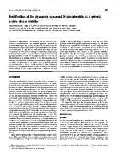
RESULTS AND DISCUSSION Glycinamidylation of D-xylose Isomerase Each subunit of D-xylose isomerase contains 58 Asp+ Glu residues and one C-terminal carboxy group (Loviny-Anderton et al., 1991). These were first modified under denaturing conditions (6 M guanidinium chloride) using 0.1 M EDC as the activating agent and 0.01-1.0 M glycine methyl ester or glycinamide as the nucleophile at 30 °C for 4 h in phosphate buffer of the desired pH. Guanidinium chloride does not interfere with the modification of carboxy groups by carbodi-imides (Hoare and Koshland, 1967). Table 1 shows that the degree of modification increases with increasing concentration of the nucleophile and with decreasing pH. A maximum of 58-60 glycine residues are incorporated at pH 4.75 with > 0.75 M glycine methyl ester or glycinamide. Results with glycine methyl ester or glycinamide were identical, since the PKa values of their a-amino groups are very similar (7.66 and 7.93 respectively at 25 °C; Edsall and Wyman, 1958). Use of glycine methyl ester as the nucleophile has the advantage that the original charge on modified peptides can be restored by alkaline hydrolysis of the coupled methyl ester, allowing purification by 'diagonal paper electrophoresis' according to the principles of Hartley (1970). However, glycinamidewas used in subsequent experiments with native enzyme to avoid hydrolysis of the methyl ester groups during purification and other manipulations, and at pH 5.7, since the enzyme is not very soluble at pH 4.75. The native enzyme in either apo- (Ml), Mg2+- (5 mM MgCl2; M2) or Mg2+-xylitol- forms (5 mM MgCl2 + 5 mM xylitol; M3) was allowed to react with 1 M glycinamide containing ['4C]glycine methyl ester at pH 5.7 and three 2 h additions of solid EDC (to 0.1 M). Figure 1 shows the rate of uptake of glycinamide residues into the Mg2+-xylitol complex; the results with apoenzyme and Mg2+-enzyme were almost identical. About 12 or 13 carboxy groups per subunit were modified within 1 min after the first addition of EDC, and a total of about 30 carboxy groups per subunit were modified shortly after the second addition of EDC, with no further incorporation thereafter. Enzyme activity remained constant throughout the experiment. Of the 58 Asp + Glu residues of D-xylose isomerase, 21 are fully exposed, 13 are partially buried and 24 fully buried as shown by solvent accessible area (A2) (Henrick et al., 1989; Collyer et al., 1990). The carboxy groups in the active site are clearly not modified, since the enzyme activity is unchanged during chemical modification. Excluding these and others involved in intra- or inter-subunit salt bridges or that lie at the A-A* interface (D149 and D189), we are left with only 30 carboxy groups that are potentially modifiable (D8, E44, D61, E64, E66, E68, D74, D80, D124, E127, E131, E140, D162, D170,
Table 1 Carboxy-group
denaturing
conditons
modificaton
of D-xylose isomerase under
No. of glycine residues incorporated/ subunit of D-xylose isomerase
[Glycine methyl ester] or
[glycinamide] (M)
0.01 0.10
0.50 0.75 1
pH ...
4.75
5.7
6.5
10
12 33 45 47 52
8 18 20 26 38
41 49 60 58
K. S Siddiqui and others
688
(b)
(a)
om r-
100 CO) &
.a
EE 80
..
..
....
.... ..
Ci..
U3 0
10,
:
..... ...
....
.-.
:::
L._
E 60 t 40 0, E> 20
E-C o0 ._ o
O._0
N
w Time (min)
Figure 1 Rate
of
modfflcation of carboxy groups
in
D-xylose
isomerase
D-Xylose isomerase (1 mg/ml) in 0.05 M KH2P04/K2HP04 buffer, pH 5.7, containing 5 mM MgCI2, 5 mM xylitol, 1 M glycinamide and [14C]glycine methyl ester (sp. radioactivity 35 uCi/mmol) was incubated at 25 OC. Three additions of EDC to final concentration of 0.1 M were made at 2 h intervals, as indicated by arrows. At different times, aliquots were withdrawn in duplicate. Enzyme activity remaining was measured using D-xylose as substrate; aliquots were diluted 100-fold with 0.1 M Tris/HCI buffer, pH 7.5, to stop the reaction and subjected to exhaustive dialysis against 0.1 M Tris/HCI (pH 7.5)/0.01 M MgCI2. Total protein in the dialysis residue was determined by total hydrolysis followed by amino acid analysis. 14C radioactivity in the residue was measured by liquid-scintillation counting. *, D-Xylose isomerase activity remaining; *, Number of glycinamide residues incorporated into each D-xylose isomerase subunit.
Figure 2 isomerases
Gel 8electrophoresis M In
_
E 80 (U
E 4-
6060
>
40
< 20 -3
Characterization of glycinamidylated D-xylose isomerase Native or modified D-xylose isomerases Ml, M2 and M3 were subjected to f.p.l.c. anion-exchange chromatography. The native enzyme was eluted at an NaCl concentration of 0.56 M, whereas MI, M2 and M3 were eluted at a much lower concentration of NaCl (0.2 M), indicating that the pl had increased considerably after carboxy-group modification. Only one broad symmetrical peak was obtained for all three proteins suggesting that they are fairly homogeneous species and confirming that an identical number of acidic groups have been modified in each case. SDS/PAGE of native and modified D-xylose isomerases showed only one band of protein, in all cases migrating to the same position as native monomer. H.p.l.c. gel-filtration chromatography of Ml, M2 and M3 was performed either in columns equilibrated in 0.2 M Tris/acetate (pH 7.5)/0.01 M magnesium acetate or in 0.05 M Tris/acetate (pH 7.5)/0.01 M magnesium acetate/40 % ethylene glycol (Rangarajan et al., 1992). However, in no case was it possible to elute the proteins off the column, suggesting that the modified proteins are very hydrophobic. Gel electrophoresis, in 8 M urea at pH 8.8, of native D-xylose isomerase and proteins Ml and M2 is shown in Figure 2. Native enzyme gives the expected pattern (Siddiqui et al., 1993) of an active dimer band and an inactive denatured monomer band. However, both Ml and M2 gave only a very slow moving band which did not show enzyme activity and probably corresponds to the denatured modified monomer; no band of enzymatically
glycinamidylated o-xylose
Samples of native and purified modified D-xylose isomerases (Ml and M2) were treated with 8 M urea/0.025 M Tris/HCI buffer, pH 6.5, at 25 0C for 30 min or at 60 0C for 1.5 h prior to electrophoresis in 8 M urea gels, pH 8.8. The gel was cut in half and stained for protein (a) or D-xylose isomerase activity (b). Abbreviations: m, denatured monomers; D, active dimers; N, native enzyme; Ml, modified apoenzyme; M2, D-xylose isomerase modified with EDC-glycinimide in the presence of 5 mM MgCI2. N*, Ml*, and M2* refer to samples heated at 60 0C for 1.5 h prior to electrophoresis.
100
E203, E206, E238, D264, D299, D302, D306, D328, E233, E345, E352, D356, D364, E370, E374 and the C-terminal carboxy group). The results suggest that all, and only, these 30 have been modified and that the other 28 are inaccessible. Hence all activesite carboxy groups are protected, even in the apoenzyme, indicating that the bulky EDC molecule cannot enter the rather narrow substrate-binding pocket. Therefore the glycinamidylated apoenzyme (Ml), Mg2"-enzyme (M2) and ternary complex (M3) are identical.
of native and
urea
-2
-1
0
1
2
log{[Mg2+J (mM)}
Figure 3 Effect of Mg2+ on D-fructose isomerase activity of native and modffied D-xylose isomerases Native and modified D-xylose isomerase (M2) apoenzymes (0.8 mg/ml) were preincubated at 30 OC for 5 h in 0.05 M Tris/HCI buffer, pH 8.0, containing 5 ,uM-20 mM Mg2+ and then assayed against 3.2 M D-fructose in similar buffers. Activity is expressed as a percentage of the maximum activity obtained. *, Native enzyme; 0, Modified enzyme.
active modified dimer band was seen. Hence the modified proteins are much less stable to urea. They are also less thermostable. The melting point of the apoand Mg24+-forms of glycinamidylated D-xylose isomerase and native D-xylose isomerase were compared at pH 6.5 as described in Rangarajan et al. (1992). The melting points of the modified enzyme were considerably lower (590 versus 73 °C for the apoenzymes and 670 versus 81.5 °C for the Mg2+-enzymes).
Enzyme kinetics of glycinamidylated D-xylose isomerase Elimination of 30 surface carboxy groups/subunit does not affect Mg2` binding. Figure 3 shows the effect of Mg2+ on D-fructose isomerase activity of native and modified D-xylose isomerase (M2) at 30 °C at pH 8.0 with a substrate concentration of 3.2 M that is well in excess of the observed
KmC(app)
for the
Modified carboxy
1210 _
4/ 2
o
-
6
5
7
9
8
pH
Figure 4 pH-dependence of Km for D-tructose of the modified Mg2+-enzyme The Mg2+-enzyme (M2) was assayed at 30 OC against 0.04-3 M D-fructose in 0.1 M buffers (pH 5.80-6.50, Mes; pH 6.60-7.80, Mops; pH 7.904.30, Hepes) containing 10 mM Mg2+. Values of Km were obtained by non-linear regression analysis. The broken line refers to data for the native enzyme.
-c3 E -
C
' 2
.0-
/
-
.
-
x E
/
1
-
in D-xylose isomerase
689
whereas it depends on ionization of His2"9 with pKa 6.0 in the native enzyme (Rangarajan and Hartley, 1992). This suggests that the pK8 of His219 is much reduced by the surface charge modification. These kinetic data with D-fructose should not be overinterpreted, since Km(app ) for fructose is very high compared with that for D-xylose, or even D-glucose, and is a complex constant reflecting both substrate binding and the kinetics of the ringopening step (Rangarajan and Hartley, 1992). The fact that Dxylose isomerase activity remained unchanged during glycinamidylation implies very little effect on enzymic activity (Figure 1). Moreover, the modified and native enzymes are similar at pH 7.3, and the apparently lower Vax. (Figure 5) may reflect some inactivation during purification of the modified enzyme. Nevertheless, the sharp increase in Km(app ) for fructose above pH 7.3 deserves comment. None of the carboxy groups in the immediate vicinity of the active site seem to have been glycinamidylated, as argued above, so all active-site carboxy groups and both histidine residues will be unprotonated at pH 8. The long-distance effects of reducing negative surface changes on Km(app ) imply a change in conformation of the Mg2+-enzymeD-fructose complex at alkaline pH.
Thermolysin nicking of glycinamidylated D-xyiose isomerase The reduced thermostability of the modified enzyme prompted a study of the pattern and kinetics of its nicking by thermolysin, since this technique reveals the location and degree of flexibility of surface loops (Siddiqui et al., 1993). The pattern of thermolysin nicking of the glycinamidylated enzyme was similar, but both
4
0
groups
Ll 5
6
8
7
9
pH
Figure 5 pH-dependence of
L(,, for D-fructose for modified Mg2+-enzyme
The Mg2+-enzyme (M2) was assayed at 30 OC against 3.2 M D-fructose in buffers of various pH values as in Figure 4, containing 10 mM Mg2+. The apparent values of Vmax at various pH values were corrected by using the values of Km shown in Figure 4. The broken line is the curve obtained with the native enzyme.
native Mg2+-enzyme. In both cases, Mg2+ shows an apparent activation constant of 60-80 ,uM (Rangarajan and Hartley, 1992). The Km(app) of the enzyme is affected by surface glycinamidylation, particularly at alkaline pH. Figure 4 shows values from fructose isomerization assays at 30 °C in saturating Mg2` (10 mM) at various substrate concentrations over a range of pH values. The value of 1/Km at pH 7.3 is similar to that of the native enzyme, but decreases at acid pH and drastically above 7.30. The data do not fit a simple ionization curve (non-linear ENZFITTER program; Biosoft), in contrast with native Mg2+D-xylOse isomerase, where Km depends on a single ionizing group of PKa 6.80. The pH-dependence of Vmax for the modified and native Mg2+enzymes was compared using 3.2 M D-fructose [which is in large excess of the native Km(app,) 0.12-0.3 MI and similar Mg2+ concentrations at 30 'C. The data in Figure 5 show that Vm.ax remains constant between pH 6.0 and 8.0 in the modified enzyme,
nicking sites appeared to be more labile. There was no loss of isomerase activity of the modified enzyme over 10 h during incubation with thermolysin at 37 °C in 5 mM Mg2+/2.5 mM Ca2+/0.1 M Tris/HCl, pH 8.1, in the presence or absence of 10 mM xylitol (the enzyme will be in the Ca2+-form under these conditions; Siddiqui et al., 1993). Analysis ofthe reaction mixture by SDS/PAGE during the incubation showed disappearance of the 43.3 kDa monomer and the transitory appearance of a 38 kDa fragment which was rapidly converted into a 36 kDa subunit. The rate of conversion of the 43.3 kDa band into the 38 kDa band was unaffected by the presence of 10 mM xylitol, and the first-order rate constant (420 x 10-3 h-') was about twice the rate of nicking of the native enzyme under identical conditions (190 X 10-3 h-1; Siddiqui et al., 1993). The results suggest that the primary nicking site (Thr347-Leu348) is more mobile in the modified enzyme, and the secondary site between helices H8 and H9 even more so. However, the drastic modification in surface charge of the tetramer did not cause mobility in any other surface strands that are cleavable by the broad-specificity proteinase.
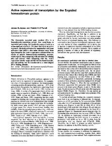
Protein-engineered mutants to alter pH optimum It seemed possible that protein-engineered mutations to convert some of the surface carboxy residues into lysine or arginine might decrease the pH optimum of enzyme activity so as to prevent 'browning reactions' in commercial production of highfructose syrups. Mutants in which Glu140 and Asp'89 were converted to Lys were constructed, since a mutation where a non-essential negative charge is replaced by a positive charge should double the electrostatic effect at the active site. The procedure used to purify these proteins was essentially identical with that described by Smith et al. (1991) for purification of the wild-type enzyme from Arthrobacter cells. Column fractions after ion-exchange chromatography were monitored for the presence of mutant D-xylose isomerase protein by A280 and by
690
K. S Siddiqui and others (b)
(a) 45 6 7 8 9
0
.+
-_. 1 2 3 4 5 6 7 8 9
..:
Figure 6 Urea-gradIent (0-9 M)-gel electrophoresis of D-xylose
Iso-
merases
Samples of native (a) and Trp136-+Glu mutant D-xylose isomerases (b) were subjected to 0-9 M urea-gradient electrophoresis at pH 8.8 in 7.5% acrylamide gels at 25 OC, and the gels were stained for protein. Arrows alongside the gels indicate the direction of migration of protein and the numbers indicate the molarity of urea across the gradient.
SDS/PAGE. No band corresponding to the 43.3 kDa monomer of D-xylose isomerase was present in column fractions of extracts of mutant proteins (Glu'40-+Lys and Asp189-+Lys). Hence both the Glu140-+Lys and Asp'89-.Lys mutant proteins appear to be synthesized in the E. coli host, but incapable of folding correctly. In hindsight this can be explained (C. A. Collyer and D. M. Blow, unpublished work), since Glu'40 is involved in tight H-bonds to the main chain-NH-groups of residues 102 and 103, and conversion into Lys may disrupt the 100-107 loop between Strand 3 and Helix 3. Asp"89 is close to Lys252 and Arg249 of a neighbouring subunit, though not salt-bridged to these. Conversion of the negative charge to a positive charge may therefore cause repulsions between the subunits.
The
Trpl3"-+GIu
mutant
indicating reversible dissociation between tetramers and dimers (Rangarajan et al., 1992). Attempts to reduce the pH optimum of other glucose isomerases by protein engineering have not been conspicuously more successful. Modifications of the cluster of active-site carboxy groups in the homologous D-xylose isomerase from Actinoplanes missouriensis involved in binding metal ions (Jenkins et al., 1992) predictably either prevented expression or produced inactive enzyme. Some mutants, such as Glu181-.Gln, Glu217-+Ser, Asp255-.Asn or Asp257 -+Asn retained activity, but this was very low. However, a mutation of Asp57-+Asn, whose carboxy group intacts with N41 or His54 which catalyses ringopening, somewhat surprisingly retained almost 60% activity towards xylose and 30% activity towards glucose, but the pH optimum was reduced by only 0.1 pH unit (Lambeir et al., 1992). Another mutant, Glu'86-Gln, did have an altered pH optimum in the Mn2+-enzyme form. However, this appears to be due to a rapid decline in activity towards xylose above pH 6; the activities at lower pH are similar to those of the wild-type. Moreover, the activity of the Mg2+-enzyme was negligible at all pH values. As suggested by Varsani et al. (1993), these apparently confusing results can be explained by a conformational change in the mutant enzyme stimulated by electrostatic repulsions at alkaline pH. These results suggest that it will be difficult to achieve a significant change in pH optimum by changing a small number of carboxylic acid groups by protein engineering. There is a large number of charged residues at the active site that may act as a buffer against long distance electrostatic effects. However this work shows that a decrease in pH optimum can be achieved by glycinamidylating 30 surface carboxy groups, so this may prove to be commercially useful. The research was funded by the Science and Engineering Research Council Protein Engineering Initiative in the U.K. We thank V. Thirunavakkarasu for performing some experiments as part of a short M.Sc. research project.
enzyme
as above in order to test the mechanistic hypothesis of Collyer et al. (1990). It gave a peak of protein that was eluted from the ionexchange column at the same concentration of NaCl as the wildtype enzyme. SDS/PAGE of the peak fractions showed the presence of a band of protein corresponding to the 43.3 kDa monomer band. After ACA-34 gel filtration, the mutant protein was essentially pure, as shown by SDS/PAGE and N-terminal sequencing. The yield of Trpl36-Glu protein was comparable with that obtained for wild-type enzyme starting from a similar amount of cell paste. However, the mutant protein was completely inactive. SDS/ PAGE of mutant protein showed that the subunit was identical in size to the native 43.3 kDa monomer. H.p.l.c. gel filtration performed as described in Rangarajan et al. (1992) confirmed that the mutant enzyme was a tetramer (results not shown). The Trp 138-Glu D-xylose isomerase was as stable as native enzyme to SDS treatment. Treatment of either with 1 % SDS in Tris/HCl buffer, pH 6.5, at 25 'C for 30 min gave a mixture of dimers and monomers, whereas heating at 100 'C for 2 min gave only monomers (results not shown). Heating at 60 'C for different lengths of time gave a mixture of monomers and dimers, the proportion ofdimers decreasing as incubation time was increased. However, it was less stable than native enzyme in urea. Electrophoresis in 0-9 M urea gradient gels at 25 IC (Figure 6) shows that the mutant enzyme is irreversibly denatured between 6 and 7 M urea to give monomer, whereas wild-type enzyme gives a smooth continuous band of protein under these conditions,
This mutant was constructed, expressed and purified
REFERENCES Carraway, K. L. and Koshland, D. E., Jr. (1968) Biochim. Biophys. Acta 160, 272 Collyer, C. A., Henrick, K. and Blow, D. M. (1990) J. Mol. Biol. 212, 211-235 Darbre, A. and Islam, A. (1968) Biochem. J. 106, 923-925 Edsall, J. T. and Wyman, J. (1958) Biophysical Chemistry, vol. 1, pp. 136-240 and 477-549, Academic Press, New York, San Francisco and London Fersht, A. R. (1985) Enzyme Structure and Mechanism, 2nd edn., pp. 155-175, W. H. Freeman and Co., New York Fontana, A., Fassina, G., Vita, C., Dalzoppo, D., Zamai, M. and Zambonin, M. (1986) Biochemistry 25, 1847-1851 Hartley, B. S. (1970) Biochem J. 119, 805-822 Hartley, B. S. (1988) Abstr. Int. Congr. Biochem. 14th Tu, S5-1, p. 12. Henrick, K., Collyer, C. A. and Blow, D. M. (1989) J. Mol. Biol. 208, 129-157 Hoare, D. G. and Koshland, Jr., D. E. (1967) J. Biol. Chem. 242, 2447-2453 Islam, A. and Darbre, A. (1969) J. Chromatogr. 43,11-24 Jenkins, J., Janin, J., Rey, F., Chiadmi, M., van Tilbeurgh, H., Lasters, I., De Maeyer, M., Van Belle, D., Wodak, S. J., Lauwereys, M., Stanssens, P., Mrabet, N. T., Snauwert, J., Matthyssens, G. and Lambeir, A.-M. (1992) Biochemistry 31, 5449-5458 Kunkel, T. A. (1985) Proc. Natl. Acad. Sci. U.S.A. 82, 488-492 Lambeir, A. M., Lauwereys, M., Stanssens, P, Mrabet, N. T., Snauwaert, J., van Tilbeurgh, H., Matthyssens, G., Lasters, I., Wodak, S. J., Jenkins, J., Chiadmi, M. and Janin, J. (1992) Biochemistry 31, 5459-5466 Loviny-Anderton, T., Shaw, P.-C., Shin, M.-K. and Hartley, B. S. (1991) Biochem. J. 277, 263-271 Marshall, R. C. and Inglis, A. S. (1986) in Practical Protein Chemistry (Darbre, A., ed.), pp. 1-66, John Wiley and Sons, Chichester, New York, Brisbane, Toronto and Singapore Mead, D. A., Szczesna-Skorupa, E. and Kemper, B. (1986) Protein Eng. 1, 67-74 Rangarajan, M. and Hartley, B. S. (1992) Biochem. J. 283, 223-233 Rangarajan, M., Asboth, B. and Hartley, B. S. (1992) Biochem. J. 285, 889-898 Russell, A. J. and Fersht, A. R. (1987) Nature (London) 328, 496-500 Sanger, F., Nicklen, S. and Coulson, A. R. (1977) Proc. Natl. Acad. Sci. U.S.A. 74, 643-657
Modified carboxy groups in D-xylose isomerase Siddiqui, K. S., Rangarajan, M., Hartley, B. S., Kitmitto, A., Panico, M., Blench, I. P. and Morris, H. R. (1993) Biochem. J. 289, 201-208 Smart, 0. S., Akins, J. and Blow, D. M. (1992) Proteins Struct. Funct. Genet. 13, 100-111 Smith, C. A., Rangarajan, M. and Hartley, B. S. (1991) Biochem. J. 277, 255-261 Spomer, W. E. and Wootton, J. F. (1971) Biochim. Biophys. Acta 235,164-171 Received 23 March 1993/30 July 1993; accepted 6 August 1993
691
Thomas, P. G., Russell, A. J. and Fersht, A. R. (1985) Nature (London) 318, 375-376 Valenzuela, P. and Bender, M. L. (1971) Biochim. Biophys. Acta 250, 538-548 Varsani, L., Cui, T., Rangarajan, M., Hartley, B. S., Goldberg, J., Collyer, C. A. and Blow, D. M. (1993) Biochem. J. 291, 575-583 Zoller, M. Y. and Smith, M. (1982) Nucleic Acids Res. 10, 6487-6500