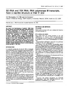
1086±1096 Nucleic Acids Research, 2003, Vol. 31, No. 3 DOI: 10.1093/nar/gkg196
DNA tri- and tetra-loops and RNA tetra-loops hairpins fold as elastic biopolymer chains in agreement with PDB coordinates Guillaume P. H. Santini, Christophe Pakleza and Jean A. H. Cognet* Laboratoire de Physico-chimie BiomoleÂculaire et Cellulaire, UMR 7033 CNRS, T22-12, Universite Pierre et Marie Curie, 4 place Jussieu, 75252 Paris cedex 05, France Received July 24, 2002; Revised November 7, 2002; Accepted November 23, 2002
ABSTRACT The biopolymer chain elasticity (BCE) approach and the new molecular modelling methodology presented previously are used to predict the tridimensional backbones of DNA and RNA hairpin loops. The structures of eight remarkably stable DNA or RNA hairpin molecules closed by a mispair, recently determined in solution by NMR and deposited in the PDB, are shown to verify the predicted trajectories by an analysis automated for large numbers of PDB conformations. They encompass: one DNA tetraloop, -GTTA-; three DNA triloops, -AAA- or -GCA-; and four RNA tetraloops, -UUCG-. Folding generates no distortions and bond lengths and bond angles of main atoms of the sugar±phosphate backbone are well restored upon energy re®nement. Three different methods (superpositions, distance of main chain atoms to the elastic line and RMSd) are used to show a very good agreement between the trajectories of sugar±phosphate backbones and between entire molecules of theoretical models and of PDB conformations. The geometry of end conditions imposed by the stem is suf®cient to dictate the different characteristic DNA or RNA folding shapes. The reduced angular space, consisting of the new parameter, angle W, together with the c angle offers a simple, coherent and quantitative description of hairpin loops. INTRODUCTION In the preceding article in this issue, we have postulated that the backbone of single-stranded DNA hairpin loops behaves as a continuous, inextensible and ¯exible thin rod. With this simple hypothesis, the tri-dimensional trajectory of this elastic line was derived from the theory of elasticity and we have shown how it can be used to predict the structures of the sugar±phosphate backbone of DNA hairpins. We have shown also how single-stranded trinucleotide B-DNA TTT could be folded into hairpin loops, G-TTT-C or C-TTT-G, where most
torsion angles are preserved, to match four different sets of NMR data or ®ve different molecular conformations (1±4). In this approach, called biopolymer chain elasticity (BCE), the trajectories of the two helical backbones of the hairpin stem de®ne the geometry of the extremities of the hairpin loop. In the theory of elasticity of thin rods, the geometry of end conditions dictates the shape of the trajectories. Therefore the different shapes of DNA and RNA hairpin loops should be predicted or should result from the different geometries imposed by the stem structures. Double helical B-DNA and A-RNA differ in two respects. Firstly the planes of base pairs and of helical extremities are perpendicular to the helical axis in B-DNA whereas they are tilted in A-RNA. Secondly B-DNA helix has a smaller radius. As shown in Figure 1, when the backbones trajectories of the loops (in dark red or blue) are in perfect continuity with the backbones trajectories of the double helix (in light colour), we predict that the backbone of the DNA tetraloop must go over the surface of the DNA cylinder, whereas the trajectory of RNA backbone is circumscribed on or slightly outside the RNA cylinder as shown in Figure 1. In this article, we investigate whether the BCE methodology and its tri-dimensional predictions that were presented previously for DNA triloops (5) and build on previous ideas (6,7) can be applied not only to other hairpins of different structures, of different lengths, tri- and tetra-loops of DNA, but also to RNA tetraloops. To this end, we have selected eight different molecules of which structures have been recently determined in solution by NMR and deposited in the Protein Data Bank (PDB) (8). They encompass: one DNA tetraloop, -GTTA-; three DNA triloops, -AAA-, -GCA-, -GCA-; and four RNA tetraloops, -UUCG-, as presented in Table 1. The DNA triloops and RNA tetraloops have been the subject of many determinations over the last decade and have given rise to well de®ned solution structures since early NMR studies (9,10). They have been used as test systems for theoretical studies (11±13). For brevity and convenience, the molecules are referred by their PDB identi®cations. These eight molecules have several features in common. They are all remarkably stable (7,14) and the loop is closed by a side-by-side sheared mispair (15,16) G´A (1ac7), A´A (1bjh), G´A (1xue, 1zhu), or by a head to side U´G mispair (17) (1aud, 1b36, 1c0o, 1hlx). Note that the DNA triloops studied here are structurally different from the TTT hairpins studied previously (5). In the latter, the ®rst and last
*To whom correspondence should be addressed. Tel: +33 1 44 27 27 50; Fax: +33 1 44 27 75 60; Email:
[email protected]
Nucleic Acids Research, Vol. 31 No. 3 ã Oxford University Press 2003; all rights reserved
Nucleic Acids Research, 2003, Vol. 31, No. 3
Figure 1. Superimposed stereo views into the minor groove of the computed elastic rod curve for a DNA tetraloop hairpin, shown in red, and for an RNA tetraloop hairpin shown in blue. The radii of cylinders and circles are those of the sugar±phosphate backbones, DNA (red) and RNA (blue). The mean planes of base pairs are indicated by the top sections of the cylinders.
nucleoside of the loop were not stacked on the top base pair of the stem. They never formed a mispair. They were located in the minor groove, in the major groove or in the solvent and did not interact with each other. Well de®ned DNA and RNA tetraloops come essentially in three different folds (7,18). Type-I loops are only observed for DNA. They adopt a conformation with the ®rst three bases at the 5¢-end of the loop forming a more or less continuous stack on the 3¢-end of the stem (1ac7) (15). Type-II loops are found in both DNA (CTTG) (18) and RNA (1aud, 1b36, 1c0o, 1hlx). As indicated in Table 1, Nb is turned into or towards the minor groove and Nc lies over the closing base pair NaNd. A third fold, type-III, is only observed in RNA and is described by a continuous stacking of Nd, Nc and Nb on the 5¢-end of the stem. DNA hairpins can perform many important and diverse biological functions as recently established by numerous
1087
experiments and as brie¯y reviewed in the previous article in this issue. The DNA tetraloop -GTTA- (1ac7) is related to telomeric and centromeric structures (15). The DNA triloops -AAA- (1bjh) and -TTT- are important components of the adenoassociated virus 2 (4,5). The two DNA triloops -GCAare encountered in human centromere repeats (1xue) and in centromeric GNA triplets (1zhu) and are important to account for the observed expansion of triplet repeats (5). RNA hairpins have been known to play essential structural and biological roles for several decades (14,19). In particular, the hairpin contained in 1aud is part of the polyadenylation inhibition element bound to the RNP domain of the human U1A protein (20). In 1b36, the -UUCG- tetraloop was added to stabilise the structure of one of the two domains essential for catalysis in a ribozyme molecule (21). Similarly in 1c0o, the stable -UUCGloop was added to close an RNA metal hexammine binding site from the P5 helix of the catalytic core of the Tetrahymena group I intron ribozyme (22). In 1hlx, the tetraloop is the capping part of the P1 helix from group I self-splicing introns (17). Advances in synthetic and spectroscopic techniques have recently extended the size and the accuracy of RNA molecular structures that can now be solved by NMR (20). The solution structures retained here for analysis were determined between 1995 and 1999, from large collections of NMR data. Due to their sizes, to the different complex protocols used, and to the rapid evolution of computer programs, the complete data may be only partly available and it may be dif®cult to analyse in an identical way to that of the original authors and as we have done in the previous article in this issue. For these reasons, the theoretical molecular structures built with the BCE approach were not compared with NMR-derived distances and to a single molecular structure as previously described (5), but directly to available PDB solution structures. Note that the eight corresponding PDB ®les contain many molecular conformations, in numbers from 10 up to 31 per coordinates ®le (Table 1), because solution structures were derived from NMR. Their total number is 126. Due to this large number, the computer program introduced previously, S-mol (5), was
Table 1. Molecular structures selected from the PDB, with PDB identi®cation, number of structures in square brackets and tetraloop folding type (7,18), original authors, DNA or RNA sequences of PDB structures and of theoretical BCE models, and locations of the loop bases in the Major (M) groove, in the minor (m) groove, stacked in the central part of the helix (c) or in the solvent outside the structure (solv.) PDB identi®cation DNA 1ac7 [10] Type I 1bjh [16] 1xue [10] 1zhu [10] RNA 1aud [31] Type II 1b36 [10] Type II 1c0o [19] Type II 1hlx [20] Type II
Authors
DNA or RNA sequence solved experimentally & sequence used in theoretical models
Na
Nb
Nc
Nd
van Dongen et al. 1997 (15)
d(¼ccta-GTTA-tagg¼) & d(gcta-GTTA-tagc)
c/M
M
M / solv
c/m
Chou et al. 1996 (16) Zhu et al. 1996 (30) Zhu et al. 1995 (38)
d(gtac-AAA-gtac) & d(gcac-AAA-gtgc) d(...gaat-GCA-atgg...) & d(gcat-GCA-atgc) d(caat-GCA-atg) & d(gcat-GCA-atgc)
c/M c/M c/M
M M M
c c c
-
Allain et al. 1997 (20)
r(¼gucc-UUCG-ggac¼) & r(gccc-UUCG-gggc)
c/M
m / solv
M
c
Butcher et al. 1999 (21)
r(¼gcgc-UUCG-gcgc¼) & r(gcgc-UUCG-gcgc)
c/M
m / solv
M
c
Colmenarejo and Tinoco 1999 (22)
r(¼gguc-UUCG-gguc¼) & r(gcuc-UUCG-gggc)
c/M
m / solv
M
c
Allain and Varani 1995 (17)
r(¼uaac-UUCG-guug¼) & r(gcac-UUCG-gugc)
c/M
m / solv
M
c
Absence of fourth nucleotide in the loop is denoted (-). The loop bases are marked Na, Nb, Nc and Nd in the 5¢ to 3¢ direction.
1088
Nucleic Acids Research, 2003, Vol. 31, No. 3
enhanced to deal with automatic comparisons. This is an important change that required speci®c modi®cations of the BCE methodology as explained in Materials and Methods and below. In this article, our main focus is to search a general theoretical approach, which is capable of: (i) predicting a priori the tri-dimensional course of the sugar±phosphate chains, not only of DNA hairpin molecules structurally different from TTT hairpins, but also of RNA hairpins, (ii) generating models close to solution structures from these predictions and from large numbers of given PDB conformations, and (iii) characterising the importance of the sugar± phosphate chain and of its elastic properties in the folding process.
Original molecular structures, PDBid Original molecular conformations are from the PDB (8) and are referred by their PDBid: 1ac7, 1b36, 1xue, 1zhu, 1aud, 1bjh, 1c0o and 1hlx. Initial stem and loop model building by molecular mechanics All initial structures were generated from canonical B-DNA or A-RNA (23). Theoretical molecular structures, BCE A registered software Smolã (5) was extended under UNIX and Linux environments using Mathematica (24), Geomview (25) and C languages to build and to compare BCE models with solution conformations of PDB ®les. The complete DNA or RNA sequences of the theoretical molecular structures, given in Table 1, were simpli®ed with the two following rules. The sequence of the loop and of the ®rst two base pairs in the stem is identical to original PDB molecular structures. The length of stems is reduced to four base pairs and the remaining sequence of the stem is set to d(GC).d(GC) or r(GC).r(GC). Note that all PDB conformations proposed under a given PDB identi®cation were used for building the theoretical structures. The length, L, of the capping rod was obtained as previously described (5) by ®tting a helical line to the atoms of the main sugar±phosphate backbone (O5¢, C5¢, C4¢, C3¢, O3¢, P) of a single-stranded helical A-RNA or B-DNA and by minimising the root-meansquare of the sum of squared distances to the helical line. For Ê and its pitch was A-RNA radius of helical line was 9.35 A Ê /turn. For B-DNA, values were respectively 8.35 A Ê 30.85 A Ê /turn. Molecules were folded into hairpin loops and 33.74 A using prescribed geometric boundary conditions. Setting all PDB conformations in the laboratory reference frame PDB conformations are moved onto BCE molecular models by a translation-rotation coordinate transformation. ®
R
the global reference frame
rotation matrix
®
. OM in
=
local reference frame
Molecular structures provided in PDB ®les and summarised in Table 1 were used at the third step of theoretical molecular modelling to optimise the rotation angles about the elastic line, W, and the glycosidic torsion angles, c, of each nucleoside in the loop independently from other nucleosides. This was performed by a least square ®t on homologous atom positions to give optimised BCE models, BCE3Wopt. Final theoretical molecular structures, `BCE4®nalm' BCE molecular models were energy re®ned without restraints. Energy re®nements were carried out with the program AMBER (5,26,27) without any restraints and with a large stopping root-mean-square energy gradient criterion 0.5 kcal/ Ê ) to yield ®nal molecular models, BCE4®nalm. (mol.A RMSd analysis
MATERIALS AND METHODS
OM in
Optimised molecular structures, `BCE3Wopt'
®
+V
vector of translation
RMSd are computed after superposing the two sets of matching atoms by a translation-rotation coordinate transformation.
RESULTS The BCE approach enhanced to treat multiple PDB conformations Folding a DNA or an RNA hairpin loop with the BCE approach can be described as a three-step procedure, completed by a short energy re®nement step to restore backbone bond lengths and bond angles (5). A short and intuitive account of the procedure modi®ed to treat multiple PDB conformations is given below and in Figure 2. (i) Singlestranded A-RNA or B-DNA are basically considered as a continuous and ¯exible thin rod in the following practical manner. These polymers are generated along helical lines, which are also viewed as elastic lines. The main atoms of the sugar±phosphate backbone (O5¢, C5¢, C4¢, C3¢, O3¢, P) play a key role because they are attached to this line and because they are used to de®ne the origins of local reference frames for all remaining atoms in the nucleotide. As a result, there are six different groups of atoms per nucleotide. The polymer may thus be viewed as a succession of individual solid blocks of atoms attached to the elastic line. Using this basic framework where all backbone atoms are made part of the elastic line as shown for A-RNA in Figure 2A, the biopolymer chain can be bent and twisted smoothly using elasticity theory of thin rods into a given loop with prescribed end conditions (Fig. 2B and C). This step yields a elastic curve, BCE1curve (Fig. 2D), which can be ®tted onto the double helical stem (Fig. 2C). Note that the tri-dimensional trajectory of the elastic line is uniquely determined for end conditions of Figure 2B and C. (ii) Transportation of the biopolymer chain onto the elastic line step yields a molecular model, BCE2basicxyz (Fig. 2A, D and E). Crucial parameters are the length of the loop, tri- or tetra-loop, and the geometry of end conditions imposed by the A-RNA or B-DNA helices. (iii) A useful feature provided by this formalism is that each block of atoms, and consequently an entire nucleoside, can be rotated about the elastic line with an angle, W. Each nucleoside block can be rotated independently to match NMR-derived distances as in the previous
Nucleic Acids Research, 2003, Vol. 31, No. 3
1089
Multiplicity and heterogeneity of the PDB structures are overcome by setting each conformation in an absolute coordinate frame
Figure 2. Schematic overview of the construction process of an RNA tetraloop hairpin molecular structure using the BCE approach proposed in the previous article in this issue (5). (A) A continuous and ¯exible thin rod, represented by a ribbon for better visibility, and shown in red is associated to a four nucleotides helical segment; (B and C) two helices of RNA are generated along helical lines, shown in yellow; (D) the ¯exible rod is bent smoothly into the capping elastic solution curve so that the tangents at its extremities, shown as blue and green arrows, match those of the two helices; helical segments and the capping rod are dissociated for clarity; (E) they are shown fully assembled; the complete molecular structure is the basic BCE molecular model and is computed after global deformation by keeping track of the translations and the rotations required to leave unchanged atoms in their nearest local reference frames along the helical segment.
article in this issue, or in order to match each one of the molecular conformations given in a PDB ®le as in this study. This step is de®ned here as an automated optimisation of angles, W, and of glycosidic torsion angles, c, and yields optimised BCE molecular models, BCE3Wopt. (iv) Individual molecular blocks are displaced by the folding procedure without internal deformation. However the chemical bonds and bond angles of the main atoms of the sugar±phosphate backbone (O5¢, C5¢, C4¢, C3¢, O3¢, P) are modi®ed by the BCE folding procedure. This is why each molecular structure is very shortly energy re®ned without restraints to restore backbone bond length and bond angles. This step yields the ®nal theoretical molecular model, BCE4®nalm. In this article, we compare the large number of original molecular conformations supplied by a PDB ®le to their corresponding BCE4®nalm molecular models. As summarised in Table 1, these molecules differ from one another in nature, DNA or RNA, in sequence, in length, and in protocol used to determine their solution structures. This may be a source of heterogeneity that is observed in the PDB ®les. To circumvent this dif®culty each original molecular conformation in a PDB ®le is ®rst translated in an absolute reference frame where all theoretical molecular models are folded. Deformations introduced in the sugar±phosphate backbone are examined at important steps of the folding. We are then in a position to compare each original model conformation of the PDB ®le to the model structure derived from our theoretical approach.
As all structures under study were derived from NMR data, their corresponding PDB ®les contain many proposed solution conformations (Table 1). A direct view of the ®rst ten conformations of different PDB ®les demonstrates a wide heterogeneity as shown in Figure 3. For 1ac7 (Fig. 3A), the loop appears either very well determined or very rigid, whereas the stem appears either less well determined or more ¯exible. This view has the advantage of focusing on detailed features of the loop structures (15). The situation is reversed with 1b36 where the main focus is on the central region (Fig. 3B) (21). With the PDB ®le, 1xue (Fig. 3C), the molecule appears well determined or rigid at every atom positions, whereas with 1c0o, it appears homogeneously underdetermined or ¯exible (Fig. 3D). This heterogeneity in the PDB structures originates from the arbitrary choice of presentation of superposed molecules. It depends on the molecule and its properties (DNA or RNA, size and sequence, free or bound to a protein) and on the local nature of the two types of information derived from NMR data (torsion angle values Ê from NOE data). from J-couplings and short distances