Villareal et al. BMC Complementary and Alternative Medicine (2017) 17:549 DOI 10.1186/s12906-017-2060-1
RESEARCH ARTICLE
Open Access
Anti-stress and neuronal cell differentiation induction effects of Rosmarinus officinalis L. essential oil Myra O. Villareal1,2, Ayumi Ikeya3, Kazunori Sasaki4,5, Abdelkarim Ben Arfa6, Mohamed Neffati6 and Hiroko Isoda1,2*
Abstract Background: Mood disorder accounts for 13 % of global disease burden. And while therapeutic agents are available, usually orally administered, most have unwanted side effects, and thus making the inhalation of essential oils (EOs) an attractive alternative therapy. Rosmarinus officinalis EO (ROEO), Mediterranean ROEO reported to improve cognition, mood, and memory, the effect on stress of which has not yet been determined. Here, the anti-stress effect of ROEO on stress was evaluated in vivo and in vitro. Methods: Six-week-old male ICR mice were made to inhale ROEO and subjected to tail suspension test (TST). To determine the neuronal differentiation effect of ROEO in vitro, induction of ROEO-treated PC12 cells differentiation was observed. Intracellular acetylcholine and choline, as well as the Gap43 gene expression levels were also determined. Results: Inhalation of ROEO significantly decreased the immobility time of ICR mice and serum corticosterone level, accompanied by increased brain dopamine level. Determination of the underlying mechanism in vitro revealed a PC12 differentiation-induction effect through the modulation of intracellular acetylcholine, choline, and Gap43 gene expression levels. ROEO activates the stress response system through the NGF pathway and the hypothalamus-pituitary-adrenal axis, promoting dopamine production and secretion. The effect of ROEO may be attributed to its bioactive components, specifically to α-pinene, one of its major compounds that has anxiolytic property. Conclusions: The results of this study suggest that ROEO inhalation has therapeutic potential against stress-related psychiatric disorders. Keywords: Catecholamines, Cell differentiation, Inhalation, Nerve growth factor, Rosemary essential oil
Background Emotional disorders, including depression, cause a huge burden on health worldwide. Several theories to explain the pathogenesis of depression have been proposed but because of the complexity, identification of specific mechanism of depression has been difficult. However, evidences suggest that stress and depression are associated with atrophy and loss of neuronal cells, as well as reduction in the volume of key brain structures. Chronic exposure to stress, for example, induces neuronal inflammation, neuronal degeneration, and brain micro* Correspondence:
[email protected] 1 Faculty of Life and Environmental Sciences, University of Tsukuba, 1-1-1 Tennodai, Tsukuba City, Ibaraki 305-8572, Japan 2 Alliance for Research on North Africa (ARENA), University of Tsukuba, 1-1-1 Tennodai, Tsukuba City, Ibaraki 305-8572, Japan Full list of author information is available at the end of the article
damage [1]. Recently approved antidepressants, such as serotonin selective reuptake inhibitors (SSRIs) or anxiolytics that are mostly administrated orally, are found to exert better therapeutic efficacy than previous medications. However, the unwanted side effects of these drugs, such as the amnestic effect of benzodiazepine [2], have not yet been entirely eradicated and their molecular and therapeutic mechanisms are not fully understood [3]. To resolve concerns on limited efficacy of existing drugs, new antidepressant agents or alternative agents with novel mechanisms that are more effective and without or fewer side effects are sought. Essential oils (EOs) have a long history of use as folk medicine and are still attractive as a therapeutic agent against oxidative stress [4], tumorigenesis [5], stressrelated disorders including depression and anxiety [6].
© The Author(s). 2017 Open Access This article is distributed under the terms of the Creative Commons Attribution 4.0 International License (http://creativecommons.org/licenses/by/4.0/), which permits unrestricted use, distribution, and reproduction in any medium, provided you give appropriate credit to the original author(s) and the source, provide a link to the Creative Commons license, and indicate if changes were made. The Creative Commons Public Domain Dedication waiver (http://creativecommons.org/publicdomain/zero/1.0/) applies to the data made available in this article, unless otherwise stated.
Villareal et al. BMC Complementary and Alternative Medicine (2017) 17:549
Both anecdotal and empirical literature reports some major EOs, on the psychological or behavioral effects, however the effect of many EOs remained largely undiscovered. Rosmarinus officinalis is herb used for culinary and medicinal purposes that contains polyphenols such as rosmarinic acid, carnosic acid, and luteolin and other water-soluble phytochemicals that exert several effects on psychiatric diseases or neurological function including neuroprotective, cognitive properties, anti-depressive and anti-anxiety effects [7–9]. R. officinalis EO (ROEO) has been reported to exhibit anti-proliferative, antioxidant and antibacterial activities [10], as well as improvement of cognition, mood, and memory function in healthy adults [11] and improvement of locomotor activity in mice [12]. Oral intake of ROEO showed antidepressant-like effect in mice [13] but EOs terpenoids and ketones often exhibit acute toxicity even in very low concentration [14]. Inhalation of EO as a sedative [15–17], anxiolytic [18–20] and anti-stress [6, 21] therapy ensures effectivity without unwanted side effects and has an advantage in terms of the absence of pathological consequences on organs and tissues [14]. The effectiveness and detailed mechanism of inhalation of ROEO on psychological and neurological function however, remains unknown. Here, we evaluated the effect of ROEO inhalation on the molecular mechanism by which ROEO alleviates stress in vitro using PC12 cells and in vivo using ICR mice, a mice strain widely used for behavioral tests. Psychiatric functions and neurophysiological mechanism were determined in vivo using tail suspension test (TST), and measurement of the catecholamine and corticosterone level, while the effect on neuronal differentiation was determined using activity of acetylcholinesterase (AChE) and neuronal elongation as indicators, in vitro. We hypothesized that ROEO actively enhance the neuronal activity in mice by promoting cell differentiation.
Methods Preparation of essential oil (EO) samples
The Rosmarinus officinalis L. leaves collected from Matmata, Tunisia that were used in the extraction of the R. officinalis essential oil (ROEO) used in this study and authenticated by Prof. Mohamed Neffati (Arid Land Institute, Tunisia). Voucher specimens of the Rosemary leaf samples (UT-ARENA-00323) used in this study were deposited in the Alliance for Research on North Africa, University of Tsukuba, Japan. ROEO was extracted from dried plant material (leaves) by hydrodistillation method, and kept in the dark at 4 °C until use. Pure EO of lavender (Lavandula angustifolia) (LAEO) and almond (Prunus dulcis) oil (AL) were purchased from Sigma
Page 2 of 10
(St Louis, MO, USA) and Wako Pure Chemical Industries, Ltd., (Osaka, Japan), respectively. ROEO components were analyzed using Agilent 6890 N Network gas chromatographic (GC) system gas chromatograph equipped with a HPMS 5973 mass spectrometer with a HP-5MS fused silica column (30 m × 0.25 mm ID, film thickness 0.25 μm, Hewlett-Packard; carrier gas was helium adjusted to a linear velocity of 34 cm/s). The oven temperature program was the same as that used for the HP-5 column for GC-FID analysis: source temperature was 230 °C, quadrupole temperature was 150 °C, injector and detector was 250 °C and 280 °C respectively. Samples (0.1 μl) were injected with a split ratio of 1:100. The composition of the ROEO was identified by GCMS and the main components of the ROEO were identified to be camphor (20.42%), alpha-pinene (10.49%), beta-pinene (10.21%) (Table 1).
Animals
Six-week-old male ICR mice were used in this study (Charles River Laboratories Japan Inc., Yokohama, Kanagawa, Japan) and were housed individually in polycarbonate cage lined with paper bedding (Palsoft, Oriental Yeast Co., Ltd., Tokyo, Japan) and with a stainless steel wire cover. The mice were given access to water and food ad libitum under a 12/12 h light/dark cycle in a temperature- and humidity-controlled animal facility of the Gene research Center of the University of Tsukuba. Before the experiments, animals were allowed to acclimatize to laboratory condition for 1 week and were randomly assigned to experimental groups. Each mouse was sacrificed by cervical spine dislocation. Blood was collected and blood serum was collected by centrifugation at 2000×g for 10 min. Brain samples were collected and washed with PBS (−), then immediately immersed in liquid nitrogen and kept at −80 °C until use. The experiments performed in this study were Table 1 GCMS analysis of the components of the R. officinalis essential oil (ROEO) Compound
RT
Amount present ROEO (%)
Alpha-pinene
6.449
10.49
Beta-pinene
6.788
10.21
Camphene
7.360
3.13
O-cymene
8.515
3.56
Camphor
11.258
20.42
Borneol
11.644
5.57
Terpineol-4
11.835
2.25
Bornyl acetate
14.011
4.46
Alpha-eudesmol
20.662
2.38
Villareal et al. BMC Complementary and Alternative Medicine (2017) 17:549
Page 3 of 10
approved by the Animal Care and Use Committee of the University of Tsukuba.
Niedersachsen, Germany) was used following the manufacturer’s instructions.
EO inhalation method
Cell culture and sample preparation
EOs were dropped on the piece of cotton (Chiyoda, Osaka, Japan) placed on the upper part of the chamber before putting the mice inside the chamber (30 cm × 23 cm × 15 cm) [6] and allowed to inhale the EO for 30 min. Control group mice (n = 6) were placed in the chamber without the EOs. The volume of EOs that diffused in the chamber are as follows: 100 μl LAEO (n = 8), 100 μl AL (n = 8) and 50 μl ROEO (n = 8) or 100 μl ROEO (n = 8). AL was used as a negative control while LAEO was the positive control. The mice inhaled the EOs every day for 14 days. All mice have never been exposed to EO prior to this study.
PC12 cells (Riken, Tsukuba, Ibaraki, Japan) were maintained in Dulbecco’s Modified Eagle Medium (DMEM) (Sigma) supplemented with 5% heat-inactivated fetal bovine serum (FBS) (Bio West, USA), 10% heat-inactivated horse serum (HS) (Invitrogen, New Zealand) and 50 U/ml penicillin, 50 μg/ml streptomycin (Lonza Inc., Walkersville, MD, USA) and incubated at 37 °C in a humidified incubator with 5% CO2. The cells between passages 3 and 8 were used for the experiments. ROEO was sterilized by filtration (0.22 μm filter) and emulsified in growth medium, diluted with 10% volume of ethanol, and homogenized on ice prior to use. Nerve growth factor 7 s (NGF) (Sigma) was diluted in the growth medium at 50 ng/ml.
Tail suspension test (TST)
TST, a widely accepted method used for screening of antidepressants or anxiolytics in rodents, was performed following the method described by [22] Cavanagh et al. (2002). After EO inhalation, the mice were suspended for 6 mins by taping the tail to the hooks attached to the ceiling of the apparatus (30 × 15 × 50 cm; L × W × H) and their immobility time in the last 4 min was recorded, as described previously [8]. The immobility time measurement starts when the mice show no movement without postural distortion. TST was performed every other day for 2 weeks, the order of the treatment and testing was also done not in any particular order (random) until all the mice have been treated or tested. Serum corticosterone assay
Serum corticosterone (ng/ml) level was determined using AssayMax Corticosterone ELISA kit (AssayPro LLC, Saint Charles, MO, USA) following the manufacturer’s instructions. The serum was prepared as described previously [9]. Briefly, blood was centrifuged for 10 min at 2000×g to collect the serum samples which were stored in -80 °C prior to analysis. The corticosterone level was calculated based on the standard curve prepared using corticosterone standard reagent included in the kit.
Assessment of cell viability
Cell viability was assessed using 3-(4, 5-dimethylthiazol2-yl)-2, 5- diphenyltetrazolium bromide (MTT) assay. PC12 cells were seeded at 1 × 10⁵ cells/ml into poly L-lysine-coated 96-well plates (Wako). After overnight incubation, cells were treated with or without ROEO at 5, 10, 50 and 100 μg/ml for 48 h. MTT assay was then performed by adding 10 μl of MTT (Dojindo, Japan) solution and incubating the cells further for 24 h at 37 °C in the dark., followed by addition of 100 μl of 10% SDS (Wako) and incubation for 24 h. The absorbance at 570 nm was determined using a multidetection microplate reader (Powerscan HT, Dainippon Pharmaceutical, Osaka, Japan). The viability of PC12 cells was calculated as percentage of control. Acetylcholinesterase (AChE) assay
Differentiating PC12 cells synthesize neurotransmitters that can be used as a marker of neuronal differentiation. PC12 cells were cultured as described above and treated without or with nerve growth factor (NGF), the positive control, or 10 μg/ml ROEO and incubated further for 48 h. The effect of ROEO on neuronal differentiation, using AChE assay, was performed as previously reported [23].
Dopamine (DOP), noradrenaline (NAD) and adrenaline (ADR) assay
Determination of intracellular choline (Ch) and acetylcholine (ACh) levels
Mice brains were collected and 100 mg sections of hippocampus and cerebral cortex were taken for analysis. To prepare the samples for analysis, 100 μl of distilled water was added to each sample, followed by homogenization for 1 min on ice. For the analysis of dopamine (DOP), noradrenaline (NAD) and adrenaline (ADR) levels, 3-CAT Research ELISA (LDN, Nordhorn,
The intracellular concentration of ACh and Ch in PC12 cells was measured using Ultimate 3000 Dionex HPLC coupled with electro-chemical detector (ECD). PC12 cells were seeded at 1 × 10⁵ cells/ml into 10-cm dish and incubated overnight. Then, cells were treated as described above and washed with PBS(−) twice and collected by trypsination. Cells were homogenized in 15%
Villareal et al. BMC Complementary and Alternative Medicine (2017) 17:549
To reveal the effect of ROEO on neuronal differentiation at the transcription level, the expression of Gap43 gene was quantified using real-time PCR. PC12 cells were seeded and treated as described above. After treatment, the cells were washed with PBS(−) twice and detached by gently scraping the cells. RNA was extracted using ISOGEN (NIPPON GENE CO. Ltd) following the manufacture’s instruments. Reverse transcription was performed using Superscript III reverse transcriptase kit (Invitrogen, Carlsbad, CA, USA) and amplified using 2720 Thermal Cycler (Applied Biosystems, USA). Real-time PCR amplification reactions were performed using an AB7500 Fast real-time system (Applied Biosystems). Using the following cycling conditions: 2 min at 50 °C, 10 min at 95 °C, and 40 cycles of 15 s at 95 °C/1 min at 60 °C. Specific for rat growth associated protein 43 (Gap43) (Rn01474579_m1) and Gapdh (Rn01775763_g1) were purchased from Applied Biosystems.
Effect of ROEO on differentiated neuronal cells
To determine the effect of ROEO on neuronal cells, PC12 cells were treated with 50 ng/ml NGF (NGF) to induce the cells to differentiate while untreated cells served as the control. The cells were then incubated further for 24 h, followed by treatment with (NGF + ROEO) or without 10 μg/ml ROEO (ROEO) for 48 h. The level of AChE, which indicates the induction activity of neuronal differentiation of PC12 cells, was then quantified using the AChE assay. AChE activity was assessed using the protocol described above.
Statistical analysis
All the results were expressed as mean ± standard deviation. Statistical analysis was performed using Student’s t-test. Comparisons between groups were carried out using one way analysis of variance (ANOVA) (One-Way ANOVA, Inferential Statistics Computational Suite version 10.4.0) followed by a pairwise comparison using the JMP Statistical Discovery software from SAS version 13.2.0. P ≤ 0.05 was considered to be statistically significant.
ROEO alleviated TST-induced stress in vivo
To assess the effect of ROEO inhalation on mice behavior, specifically on stress, TST was performed. TST is a well-accepted method for measuring stress in mice as well as their reaction to anti-depressants, with the immobility time considered as the behavioral index of animal’s ability for coping with stressful stimuli [24]. As shown in Fig. 1a, ROEO treatment at 50 μl/day and 100 μl/day decreased the immobility time to 20.13 s and 8.92 s, respectively, in comparison with the control (76.17 s). The average immobility time of the almond oil (AL) group was not significantly different compared to
a 100 90 Immobility time (s)
Measurement of growth associated factor 43 (Gap43) gene expression
Results
80 70 60
50 40 30 20 10 0 Control
b
AL
LAEO
50 µL
100 µL
ROEO treatment 50 45
Serum corticosterone (ng/ml)
formic acid in acetone and centrifuged at 12,000×g, 4 °C for 15 min. The supernatant was evaporated using a vacuum centrifuge (SCRUM Inc., Japan) and the precipitate dissolved in 100 μl of milliQ water and filtered and analyzed for intracellular level of ACh and Ch using AFpack ACH-494 column (4.6 mm ID ×10 mm L) (Shodex, Japan) was used. The amount of ACh and Ch was measured as described previously [9].
Page 4 of 10
40 35 30 25 20 15 10 5
0 Control
AL
LAEO
50 µl
100 µl
ROEO treatment
Fig. 1 Effect of Rosmarinus officinalis essential oil (ROEO) on mice immobility time in tail suspension test (TST), and serum corticosterone level. a Effect of ROEO on the immobility time of mice in tail suspension test (TST). Mice were made to inhale the EOs for 30 min daily for 2 weeks. The volume of EO for each experiment was 100 μl for almond oil (AL) and lavender EO (LAEO), and 50 or 100 μl for ROEO. During the two-week inhalation of EO, TST was performed every other day measuring the immobility time at the last 4 min of the six-min TST session. Data represent the average result of the TST performed on the last day of the experiment. Each result represents the mean ± .SD (n = 5). *P≤0.05, **P≤0.01 vs. control. b Effect of ROEO on mice serum corticosterone level. At the end of the two-week EO inhalation, mice were sacrificed and serum was collected. Serum corticosterone level was quantified using an ELISA kit (AssayMax Corticosterone ELISA Kit, AssayPro LLC, Saint Charles, MO, USA) as described in Materials and Methods. Data represent the mean of ±.SD (n = 3). *P≤0.05, **P≤0.01 vs. control
Villareal et al. BMC Complementary and Alternative Medicine (2017) 17:549
the control (61.75 s) while L. angustifolia EO (LAEO) decreased it to 17.36 s. Pairwise comparison analysis results revealed that LAEO, the positive control, was not was not significantly different to 50 and 100 μL ROEO (P = 0.59 and 0.53, respectively). Analysis of variance results showed significant differences in the immobility time between treatment groups (P = 0.0076). The body weight and the overall health condition of the animals and the amount of feeds consumed were measured throughout the duration of the study and no significant difference among all groups was recorded (data not shown). ROEO modulated the activity of HPA axis and brain catecholamine level in vivo
With the observed decrease in immobility time, we then wanted to verify if this was a result of any effect of ROEO on the HPA axis activity. Chronic elevated serum corticosterone level is an indication of hyperactivity of the HPA axis, which may lead to depressive-like behaviors, which increases immobility time in TST [24]. Corticosterone is a hormone associated with stress and is secreted when the hypothalamus-pituitary-adrenal (HPA) axis is activated [25]. The corticosterone levels of mice groups treated with 50 μl/day or 100 μl/day ROEO were 33.8 ng/ml and 35.3 ng/ml, respectively (Fig. 1b). The effect was not significant for the AL mice group (43.1 ng/ml) compared with the control group (44. 2 ng/ml). For LAEO group, the corticosterone level was 31.83 ng/ml. Mice that inhaled 50 μl/day and 100 μl/ day ROEO and LAEO had significantly lower corticosterone level compared to the AL group. ROEO at 50 μl/day treatment showed lower corticosterone level compared to LAEO and was the most effective among these groups. The level of dopamine (DOP), noradrenaline (NAD) and adrenaline (ADR) in the mice brain were also evaluated and presented in Table 2. ROEO at 50 μl/day and 100 μl/day had significantly improved DOP level compared to the control (333.86 ng/100 mg). AL did not have significant effect on the levels of the neurotransmitters. Like ROEO, LAEO also improved the DOP level. Compared with LAEO (792.03 ng/g), both ROEO treatment groups Table 2 Catecholamine, dopamine (DOP), noradrenaline (NAD) and adrenaline (ADR) levels in mice brain (ng/100 mg brain tissue)1 Group
DOPa
NADb
ADRb
Control
333.86 ± 12.32
44.31 ± 2.73
0.33 ± 0.057
AL
443.1 ± 5.65
48.81 ± 2.64
0.31 ± 0.051
LAEO
792.03 ± 14.49 *
55.29 ± 3.18 *
0.44 ± 0.042
ROEO 50
653.56 ± 8.22 *
50.74 ± 4.86
0.35 ± 0.071
ROEO 100
765.52 ± 12.92 *
46.64 ± 4.51
0.34 ± 0.067
Data represent the mean ± SD. *P ≤ 0.05, **P ≤ 0.01 vs. control; aP ≤ 0.01 significant differences between groups, bnot significant differences between groups (ANOVA) 1
Page 5 of 10
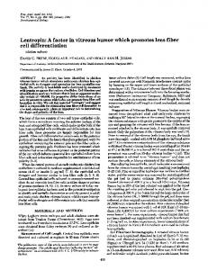
showed lower DOP concentration (653.56 ng/100 mg and 765.52 ng/100 mg for 50 μl/day and 100 μl/day, respectively). With regards to the NAD levels, no significant effect was observed in ROEO (507.36 ng/100 mg and 46.64 ng/100 mg in 50 μl/day and 100 μl/day, respectively) group but a significant improvement was observed in LAEO group (55.29 ng/100 mg) compared with control group (44.31 ng/100 mg). For ADR, ROEO group did not show significant change (0.35 ng/100 mg and 0.34 ng/100 mg, in 50 μl/day and 100 μl/day ROEO, respectively), as with LAEO group (0.44 ng/100 mg LAEO) group compared to the control group (0.33 ng/ 100 mg). Both ROEO treatment groups showed lower level of ADR compared with LAEO. Inhalation of ROEO (50 μl/day and 100 μl/day) showed significant effects on serum brain DOP level. ANOVA results showed significant differences in the DOP levels between groups (Table 2). ROEO promoted acetylcholinesterase (AChE) activity and upregulated Gap43 mRNA expression in PC12 cells
Observed improved corticosterone and dopamine levels, which could be due to neuronal differentiation, was observed following ROEO inhalation. To verify if ROEO induced neuronal differentiation, acetylcholinesterase (AChE) activity and the expression of Gap43 mRNA expression in PC12 cells were determined. First, the non-cytotoxic concentration of ROEO was determined by performing MTT assay. PC12 cells were treated with or without ROEO (0, 5, 10, 50, and 100 μg/ml) for 48 h. MTT assay results showed that ROEO was not cytotoxic at low concentrations (5 and 10 μg/ml) but was slightly cytotoxic at 50 and 100 μg/ml, decreasing the cell viability to 97% and 81.41% for 50 and 100 μg/ml ROEO, respectively. The synthesis and secretion of neurotransmitters are significant events associated with neuronal differentiation. In PC12 cells, the secretion of acetylcholine (ACh) is induced following induction of PC12 cell differentiation. Treatment with μg/ml ROEO increased the AChE activity in PC12 (124.16%) compared both the control (100%) and 50 ng/ml NGF (116.36%) (Fig. 2b). Since ACh is produced from ingested choline (Ch) and from recycled choline in the synapse, we then determined the intracellular concentration of ACh and its substrate Ch using HPLC-ECD. As presented in Table 3, ROEO increased the intracellular ACh and Ch levels to 11.88 ng/ mg and 2645.49 ng/mg, respectively. As expected, NGF increased the intracellular ACh (11.99 ng/mg) and Ch levels (2660.6 ng/mg) compared to the control. The control had ACh and Ch levels of 9.51 ng/mg and 2156.21 ng/mg, respectively. To further investigate the effects of ROEO on neuronal differentiation, the gene expression level of growth-
Villareal et al. BMC Complementary and Alternative Medicine (2017) 17:549
a Cell viability (% of control)
140
*
120
*
100
*
80 60 40 20 0 0
b
5
10 ROEO (µg/ml)
AChE activity (% of control)
140
50
*
**
NGF 50 ng/ml
ROEO 10 µg/ml
100 80 60 40 20 0
Fig. 2 Effect of Rosmarinus officinalis essential oil (ROEO) on PC12 cell proliferation and acetylcholinesterase activity. a Effect of ROEO on PC12 cells viability. PC12 cells were seeded onto 96-well plates at a density of 1×10⁵ cells/ml. After overnight incubation, cells were treated with 0, 5, 10, 50 or 100 μg/ml ROEO for 48 h and the cell viability was evaluated using MTT assay. Data are presented as a percentage of the control and represent the mean of three independent trials ±.SD *P≤0.05 vs. control. b Effect of ROEO on the acetylcholinesterase (AChE) activity in PC12 cells. PC12 cells were seeded onto 96-well plates at a density of 1×10⁵ cells/ml. After overnight incubation, cells were treated with 50 ng/ml of nerve growth factor (NGF) or 10 μg/ml of ROEO for 48 h. The control indicates cells incubated for 48 h without NGF or ROEO treatment. AChE activity was expressed in percentage of control. Data represent the mean of three independent trials ±.SD *P≤0.05, **P