The level of circulating antibody to phenolic glycolipid I of Mycobacterium leprae was determined in 18 inbred strains of mice after immunization withM. leprae ...
INFECTION AND IMMUNITY, May 1985, p. 474-479
Vol. 48, No. 2
0019-9567/85/050474-06$02.00/0 Copyright C) 1985, American Society for Microbiology
Antibody Response to Phenolic Glycolipid I in Inbred Mice Immunized with Mycobacterium leprae CORY TEUSCHER,'t DONNA YANAGIHARA,2 PATRICK J. BRENNAN,2 FREDERICK T. KOSTER,' AND KENNETH S. K. TUNG'* Departments of Pathology and Medicine, The University of New Mexico, Albuquerque, New Mexico 87131,1 and
Department of Microbiology, Colorado State University, Fort Collins, Colorado 805232 Received 19 October 1984/Accepted 23 January 1985
The level of circulating antibody to phenolic glycolipid I of Mycobacterium leprae was determined in 18 inbred strains of mice after immunization with M. leprae organisms. By usinlg a solid-phase radioimmunoassay with phenolic glycolipid I as test antigen, a continuous distribution of antibody levels ranging from high to low was observed. The level was found to be controlled by multiple genes, including both H-2 complex- and Igh allotype complex-linked genes. Low antibody response to phenolic glycolipid I was shown to be inherited as a dominant trait in three combinations of high x low responder F1 progeny.
Mycobacterium leprae, the causative agent of leprosy, is a remarkably nontoxic intracellular parasite capable of reaching extremely high numbers in various tissues without resulting in damage or inciting an inflammatory response (28, 30). In human infection, polar tuberculoid leprosy, the high-resistance form, is characterized by few lesions, low numbers of bacilli, high lymphocyte transformation tests, and low titers of anti-M. leprae antibodies (11, 12, 24, 40). At the other end of the clinical spectrum, polar lepromatous leprosy, the low-resistance form, is exemplified by large numbers of disseminated bacilli, reduced reactions in lymphocyte transformation tests, and high levels of anti-M. leprae antibodies. Between the two ends of this spectrum is a wide range of clinical-pathological symptoms with various cutaneous and immunological manifestations known as borderline leprosy. The results of host immune reactions to bacillary antigens (30), rather than variations in the M. leprae organism itself (27, 32), are believed to be responsible for most of the clinical-pathological spectrum of the disease. Furthermore, the expression of the clinical spectrum may be due in part to human major histocompatibility complex (HLA) and non-HLA controlled host immune responses to M. leprae (31, 38). Studies examining immune responses to whole M. leprae and M. Ieprae antigens in humans may not discriminate between naive responses and altered responses secondary to the infectious process. On the other hand, studies in noninfected inbred mice, with well-defined genetic compositions, allow meaningful analysis of the host genetic factors involved in governing immune responsiveness to antigens of M. leprae. Comparisons between the magnitude of genetically controlled humoral and cellular immune responses in different infected and uninfected mouse strains also provide data pertinent to the hypothesis that the different clinicalpathological expressions of leprosy result from differences in the magnitude of the two types of immune response, a consequence of imbalanced T-cell immunoregulation (21-23, 36).
An M. leprae-specific glycolipid antigen, triglycosyl phenolic diacyl phthiocerol (phenolic glycolipid I [PGL-I]), has recently been isolated, purified, and structurally characterized (13, 14). It is distinguished by a species-specific trisaccharide [3,6-di-O-methyl-3-D-glucopyranosyl(1->4)2,3di-O-methyl-o-L-rhamnopyranosil (1--2) 3 -0- methyl-a-Lrhamnopyranose] glycosidically linked to a genus-specific phenolic diacylphthiocerol. PGL-I has been shown to be antigenic in animals (1, 13, 14) and leprosy patients (2, 4, 14, 25, 43). By using solid-phase immunoassays, immunoglobulin G (IgG) and IgM antibodies to PGL-I were demonstrated in the majority of untreated leprosy patients of all clinicalpathological classifications. Patients with lepromatous leprosy had higher levels of anti-PGL-I antibodies than did patients with tuberculoid leprosy (2, 4, 43). PGL-I was also found to induce nonspecific suppressor T-cell function in vitro (21). PGL-I represents ca. 2% of the organism mass and is present in large quantities in infected tissues from which the bacilli have been removed (13). Since it may represent the major interface between host defense mechanisms and the microorganisms, we have undertaken a series of studies to examine the genetic control of immune responses to PGL-I in mice immunized with M. leprae organisms.
MATERIALS AND METHODS Mice. The following strains of inbred female mice and F, hybrids were used: A/J, A.SW/SnJ, B10.BRISgSnJ, BALB/cByJ, CBA/J, C57BL/6J, C57BL/10J, DBA/1J, DBA/2J, SJL/J, SWR/J, (C57BL/6J x A/J)Fl, (SWR/J x A/J)Fl, (SJL/J x CBA/J)F1 (Jackson Laboratory, Bar Harbor, Maine) and BALB.10, BALB.K, BALB.Igc (N-20), BALB.1ge (N-10), BALB.Igf (N-18), BALB.Igg (N-11,F-3), BALB.Ig" (N-11) (Noel Warner and Ed Walker, University of New Mexico School of Medicine, Albuquerque, N.M.). All mice were 8 to 20 weeks of age at the start of the experiment. Immunization. M. leprae was isolated from irradiated infected armadillo livers (13). Purified M. Ieprae was emulsified in incomplete Freund adjuvant (Marcol:Arlacel, 4:1). Primary and secondary immunizations with 0.1 ml of emulsion containing 50 p.g of M. leprae were carried out intraperi-
* Corresponding author. t Present address: Division of Reproductive Biology and Endocrinology, Department of Obstetrics and Gynecology, University of Pennsylvania, Philadelphia, PA 19104.
474
ANTIBODY RESPONSE TO M. LEPRAE PHENOLIC GLYCOLIPID I
VOL. 48, 1985
Strain SWR/J C57BL/6J
Ig H-2 No. Type Allotype Animals q/q b/b
c /c b/b
6 7
k/k k/k
7 5 5 7 7
5 6 6 9 6 7
CBA/J BALB.K DBA/IJ C57BL/IOJ BIO.BR/SgSnJ
q/q b/b k/k
a/a a/a c/c b/b b/b
A/J A.SW/SnJ DBA/2J BALB/cByJ SJL/J BALB. BIO
a/a s/s d/d d/d s/s b/b
e/e e/e c/c a/a b/b a/a
Acpm I125 SaMk (x103) 2 3 4
475
5
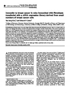
FIG. 1. Levels of circulating anti-PGL-I antibodies detected by a solid-phase radioimmunobinding assay with 1251I-SaMK in different inbred strains of mice immunized with 100 ,u.g of nonviable M. leprae organisms. A continuous distribution in the level of circulating anti-PGL-I antibodies was observed. Mouse strains are arbitrarily grouped into high, intermediate, and low responders. Data expressed as means + standard error.
toneally on day 0 and day 21. Serum was obtained by tail bleeding 2 weeks before and 30 days after primary immunization. Sera were stored at -70°C until tested. Solid-phase radioimmunoassay for anti-PGL-I antibodies. The solid-phase radioimmunoassay used to detect antibodies to PGL-I in the serum of immunized mice was essentially that of Smolarsky (35). Briefly, 50-plI samples of PGL-I suspended in 100% ethanol at a concentration of 0.04 mg/ml were coated onto flat-bottomed 96-well polyvinyl microtiter plates (Dynatech Laboratories, Inc., Alexandria, Va.) by drying. The plates were washed three times, 5 min each time, with 150 plI of 0.3% gelatin (gel) dissolved in phosphate-buffered saline (PBS; pH 7.2) containing 1 mM EDTA (gel-EDTA-PBS). Samples of 50 pA of preimmune and day-30 postimmunization sera diluted 1:5 in gel-EDTA-PBS were added to duplicate wells and incubated at room temperature
No. Iype Animals 7 b/b 7 k/k
H-2
Strain
C57BL/IOJ BIO. BR/SgSnJ BALB/cByJ BALB.BIO BALB.K
d/d b/b k/k
9 7 5
for 3 h. Plates were then washed rapidly three times with gel-EDTA-PBS, followed by a 5-min wash with gel-EDTAPBS and a 5-min wash with 1% gamma globulin-free bovine serum albumin (BSA) in PBS (BSA-PBS). An amount of 100,000 cpm of 125I-sheep anti-mouse kappa (125I-SaMK) or 125I-protein A (125I-PA) diluted in 50 ,ul of BSA-PBS was added and incubated for 3 h at room temperature. Each well was washed three times, for 5 min each time, with 150 ,ul of BSA-PBS, cut from the plate with a hot wire, and counted in an a-spectrometer. All sera were also studied in dilutions of 1:50 to ensure that the differences observed in uptake of 1251 at 1:5 dilutions was not due to a pro-zone effect. The paired t-test was used to determine the statistical significance of antibody present in serum on day 30 after immunization. The specific antibody response for each mouse represents the difference in counts per minute obtained by subtracting
Acpm I125 SaMk (xI03) 4 3 2 I
I
pa Value
~-A
NS .
I
I