ity spectrum of A. aegypti and Drosophila cecropin A showed a lower ... challenged adults 6 h after bacteria inoculation, and it continued ...... 83, 191â201. 15.
THE JOURNAL OF BIOLOGICAL CHEMISTRY © 1999 by The American Society for Biochemistry and Molecular Biology, Inc.
Vol. 274, No. 29, Issue of July 16, pp. 20092–20097, 1999 Printed in U.S.A.
Antimicrobial Activity Spectrum, cDNA Cloning, and mRNA Expression of a Newly Isolated Member of the Cecropin Family from the Mosquito Vector Aedes aegypti* (Received for publication, February 23, 1999, and in revised form, April 26, 1999)
Carl Lowenberger‡§¶, Maurice Charlet§i, Jacopo Viziolii**, Sofie Kamal‡, Adam Richman‡‡, Bruce M. Christensen‡, and Philippe Buleti From the ‡Animal Health and Biomedical Sciences University of Wisconsin, Madison, Wisconsin 53706, the iUPR CNRS 9022, Institut de Biologie Mole´culaire et Cellulaire, 15 rue Rene´ Descartes, 67084 Strasbourg Cedex, France, and the ‡‡European Molecular Biology Laboratory, Meyerhofstrasse 1, 69117 Heidelberg, Germany
An antimicrobial peptide belonging to the cecropin family was isolated from the hemolymph of bacteriachallenged adult Aedes aegypti. This new peptide, named cecropin A, was purified to homogeneity and fully characterized after cDNA cloning. The 34-residue A. aegypti cecropin A is different from the majority of reported insect cecropins in that it is devoid of a tryptophan residue and C-terminal amidation. The importance of these two structural features on the activity spectrum was investigated using a chemically synthesized peptide. A comparison of the antimicrobial activity spectrum of A. aegypti and Drosophila cecropin A showed a lower activity for the mosquito molecule. A. aegypti cecropin mRNA expression was not detected by Northern blot or reverse transcription-polymerase chain reaction analysis in any immature stage of the mosquito, nor in naı¨ve adults, but it was observed in challenged adults 6 h after bacteria inoculation, and it continued over 7–10 days.
One factor contributing to the enormous success that insects have exhibited through evolutionary time is their ability to mount a rapid and effective response against invading pathogens. This response includes the rapid production of potent antimicrobial peptides effective against bacteria and fungi; transcriptional activity may be detected within minutes of a stimulus, resulting in 1–100 mM concentrations of specific peptides in the hemolymph within 24 h (1). Since the isolation of cecropin from Hyalophora cecropia by Boman and co-workers (2), approximately 170 immune peptides have been characterized from insects and other invertebrates (for a recent review, see Ref. 1). In insects, these molecules are rapidly synthesized, easily stored, and promptly available after infections. These features, together with the low metabolic cost for the insect and a low specificity (see review in Ref. 3), make antimicrobial
* This research was funded in part by a Burroughs Welcome Fund grant and National Institutes of Health Grants AI19769 and AI28781 (to B. M. C.) and AI44966 (to C. L.). This study was also supported by the Center National de la Recherche Scientifique and the University Louis Pasteur of Strasbourg (France). The costs of publication of this article were defrayed in part by the payment of page charges. This article must therefore be hereby marked “advertisement” in accordance with 18 U.S.C. Section 1734 solely to indicate this fact. § The first two authors contributed equally to this paper. ¶ To whom correspondence should be addressed: Animal Health and Biomedical Sciences University of Wisconsin-Madison, 1655 Linden Dr., Madison, WI 53706. Tel.: 608-262-2373; Fax: 608-262-7420. ** Supported by a postdoctoral fellowship from the Fondazione Pasteur-Cenci Bolognetti.
peptides a very versatile component of the insect immune response. In mosquitoes, which serve as obligate vectors for serious debilitating human diseases such as malaria, lymphatic filariasis, and numerous arboviruses, the predominant immune peptide is a member of the insect defensin family (4 –7) Although it has been suggested that inducible antimicrobial peptides have no impact on eukaryotic parasites (8), several studies suggest otherwise. Reports have implicated cecropins or synthetic cecropin derivatives (9 –12), insect defensins (13), or a general immune activation process (14, 15) in reducing the development of parasites in mosquitoes. Because of the potential role cecropins might play in the anti-parasite responses cited above and the recent reports of synthesized cecropin-like molecules that kill Plasmodium sp. (16, 17), we have attempted to isolate and characterize cecropins from mosquitoes. Cecropins are a family of inducible antibacterial peptides of 4 kDa, devoid of cysteine residues, constituted by two a-helices linked by a short hinge, with a strongly basic N-terminal region and a long hydrophobic domain in the C-terminal half. Cecropins integrate into the bacterial cell membrane and cause lysis and cell death by a loss of cations (18). Cecropins have been characterized from Lepidoptera and Diptera (1, 19), but in mosquitoes, there has been only limited information (20, 21) until recently, when Sun et al. (22) reported the purification of a 35-amino acid cecropin from an Aedes albopictus cell line. We report herein the identification and full characterization of a new member of the cecropin family isolated from the hemolymph of immune-activated Aedes aegypti. MATERIALS AND METHODS
Insect Immunization and Hemolymph Collection—A. aegypti (Liverpool strain) mosquitoes were reared as described previously (23). Adult female mosquitoes were immunized (4), and 24 h later, the hemolymph was collected from individual mosquitoes by perfusing the hemocoel with Aedes saline as described previously (23). The hemolymph from about 2600 bacteria-challenged mosquitoes or 2000 naı¨ve mosquitoes was centrifuged at 5000 3 g for 5 min at 4 °C, and the supernatant was frozen until use. Extraction and Purification of Antibacterial Peptides—Antibacterial peptides were purified from acidified cell-free hemolymph. Acidification was performed with ammonium acetate buffer at pH 3.5 (final concentration, 25 mM), supplemented with aprotinin (Sigma) as a protease inhibitor (final concentration, 1.5 mM) and phenylthiourea (Prolabo) as a melanization inhibitor (final concentration, 20 mM). After extraction under gentle stirring for 30 min in an ice-cold water bath, the sample was centrifuged at 10,000 3 g for 20 min at 4 °C. The supernatant then was loaded onto a 12-cc Sep-Pak Vac C18 cartridge (WatersTM) equilibrated with acidified water (0.05% trifluoroacetic acid). Stepwise elution was performed with 2, 40, and 80% acetonitrile in acidified water.
20092
This paper is available on line at http://www.jbc.org
Aedes aegypti Cecropin
20093
1
The 40% Sep-Pak fraction was analyzed by reversed-phase HPLC on an Aquapore RP 300 C8 column (250 3 7 mm; BrownleeTM) using a linear gradient of 2– 60% acetonitrile in acidified water for 120 min (flow rate 1.3 ml/min). The antibacterial activity of the collected fractions was detected by liquid growth inhibition assay against Micrococcus luteus A270 and Escherichia coli SBS363, as described previously (4). The active fractions were further purified on a Delta-Pak HPIC18 column (150 3 2 mm; WatersTM) with a linear biphasic gradient of acetonitrile in acidified water from 2 to 22% over 10 min and from 22 to 32% over 50 min at a flow rate of 0.2 ml/min at 35 °C. The first step of HPLC purification was performed with a Beckman Gold HPLC system equipped with a photodiode-array detector Beckman 168. The subsequent steps were carried out using a Waters HPLC system (Waters, model 626) attached to a tunable absorbance detector (Waters, model 486) and equipped with an internal oven. For all HPLC purification steps, the effluent was monitored by its UV absorption at 225 nm. Fractions were hand collected, concentrated under vacuum, and reconstituted in MilliQ water (MilliporeTM) before antimicrobial assays. Capillary Zone Electrophoresis—The purity of the isolated peptides was ascertained by capillary zone electrophoresis as described previously (4). Analyses were conducted on a model 270A-HT capillary electrophoresis system (PEApplied Biosystems) equipped with a fused silica capillary. Mass Measurement by Matrix-assisted Laser Desorption Ionization Time-of-flight Mass Spectrometry (MALDI-TOF MS)—Measurements were carried out on a Bruker (BIFLEXTM) mass spectrometer equipped with a delayed extraction ion source. Mass spectrometry analysis was performed as described previously (24), using a-cyano-4-hydroxycinnamic acid (Sigma) as matrix. Microsequence Analysis—The isolated peptides were subjected to Edman degradation on a pulse liquid automatic sequenator (PEApplied Biosystems, model 473A). Cecropin cDNA Isolation—Total RNA was extracted from whole bodies of immune-activated mosquitoes 1–24 h after inoculation and reverse-transcribed as described previously (25), using the primer 59CGGGCAGTGAGCGCAACGT14-39 for the RT reaction. PCR amplification of cecropin cDNA was performed using a reverse primer, consisting of the initial 18 nucleotides of the RT primer, and two forward degenerate primers (59-GGIAARAARYTIGARGGIGC-39 and 59 AAIATHTTYGTITTYGTIGC 39) designed to the cecropin sequence obtained by Edman degradation. PCR conditions used were 94 °C (2 min) and 30 cycles of 94 °C (1 min), 50 °C (1 min), and 72 °C (2 min), followed by one 10-min extension period at 72 °C. PCR products were cloned and sequenced as previously described (25). Full-length cDNAs were obtained using the Marathon cDNA synthesis kit (CLONTECH) using specific primers designed to the partial cDNA sequence obtained and the kit primers. PCR amplification and cDNA cloning were done as described above. Phylogenetic analysis and multiple alignment of A. aegypti cecropin A with other insect cecropins were performed with the computer program DNAstar® (using Clustal method with PAM 250 matrix). Northern Analysis—Northern analysis was performed as described previously (25) using 5 mg of total RNA extracted from whole bodies of immature stages of A. aegypti and naı¨ve and immune-activated adults. 32 P probes were made using specific primers to amplify 50 ng of a 177-base pair cecropin cDNA clone in a PCR described previously (25). The ribosomal probe rpL8 was used as a loading control. RT-PCR—RNA from naı¨ve larvae, white and black pupae, and naı¨ve and immune-activated adults was extracted, reverse-transcribed, and PCR-amplified as described above. PCR products, obtained with the cecropin specific primers indicated in Fig. 2, were quantified on a 1% agarose gel using an Eagle Eye II still video system (Stratagene). PCRs were standardized using ubiquitin specific primers. Peptide Synthesis—Peptide synthesis was performed by classic Fmoc fluoren-9-ylmethoxycarbonyl methodology as described previously (26). The synthetic cecropin was prepurified by solid phase extraction on Sep-Pak C18 and by two reversed-phase HPLC. Authenticity and purity of the peptide was determined by Edman degradation, mass spectrometry and capillary zone electrophoresis. Bioassays—Antimicrobial assays were performed as described previously (27). To define the antibacterial activity spectrum we used, in addition to the bacterial strains reported in a previous study (4), the
1 The abbreviations used are: HPLC, high performance liquid chromatography; RT, reverse transcription; PCR, polymerase chain reaction; MIC, minimal inhibitory concentration; MALDI-TOF MS, matrixassisted laser desorption time-of-flight mass spectrometry.
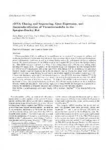
FIG. 1. Reversed-phase HPLC of an acidic hemolymph extract from bacteria-challenged adults A. aegypti. Step 1, the 40% acetonitrile fraction eluted from Sep-Pak C18 was first analyzed by reversedphase HPLC with a linear gradient of acetonitrile in acidified water. Fractions were tested against M. luteus and E. coli. The active fractions (shaded area in Step 1) were further purified and the chromatograms obtained for three of them are reported (A, B, and C in Steps 1 and 2). Step 2, purifications were performed on a Delta-Pak HPIC18 column with a same biphasic linear gradient of acetonitrile in acidified water (dotted line). Absorbance was monitored at 225 nm (solid line). Peaks 1 and 2 highlighted in dark gray were active against both M. luteus and E. coli, whereas those in light gray were only active against M. luteus. Peak 3 (hatched lines) was exclusively active against E. coli. following strains: E. coli 1106, E. coli D31, Serratia marcescens, Bacillus megaterium, Erwinia carotovora, Xanthomonas campestris, Klebsiella pneumoniae, Listeria monocytogenes, and Salmonella typhimurium. The following fungal and yeast strains were also evaluated: Aspergillus fumigatus, Candida albicans, Candida glabrata, Cryptococcus neoformans, Beauveria bassiana, Fusarium oxysporum, Fusarium culmorum, Neurospora crassa, and Saccharomyces cerevisiae TGY 48 –1. Antiyeast activity was assessed in yeast complete medium (1% yeast extract, 1% peptone, 2% glucose (w/v)), following the same procedure used for antibacterial assays. The control antibiotic peptides MSI-94 and PGLa (linear amphipathic frog antibiotics) were gifts from M. A. Zasloff (Magainin Scientific Institute, Philadelphia, PA). RESULTS
Purification of Cecropin from the Hemolymph of A. aegypti— With regard to our previous study on Aedes defensins (4), we focused the present analysis on the fraction, eluted with 40% acetonitrile, that contained the bulk of antibacterial activity. This fraction was subjected to reversed-phase HPLC analysis (Fig. 1, Step 1). A marked antibacterial activity against M. luteus and E. coli was observed in eight consecutive fractions eluted from 25 to 30% acetonitrile. Each individual active fraction (Fig. 1, Step 1, shaded area) was further subjected to a second reversed-phase chromatography. The collected fractions were tested against M. luteus and E. coli, as before. The results obtained are only reported for the three most representative fractions (Fig. 1, A-C). After the antibacterial assays, three individual active peaks were purified from fractions A, B, and C. Two peaks (Fig. 1, Step 2, peaks 1 and 2 in dark gray) eluted at 26 and 28% acetonitrile, respectively, were active against M. luteus and E. coli at high concentrations. At a lower concentration (in regards to the absorbance at 225 nm), an activity was detected only against M. luteus. In contrast, peak 3 (Fig. 1, Step 2, hatched peak) was active only against E. coli. Mass measurements of fractions 1 and 2 by MALDI-TOF MS indicated the presence of compounds with mass values similar to those of the A. aegypti defensins A, B, and C (4) (data not shown). Partial
20094
Aedes aegypti Cecropin
FIG. 2. Nucleotide sequence of cDNA and deduced amino acid sequence of the A. aegypti preprocecropin. The mature peptide sequence is in italics. The beginning of the mature peptide (arrow) and the stop codon (asterisk) are indicated. The putative polyadenylaton signal is double underlined. The nucleotide sequences used as primer sites for RT-PCR and for making 32P-labeled probes are underlined.
N-terminal sequencing by Edman degradation confirmed that peak 1 was Aedes defensin C (4155.7 Da) and that peak 2 contains the isoforms A and B, as judged by an additional capillary zone electrophoresis analysis (data not shown). To obtain structural information on the compound present in peak 3, this peak was further purified to homogeneity by an additional chromatography step using the same reversed-phase column as in step 2 (data not shown). After the antibacterial assay against E. coli, only a single active fraction was found. The purity was ascertained by capillary zone electrophoresis and MALDI-TOF-MS. Only a single mass signal at 3391.5 Da was recorded, confirming the purity of the molecule. The peptide then was subjected to Edman degradation and the following 32-amino acid sequence was obtained: GGLKKLGKKLEGAGKRVFNAAEKALPVVAGAK. The calculated molecular mass for this 32-residue peptide (3206.8 Da) was lower than the measured molecular mass (3391.5 Da) by approximately 186 Da, indicating that the obtained sequence was not complete. We used the cDNA sequence obtained (see below and Fig. 2) to determine what residues were missing. By Edman degradation, we determined that the N-terminal residue of the mature peptide was a glycine. This residue was found at position 24 in the deduced cDNA sequence. Molecular mass measurement of the mature peptide (3391.5 Da) established that it would end at the leucine residue at position 57 in the deduced cDNA sequence. The calculated average molecular mass of the predicted sequence (3391.1 Da) was in perfect agreement with the measured molecular mass by MALDI-TOF-MS at 3391.5 Da. Thus, the active compound of peak 3 (Fig. 1, Step 2) corresponded to a 34-amino acid peptide with the residues Ala-Leu following the 32-amino acid sequence obtained by Edman degradation (Fig. 2). A search in the SWISS-PROT protein data base showed high homology with cecropins, antibacterial peptides from Lepidoptera and Diptera. We designated the new isolated peptide as A. aegypti cecropin A. cDNA Isolation—The A. aegypti cecropin cDNA contains a deduced open reading frame of 177 nucleotides. The consensus sequence begins with an ATG codon and encodes a protein of 59 residues containing a deduced sequence identical to the 32 amino acid sequence previously established by Edman degradation. The coding region contains a stop codon, a 39 untranslated region of 38 nucleotides, the putative polyadenylation consensus signal (AATAAA), and 13 additional nucleotides before the poly(A) tail (Fig. 2). The deduced protein sequence
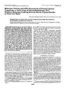
FIG. 3. Temporal expression of A. aegypti preprocecropin in naı¨ve and immune-activated mosquitoes. A, Northern analysis of naı¨ve larvae (L), white pupae (WP), black pupae (BP), adults (A0) and immune-activated adults (AI) at various times after challenge (in days). A probe generated from a ribosomal protein encoding cDNA (rpL8) was used as a loading control. B, RT-PCR analysis of cecropin expression in larvae, white and black pupae, and naı¨ve and bacteria-inoculated adults. Expression of a gene encoding the ribosomal protein ubiquitin was used as a control. PCR controls were performed with a cecropin cDNA clone as template (1) or without template (2).
contains a putative signal peptide comprising the first 23 residues and the mature cecropin (34 residues), followed by a dipeptide (Arg-Lys). The signal peptide region is highly hydrophobic and is predicted to terminate with an alanine residue (Ala23). This prediction is based on the fact that the mature cecropin peptide found in the hemolymph of immune-challenged mosquitoes started with, and did not contain any of the residues preceding, Gly24. Northern and RT-PCR Analysis—By Northern analysis, we did not detect cecropin mRNA in any immature stage of A. aegypti, nor in naı¨ve adults (Fig. 3A). Expression was found in immune-activated adults: transcription was detected 6 h postinoculation and continued for 7–10 days. Similarly, by RT-PCR we could not detect cecropin in cDNAs isolated from larvae, pupae, or naı¨ve adults of A. aegypti (Fig. 3B). However, a strong signal for cecropin was detected in immune-activated mosquitoes. Activity Spectrum of A. aegypti Cecropin—After synthesis, HPLC purification, and mass spectrometry measurement to confirm the authenticity of the molecule, 9.3 mg of pure cecropin were obtained (approximate yield, 11.6%). The activity spectrum of A. aegypti cecropin A was compared with Drosophila cecropin A, which presents the typical features of cecropins (presence of a tryptophan residue and a C-terminal amidation). The activity spectra of these two peptides are presented in Table I, compared with two control antibiotics forming a-helices, PGLa and MSI-94. In the antibacterial assays, A. aegypti and Drosophila cecropins were more effective against Gramnegative strains than against Gram-positive bacteria. Among the 11 Gram-negative strains tested, only one bacteria (S. marcescens) was not affected by A. aegypti and Drosophila cecropins at the highest concentration tested (100 mM). The insect cecropins were more active than the two control antibiotics against the Gram-negative bacteria (Table I), and the Drosophila cecropin was several times more effective than the A. aegypti cecropin. The measured difference factors were (i) 2-fold against E. coli D22 and D31, K. pneumoniae, and S. typhimurium; (ii) 10-fold against P. aeruginosa, and (iii) 100fold against E. cloacae. Gram-positive bacteria were affected almost equally by the two cecropins. A. aegypti cecropin A was essentially inactive against three of the four Bacillus species tested (B. cereus, B. thuringiensis, and B. subtilis), as well as against L. monocytogenes and S. aureus, up to a concentration of 100 mM. Against B. subtilis and S. pyogenes, however, the results differed according to the cecropin used: the growth of B. subtilis was not affected by 100 mM of A. aegypti cecropin A but
Aedes aegypti Cecropin
20095
TABLE I Activity spectrum of Aedes and Drosophila cecropins compared with two control antibiotics (PGLa and MSI-94) The MIC (28) is expressed as final concentration (mM). ND, not detected at the highest concentration tested (100 mM). MIC Microorganism
Aedes cecropin
Drosophila cecropin
PGLa
MSI-94
mM
Gram-negative bacteria E. coli D22 E. coli D31 E. coli SBS363 E. coli 1106 Enterobacter cloacae b12 E. carotovora K. pneumoniae Pseudomonas aeruginosa S. typhimurium S. marcescens Db11 X. campestris Gram-positive bacteria Aerococcus viridans Bacillus cereus B. megaterium Bacillus subtilis Bacillus thuringiensis L. monocytogenes M. luteus Staphylococcus aureus Streptococcus pyogenes Fungi A. fumigatus B. bassiana F. culmorum F. oxysporum N. crassa Yeasts C. albicans C. glabrata C. neoformans Saccharomyces cerevisiae
0.25–0.5 0.25–0.5 0.1–0.25 0.25–0.5 50–100 0.25–0.5 1–2.5 10–25 1–2.5 ND 0.5–1
0.1–0.25 0.1–0.25 0.1–0.25 0.25–0.5 0.5–1 0.5–1 0.5–1 1–2.5 0.5–1 ND 0.5–1
1–2.5 1–2.5 1–2.5 1–2.5 ND 1–2.5 10–20 20–40 10–20 ND 5–10
1–2.5 0.5–1 1–2.5 1–2.5 5–10 1–2.5 2.5–5 1–2.5 1–2.5 ND 0.5–1
2.5–5 ND 1–2.5 ND ND ND 10–25 ND 10–25
5–10 ND 1–2.5 10–25 ND ND 5–10 ND ND
0.5–1 2.5–5 0.5–1 0.5–1 5–10 1–2.5 0.5–1 1–2.5 1–2.5
0.5–1 1–2.5 0.5–1 0.5–1 1–2.5 1–2.5 0.5–1 1–2.5 1–2.5
ND ND 1–2.5 2.5–5 5–10
ND ND 1–2.5 1–2.5 5–10
10–20 ND 0.5–1 2.5–5 2.5–5
20–40 ND 0.5–1 0.5–1 5–10
25–50 ND 25–50 50–100
ND ND ND ND
was sensitive to the Drosophila molecule (MIC, 10 –25 mM). In contrast, the growth of S. pyogenes was not affected by Drosophila cecropin A but was inhibited by the A. aegypti peptide (MIC, 10 –25 mM). The two cecropins were less active against Gram-positive bacteria than PGLa and MSI-94. A. aegypti and Drosophila cecropins were generally equally active against the fungi F. culmorum, F. oxysporum, and N. crassa (MIC, 1–2.5, 2.5–5, and 5–10 mM, respectively). A. fumigatus and B. bassiana, however, were insensitive to cecropins. In antiyeast assays, only A. aegypti cecropin showed a moderate activity against C. albicans, C. neoformans (MIC, 25–50 mM), and S. cerevisiae (MIC, 50 –100 mM). DISCUSSION
The data presented here established that the cell-free hemolymph of bacteria-challenged adults of A. aegypti contains several antibacterial substances, among them the previously described A. aegypti defensins (4, 5). Following a detailed study by reversed-phase HPLC of the defensin zone, we have isolated and partially characterized a novel antibacterial peptide belonging to the cecropin family. This is the first report presenting the full primary structure of a mature mosquito cecropin isolated from the insect hemolymph. Previously, the only report in mosquitoes was the cecropin isolated from an A. albopictus cell line (22). In common with all preprocecropin sequences isolated from Diptera, the A. aegypti preprocecropin ends with an alanine residue (Fig. 4). In most of the dipteran cecropins listed in Fig. 4, the terminal region of the signal peptide has a highly conserved consensus sequence (GQSEA). All the sequences deduced from the cDNAs showed conserved residues (MNF) at the translation start site and other highly conserved areas
1–2.5 ND
0.5–1 ND
within the preproregion, showing the following overall consensus sequence: MNF-K-FIFVALIAI-GQSE. In contrast to the Diptera, preprocecropin sequences from Lepidoptera all end in a proline residue, preceded by either an alanine or a glutamic acid residue (Fig. 4). With the exception of the Bombyx mori cecropin D (39) and the Trichoplusia ni cecropin A (40), the lepidopteran preprosequences begin with the consensus MNF, suggesting that the cecropin molecules occurred in the insects before the divergence of the Diptera and Lepidoptera. Similar cecropins also have been isolated from tunicates, marine invertebrates belonging to the phylum Chordata (42), which share many of the residues in the consensus region of dipteran cecropins, suggesting the conservation of these peptides among phyla other than insects. Sequence comparison of A. aegypti mature cecropin A with other dipteran cecropins (Fig. 4) showed similarities of 82.4, 88.2, and 35.3% with the A. albopictus, Anopheles gambiae,2 and Drosophila cecropins, respectively. Comparison of A. aegypti cecropin with previously characterized dipteran and lepidopteran cecropins revealed several interesting contrasts. Of specific note is the absence of a tryptophan residue in the A. aegypti sequence, because a tryptophan residue has been reported, at amino acid position 1 or 2, in all characterized insect cecropins, except for the other mosquito cecropins (22)2 and the B. mori cecropin D (39). A. aegypti cecropin A is not amidated at the C-terminal end, as deduced from the characterization of the full cDNA sequence. This is in contrast to the common
2 J. Vizioli, P. Bulet, M. Charlet, C. A. Lowenberger, C. Bass, C., H.-M. Muller, G. Dimopoulos, J. Hoffmann, F. C. Kafatos, and A. Richman, submitted for publication.
20096
Aedes aegypti Cecropin
FIG. 4. Comparison of the amino acid sequence of Aedes preprocecropin with typical dipteran and lepidopteran cecropin precursors. The sequences are aligned for similarities and gaps were introduced for optimal alignment. The number in parentheses after the species name indicates the reference from which the sequence was obtained.
FIG. 5. Phylogenetic analysis of insect cecropins. Constructions are performed on the basis of the homology sequences calculated from the complete amino acid sequence of the mature cecropins. Sequences were selected from Hetru et al. (1) with the following, in addition: A. albopictus cecropin A (22), A. gambiae (Footnote 2), B. mori cecropin D (39), and Hyphantria cunea cecropin A (41). The homology sequences were calculated using the Clustal method with the PAM250 residue weight table.
feature of a C terminus blocked by an amine group, in all insect cecropins except the A. albopictus cecropin A (22) (Fig. 4). The A. aegypti cecropin A has more than 13 amino acids in common with the other representative dipteran cecropins over the 15 first N-terminal amino acids. This high degree of identity (87%) was not observed in the C-terminal region. It is surprising that only two amino acids (one valine and two alanine residues) are common among the 10 last residues of mosquito cecropins and the same C-terminal region of other dipteran cecropins (Fig. 4). A phylogenetic analysis of the insect cecropin family was performed at the amino acid level. The phylogenetic tree of the mature peptides (Fig. 5) shows that mosquito cecropins appear in one branch and all other dipteran and lepidopteran cecropins are grouped in another branch. This suggests an earlier divergence between the cecropins isolated from mosquito species and the other Diptera and Lepidoptera. All dipteran cecropins, except those from Aedes and Anopheles, originate from only one root, whereas the lepidopteran cecropins are separated in two rooted groups. This same pattern of separation is maintained when only the prepro se-
quences of these cecropins are compared (data not shown). Interestingly, if the phylogenetic analysis is based on a partial amino acid sequence corresponding to the hydrophobic C-terminal half of the cecropins, this pattern is altered; mosquito cecropins are more similar to lepidopteran than to dipteran cecropins and are closest to B. mori cecropin D (data not shown). Transcription for A. aegypti cecropin was restricted to immune-activated individuals (Fig. 3). In contrast with the A. aegypti defensin (25), there is no detectable transcription in any immature stage of this species. Once immune-activated, transcripts can be detected by 6 h and remain detectable for at least 7 days, which represents a large portion of the life span of a mosquito. Northern blot analysis done with whole body or fat bodies alone produce similar results (data not shown), suggesting that the majority of transcription for cecropin occurs in the fat body. The cecropins isolated from A. aegypti and D. melanogaster were generally effective against the same bacteria and filamentous fungi species tested, but the Aedes cecropin was
Aedes aegypti Cecropin generally less active than the Drosophila peptide. In addition, only the A. aegypti molecule was active against yeast. These qualitative and quantitative differences between these two peptides could be related to the absence of tryptophan residue and C terminus amidation in A. aegypti cecropin A. In H. cecropia, the activity of a synthetic analogue of cecropin was strongly reduced when the tryptophan residue was replaced by a nonaromatic residue (43, 44), and in Sarcophaga peregrina, the amidated cecropin was reported to have an antibacterial activity 3– 4-fold higher that the corresponding compound with a free carboxylic group (45). Similar results were reported for Hyalophora cecropins and analogues, in which a marked decrease in activity was observed for the nonamidated forms (43, 46). In addition, Callaway and colleagues (47) showed that only the recombinant form with an amidated C terminus had an activity similar to the native cecropin. In B. mori (39), a cecropin D precursor form showed a higher efficiency against bacteria, after a posttranslational C-terminal amidation. In our bioassays, A. aegypti cecropin exhibited a marked activity against fungi and medically important yeasts such as Candida spp. Similar anti-fungal and anti-yeast activities have been reported for Hyphantria and Hyalophora cecropins (41, 48). The activity of A. aegypti cecropin is sufficient to affect the growth of certain bacteria strains that are naturally present in mosquito midgut (i.e. Erwinia spp., Klebsiella spp., Pseudomonas spp., and Xanthomonas spp.) (29). Even if the estimated concentration of the circulating cecropin in A. aegypti (approximately 5 mM) is sufficient to affect the growth of such bacteria, it is difficult to determine the role of this peptide in protecting mosquitoes from eukaryotic pathogens. Several studies already have implicated cecropins or synthetic analogues in limiting the development of parasites (see the Introduction). In addition a possible synergistic interaction between cecropin, defensins, and other immune-induced peptides, could occur in vivo, increasing pathogen mortality. Such an aspect remains to be investigated. The characterization of the cecropin reported here and the identification of novel inducible immune peptides that is in progress will expand our knowledge of mosquito immunity. This will allow us to delve further into the role these peptides may play during insect development and protection of mosquito vectors from pathogens. Acknowledgments—We are indebted to Dr. S. Uttenweiler-Joseph for mass spectrometry determination and to Dr. J. P. Briand for A. aegypti cecropin synthesis. We thank J. K. Chiles and L. Christensen for mosquito rearing and maintenance. We also thank Dr J. A. Hoffmann for his continued interest in this work. REFERENCES 1. Hetru, C., Hoffmann, D., and Bulet, P. (1998) in Molecular Mechanisms of Immune Responses in Insects (Brey, P. T., and Hultmark, D., eds) pp. 40 – 66, Chapman & Hall, London 2. Steiner, H., Hultmark, D., Engstro¨m, A., Bennich, H., and Boman, H. G. (1981) Nature 292, 246 –248 3. Shai, Y. (1998) in Molecular Mechanisms of Immune Responses in Insects (Brey, P. T., and Hultmark, D., eds) pp. 111–134, Chapman & Hall, London 4. Lowenberger, C., Bulet, P., Charlet, M., Hetru, C., Hodgeman, B., Christensen, B. M., and Hoffmann, J. A. (1995) Insect Biochem. Mol. Biol. 25, 867– 873 5. Chalk, R., Albuquerque, C. M. R., Ham, P. J., and Townson, H. (1995) Proc. R. Soc. Lond. B. Biol. Sci. 261, 217–221 6. Cho, W. L., Fu, Y. C., Chen, C. C., and Ho, C. M. (1996) Insect Biochem. Mol. Biol. 26, 395– 402 7. Richman, A. M., Bulet, P., Hetru, C., Barillas-Mury, C., Hoffmann, J. A., and
20097
Kafatos, F. C. (1996) Insect Mol. Biol. 5, 203–210 8. Cociancich, S., Bulet, P., Hetru, C., and Hoffmann, J. A. (1994) Parasitol. Today 10, 132–139 9. Gwadz, R. W., Kaslow, D., Lee, J. Y., Maloy, W. L., Zasloff, M., and Miller, L. H. (1989) Infect. Immun. 57, 2628 –2633 10. Chalk, R., Townson, H., and Ham, P. J. (1995) Exp. Parasitol. 80, 401– 406 11. Rodriguez, M. C., Zamudio, F., Torres, J. A., Gonzalez-Ceron, L., Possani, L. D., and Rodriguez, M. H. (1995) Exp. Parasitol. 80, 596 – 604 12. Jaynes, J. M., Burton, C. A., Barr, S. B., Jeffers, G. W., Julian, G. R., White, K. L., Enright, F. M., Klei, T. R., and Laine, R. A. (1988) FASEB J. 2, 2878 –2883 13. Shahabuddin, M., Fields, I., Bulet, P., Hoffmann, J. A., and Miller, L. H. (1998) Exp. Parasitol. 89, 103–112 14. Lowenberger, C., Ferdig, M. T., Bulet, P., Khalili, S., Hoffmann, J. A., and Christensen, B. M. (1996) Exp. Parasitol. 83, 191–201 15. Lowenberger, C. A., Kamal, S., Chiles, J., Paskewitz, S., Bulet, P., Hoffmann, J. A., and Christensen, B. M. (1999) Exp. Parasitol. 91, 59 – 69 16. Boisbouvier, J., Prochnicka-Chalufour, A., Nieto, A. R., Torres, J. A., Nanard, N., Rodriguez, M. H., Possani, L. D., and Delepierre, M. (1998) Eur. J. Biochem. 257, 263–273 17. Possani, L. D., Zurita, M., Delepierre, M., Hernandez, F. H., and Rodriguez, M. H. (1998) Toxicon 36, 1683–1692 18. Durell, S. R., Raghunathan, G., and Guy, H. R. (1992) Biophys. J. 63, 1623–1631 19. Boman, H. G. (1994) in Phylogenetic Perpectives in Immunity: The Insects Host Defense. (Hoffmann, J. A., Janeway, C. A., Jr., and Natori, S., eds) pp. 3–17, Landes Company, Austin, TX 20. Knapp, T., and Crampton, J. (1990) Trans. R. Soc. Trop. Med. Hyg. 84, 459 21. Hernandez, V. P., Gerenday, A., and Fallon, A. M. (1994) Am. J. Trop. Med. Hyg. 50, 440 – 447 22. Sun, D., Eccleston, E. D., and Fallon, A. M. (1998) Biochem. Biophys. Res. Commun. 249, 410 – 415 23. Beerntsen, B. T., and Christensen, B. M. (1990) Exp. Parasitol. 71, 406 – 414 24. Uttenweiler-Joseph, S., Moniatte, M., Lagueux, M., Van Dorsselaer, A., Hoffmann, J. A., and Bulet, P. (1998) Proc. Natl. Acad. Sci. U. S. A. 95, 11342–11347 25. Lowenberger, C. A., Smartt, C. T., Bulet, P., Ferdig, M. T., Severson, D. W., Hoffmann, J. A., and Christensen, B. M. (1999) Insect Mol. Biol. 8, 107–118 26. Fehlbaum, P., Bulet, P., Chernysh, S., Briand, J. P., Roussel, J. P., Letellier, L., Hetru, C., and Hoffmann, J. A. (1996) Proc. Natl. Acad. Sci. U. S. A. 93, 1221–1225 27. Hetru, C., and Bulet, P. (1997) in Methods in Molecular Biology (Shafer, W. M., ed) Vol. 78, pp. 35– 49, Humana Press Inc., Totowa, NJ 28. Casteels, P., Ampe, C., Jacobs, F., and Tempst, P. (1993) J. Biol. Chem. 268, 7044 –7054 29. Straif, S. C., Mbogo, C. N. M., Toure, A. M., Walker, E. D., Kaufman, M., Toure, Y. T., and Beier, J. C. (1998) J. Med. Entomol. 35, 222–226 30. Zhou, X., Nguyen, T., and Kimbrell, D. A. (1997) J. Mol. Evol. 44, 272–281 31. Kylsten, P., Samakovlis, C., and Hultmark, D. (1990) EMBO J. 9, 217–224 32. Tryselius, Y., Samakovlis, C., Kimbrell, D. A., and Hultmark, D. (1992) Eur. J. Biochem. 204, 395–399 33. Okada, M., and Natori, S. (1985) J. Biol. Chem. 260, 7174 –7177 34. Rosetto, M., Manetti, A. G., Marchini, D., Dallai, R., Telford, J. L., and Baldari, C. T. (1993) Gene 134, 241–243 35. Hultmark, D., Engstrom, A., Bennich, H., Kapur, R., and Boman, H. G. (1982) Eur. J. Biochem. 127, 207–217 36. Qu, Z., Steiner, H., Engstrom, A., Bennich, H., and Boman, H. G. (1982) Eur. J. Biochem. 127, 219 –224 37. Dickinson, L., Russell, V., and Dunn, P. E. (1988) J. Biol. Chem. 263, 19424 –19429 38. Taniai, K., Kato, Y., Hirochika, H., and Yamakawa, M. (1992) Biochim. Biophys. Acta 1132, 203–206 39. Hara, S., Taniai, K., Kato, Y., and Yamakawa, M. (1994) Comp. Biochem. Physiol. 108B, 303–308 40. Kang, D., Liu, G., Gunne, H., and Steiner, H. (1996) Insect Biochem. Mol. Biol. 26, 177–184 41. Park, S. S., Shin, S. W., Park, D. S., Oh, H. W., Boo, K. S., and Park, H. Y. (1997) Insect Biochem. Mol. Biol. 27, 711–720 42. Zhao, C., Liaw, L., Lee, I. H., and Lehrer, R. I. (1997) FEBS Lett. 412, 144 –148 43. Andreu, D., Merrifield, R. B., Steiner, H., and Boman, H. G. (1983) Proc. Natl. Acad. Sci. U. S. A. 80, 6475– 6479 44. Andreu, D., Merrifield, R. B., Steiner, H., and Boman, H. G. (1985) Biochemistry 24, 1683–1688 45. Li, Z. Q., Merrifield, R. B., Boman, I. A., and Boman, H. G. (1988) FEBS Lett. 231, 299 –302 46. Merrifield, R. B., Vizioli, L. D., and Boman, H. G. (1982) Biochemistry 21, 5020 –5031 47. Callaway, J. E., Lai, J., Haselbeck, B., Baltaian, M., Bonnesen, S. P., Weickmann, J., Wilcox, G., and Lei, S. P. (1993) Antimicrob. Agents Chemother. 37, 1614 –1619 48. DeLucca, A. J., Bland, J. M., Jacks, T. J., Grimm, C., Cleveland, T. E., and Walsh, T. J. (1997) Antimicrob. Agents Chemother. 41, 481– 483