Arch Med Vet 46, 263-269 (2014) ORIGINAL ARTICLE
Aortic regurgitation associated with chronic bacterial endocarditis in one adult thoroughbred gelding Regurgitación aortica asociada a endocarditis bacteriana crónica en un equino fina sangre inglés adulto CA Dörnera, D Sáeza, J Larenasb, AF Godoya* Department of Clinical Sciences, School of Veterinary Medicine, University of Chile, Santiago, Chile.
a
Department of Animal Pathology. School of Veterinary Medicine, University of Chile, Santiago, Chile.
b
RESUMEN Este reporte describe un caso de endocarditis bacteriana crónica como causante de una regurgitación aórtica en un equino Fina Sangre Inglés de 25 años de edad utilizado para equitación. El ejemplar se presentó con signos de depresión, intolerancia al ejercicio y pérdida de peso. Se identificó un soplo pandiastólico a la auscultación, sin embargo, no se detectaron otros signos sugerentes de endocarditis bacteriana. El hemograma, perfil bioquímico y urianálisis no mostraron anormalidades de importancia para el caso. En el electrocardiograma se evidenció una arritmia fisiológica que no fue de relevancia. La ecocardiografía reveló dilatación del ventrículo izquierdo (VI), un nódulo en el velo coronario izquierdo de la válvula aortica y regurgitación aórtica. Basado en la prevalencia de las afecciones de la válvula aortica en equinos geriátricos, se sospechó de una degeneración mixomatosa no inflamatoria. De acuerdo a la información recopilada y debido a la ausencia de una insuficiencia cardiaca, se optó por un tratamiento conservador basado en la no exigencia física del ejemplar con una dieta de buena calidad. Varios meses después el ejemplar sufrió una fractura de fémur por lo que fue eutanasiado. A la necropsia se observó hipertrófía excéntrica del ventrículo izquierdo. La válvula aortica presentó pérdida de elasticidad, textura firme y presencia de 2 nódulos en el velo coronario izquierdo. Histopatológicamente la válvula reveló la presencia de bacterias, lo que llevó al diagnóstico de endocarditis bacteriana crónica. Palabras clave: equino, soplo, ecocardiografía, velo coronario izquierdo.
SUMMARY This article describes chronic bacterial endocarditis as the cause of aortic regurgitation in a 25-year-old Thoroughbred used for horseback riding. The horse presented signs of depression, exercise intolerance, and weight loss. A pandiastolic murmur was identified, but no other clinical signs of bacterial endocarditis were identified. Haematological, serum biochemical, and urine analyses did not show any particular abnormalities. Electrocardiography showed a physiological dysrhythmia that was not pertinent to this case. Echocardiography revealed left ventricle (LV) dilatation and a nodule in the left coronary cusp of the aortic valve associated with regurgitation. Based on the prevalence of aortic valve pathology in geriatric horses, a noninfectious condition with a myxomatous noninflammatory infiltrate was suspected; therefore, no special treatment was prescribed due to the absence of heart failure. Several months later, the animal was euthanised after experiencing a femur fracture. At necropsy, the horse showed an eccentric left ventricle hypertrophy and 2 nodules in the left coronary cusp of the aortic valve. Histological examination revealed the presence of bacteria, which led to the diagnosis of chronic bacterial endocarditis. Key words: horse, murmurs, echocardiography, left coronary cusp.
INTRODUCTION During routine cardiac examinations in sport horses, particularly thoroughbred horses, several abnormalities are frequently noted, but only a few represent pathological anomalies (Dörner 2009). However, this situation presents a real challenge for any veterinarian treating a horse with heart disease, who must discriminate whether the alteration corresponds to an adaptation to exercise or is a structure-based pathology. A study performed by Kriz
Accepted: 03.10.2013. * Santa Rosa 11735, La Pintana, Santiago, Chile;
[email protected]
et al (2000) showed that 81% of a population of 846 competition Thoroughbred horses presented different types of murmurs. Murmurs can be physiological due to changes in normal blood flow or valvular regurgitation in structurally healthy valves (Marr 2010, Marr and Patteson 2010). On the other hand, murmurs can occur in altered valves, a situation that can acquire a pathologic connotation. Valvular heart disease, the most common form of cardiac disease in horses, is observed in approximately 20% of horses of ≥ 16 years of age (Marr and Bowen 2006). Aortic insufficiency is more common in the older horse population and is more likely to be noninfectious with myxomatous noninflammatory infiltrate and fibrosis (Bishop et al 1966). Bacterial endocarditis, although infrequent in 263
Dörner ET AL
horses, is more commonly observed in the left side of the heart (Maxson and Reef 1997, Froehlich et al 2006). The objective of this study is to expose an uncommon presentation of a bacterial infection located in the aortic valve showing a progression and clinical manifestation that have rarely been described in the literature. MATERIAL AND METHODS History
A 25-year-old Thoroughbred gelding was admitted to the Veterinary Teaching Hospital at the University of Chile, School of Veterinary Medicine, for evaluation of exercise intolerance and weight loss despite being fed adequate pasture and, alfalfa hay, oats and vitamin and mineral supplements. At the time the owner started the horse on a training program, the animal did not show good performance as usual and after a period of training it did not achieve its previous level. The training program consisted in 30 minutes of walk, trot, and gallop without rider followed by 2.5 hours of walk, trot, gallop, and jumping, 3 times per week. The horse had been kept in the pasture for 6 months without any workout or training. Its vaccinations and deworming treatments were up to date. Clinical findings
On presentation, the horse had poor body condition that was classified as 3 on a 9-point scale (Henneke et al.
1983). It presented with depression, but was responsive to external stimuli. Clear signs of cribbing were noticed. Upon general examination, heart rate (36 beats/min.), rectal temperature (37.8ºC), pulse, and respiratory rate were within normal limits. Mucous membranes were pink and moist with a normal capillary refill time. However, on auscultation, a loud, decrescendo pandiastolic, grade 4/6 (Blissit 2010) murmur was audible over both sides of the chest but had a point of maximal intensity in the aortic valve area that radiated towards the cardiac apex. Furthermore, an irregular rhythm and an “added heart sound” audible at the end of diastole closely preceding the first cardiac tone (S1) were detected. There were no edema or alterations of the jugular vein pulse or peripheral veins. Systolic pressure was measured (average of 5 measures) using a handle located in the tail of the horse connected to a multiparameter monitor that registered a systolic pressure (NIBP) of 87 mm.Hg, diastolic pressure of 35 mm.Hg, and mean pressure of 52 mm.Hg. Pulmonary auscultation was normal. The murmur and diastolic pressure made us suspect aortic insufficiency (Reef and Spencer 1987). Diagnostic procedures
Aortic regurgitation, pulmonary regurgitation and mitral stenosis were considered the presumptive diagnoses. Serial blood samples were collected for the processing of a complete blood count and a serum biochemical profile (table 1). The first sample was collected at admission and a second sample was taken at follow up 4 months later. All
Table 1. Serial haematologic and serum biochemistry values. Valores del hemograma y perfil bioquímico seriados. 1 CBC RBC
2 SERUM BIOCHEMISTRY
7.950.000 /ul Total proteins 12.8 g/dl
6.6 g/dl
RBC Hemoglobin
Albumin
3.74 g/dl
PCV
37%
Globulins
2.86 g/dl
MCV
46 fl
A/G Relation
1.3
MCHC
35%
Bilirubin (total)
WBC
9.730 /ul
Neutrophils Lynphocytes
Hemoglobin
CBC
Albumin
3.6 g/dl
PCV
Globulins
3.4 g/dl
MCV
54 fl
A/G Relation
1.31 mg/dl
MCHC
35%
Bilirubin (total)
1.15 mg/dl
Bilirubin (Conj.)
0.41 mg/dl
WBC
8.450 /ul
Bilirubin (Conj.)
0.33 mg/dl
8.270 /ul
Creatinine
1.33 mg/dl
Neutrophils
7.521 /ul
Creatinine
1.2 mg/dl
1.168 /ul
BUN
18.2 mg/dl
Lynphocytes
845 /u #l
BUN
26 mg/dl #
Monocytes
FA
318 U/L #
Basophils
0 /ul
AST
277.67 U/L Basophils
97 /ul
GGT
14.83 U/L
CK
122 U/L
91.000 /ul
(# = values out of range). (# = valores fuera de rango).
264
7. 1 g/dl
27%
0 /ul
Platelets
5.020.000 /ul Total proteins 9.5 g/dl
Monocytes Eosinophils
SERUM BIOCHEMISTRY
Eosinophils Platelets
1.0
0 /ul
FA
270 U/L
0 /ul
AST
227 U/L
85 /ul #
GGT
13 U/L
CK
121 U/L
130.000 /ul
horse, murmurs, echocardiography, left coronary cusp
values were unremarkable. A urine sample was collected for teaching purposes, and the result was unremarkable as well. Einthoven’s system electrocardiogram (ECG) was recorded at rest and after mild exercise. Leads DI, DII, DIII, aVR, aVL, and aVF were obtained. The ECG at rest (figure 1) showed an abnormal sinus rhythm with loss of a complete cardiac cycle. The post mild exercise ECG was normal (figure 2); therefore, a physiological supraventricular dysrhythmia such as sinoatrial arrest associated with a high vagal tone was proposed (Reef and Marr 2010). Right and left parasternal color-flow Doppler ultrasound was performed according to a standardized protocol (Long et al 1992, Lightowler et al 1998). The ultrasound showed a hyperechogenic area in the left coronary cusp of the aortic valve (figure 3), whereas the color-flow Doppler showed a moderate to severe regurgitation at the same level (figure 4). Within the ultrasonographic exploration, a mild regurgitation at the tricuspid valve was also noted. This finding is considered normal and physiological in a great number of horses (Patteson and Cripps 1993, Marr and Reef 1995, Reef and Marr 2010). A study conducted by Marr and Patteson in 2010 showed that 77.5% of healthy Thoroughbred horses presented blood regurgitation at the tricuspid valve. Left ventricle Mmode echocardiography revealed increased left ventricle internal diameter in diastole (LVIDd), interventricular
septum, and left ventricle free wall thickness; in addition, the fractional shortening (FS%) suffered a considerable increment (table 2). These findings are consistent with the information collected by Reef and Spencer (1987). RESULTS TREATMENT AND OUTCOME
There was no evidence of any specific systemic alteration associated with eventual heart failure, therefore this case was managed conservatively. The age and general condition of the horse were considered to make a decision and recomend that the horse should not be used for competition or be subjected to severe exercise, instead it should be used as a companion animal only. It was recommended to improve its diet by adjusting the energy/protein relationship. The management of this horse’s condition was oriented to improve its quality of life. Periodic controls were implemented to evaluate the disease progression. The horse was successfully evaluated 2 times afterwards; auscultation, ECG and echocardiography was carried out. Each evaluation was performed every 4 months, and no deterioration or worsening of condition was noted; therefore, no changes in the management regimen were made.
Figure 1. Einthoven`s system ECG recorded at rest. Leads DI, DII and DIII were obtained. Notice the abnormal sinusal rhythm with loss of a complete cardiac cycle. P wave, QRS complex and T wave are preserved. The length of the abnormal RR interval is longer than two RR normal interval. Recorded at paper speed of 25 mm/sec., sensitivity of 10 mm = 1 mV.
ECG en reposo. Derivaciones DI, DII y DIII del sistema de Einthoven. Se evidencia ritmo sinusal anormal con pérdida de un ciclo cardiaco. Onda P, complejo QRS y onda T conservadas. Intervalo RR anormal es mayor a intervalo RR normal. Velocidad de registro de 25 mm/seg, sensibilidad de 10mm=1mV.
Figure 2. Einthoven`s system ECG recorded post mild exercise. Leads aVR, aVL and aVF were obtained. Notice the normal sinusal rhythm. P wave, QRS complex and T wave are preserved. The RR interval is normal. Recorded at paper speed of 25 mm/second, sensitivity of 10 mm = 1 mV. ECG post ejercicio moderado. Derivaciones aVR, aVL y aVF del sistema de Einthoven. Se evidencia ritmo sinusal normal. Onda P, complejo QRS y onda T conservadas. El intervalo RR es normal. Velocidad de registro de 25mm/seg., sensibilidad de 10mm = 1mV.
265
Dörner ET AL
Figure 3. Right paraesternal long-axis echocardiogram of the left ventricular outflow tract obtained from the right fourth intercostal space. The apex is on the left and the cardiac base is on the right. A nodule is present on the left coronary cusp (white arrow). LV, left ventricle; Ao, aorta. Ecocardiografía obtenida por la ventana paraesternal derecha. Eje largo. Ápice hacia la izquierda y base cardíaca hacia la derecha. Se observa la presencia de un nódulo en el velo coronario izquierdo (flecha blanca). LV, ventrículo izquierdo; Ao, aorta.
However, 1 month after the last control, the horse experienced a catastrophic femur fracture due to a kick from another horse and it was euthanised. Tissue samples were collected from the lesions encountered in the horse heart and submitted to histopathology. POSTMORTEM FINDINGS
Gross postmortem examination of the heart revealed an eccentric left ventricular hypertrophy and 2 nodules in the left coronary cusp of the aortic valve. The affected
Figure 4. A left paraesternal long-axis colour-flow Doppler echocardiogram of the left ventricular outflow tract showing abnormal forward flow in diastole representing aortic regurgita tion. The apex is on the right and the cardiac base in on the left. Ecocardiografía Doppler color del tracto de salida izquierdo obtenida desde la ventana paraesternal izquierda. Se observa alteración del flujo de salida representando regurgitación aórtica. El ápice se encuentra a la derecha y la base cardiaca hacia la izquierda.
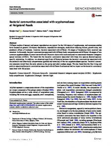
cusp of the aortic valve presented with fibrosis and loss of elasticity. No other alterations were detected macroscopically. Histologically, sections of the aortic valve tissue showed loss of endothelial surface integrity. Van Gieson staining revealed increased numbers of fibroblasts, polymorphonuclear, and mononuclear inflammatory cells. Furthermore, a large collagen connective tissue infiltrate with hyaline degeneration was identified (figure 5). Fibroblasts and inflammatory cells (mononuclear and polymorphonuclear) were also observed with hematoxylin and eosin staining. Cocci colonies were also noticed (figure 6). Gram staining indentified that these bacteria were gram positive cocci (figure 7).
Table 2. Echocardiographic parameters. Parámetros ecocardiográficos. Parameter
#
Patient
Normal Values#
Left ventricular internal diameter at end-systole (LVIDs) (cm)
5.14
6.16 ± 0.98
Left ventricular internal diameter at end-diastole (LVIDd) (cm)
14.40
10.31 ± 1.17
Shortening fracction
51.7%
39.62 ± 5.3%
Eyection fracction
80.2%
67.4 ± 6.81%
Interventricular septal thickness at end-diastole (cm)
2.48
2.07 ± 0.45
Interventricular septal thickness at end-systole (cm)
4.78
4.07 ± 0.55
Left ventricular free wall thickness at end-diastole
2.48
2.16 ± 0.46
Left ventricular free wall thickness at end-systole
5.90
3.57 ± 0.56
= Ligthowler et al 2001, Marr and Patteson 2010.
266
horse, murmurs, echocardiography, left coronary cusp
Figure 5. Photomicrographs of the aortic valve demonstrating hyaline degeneration (white arrow), polymorphonuclear, mononuclear cells (arrow heads) and fibroblast (black arrow). Van Gieson stain, x40 objective. Microfotografía de la válvula aórtica evidenciándose degeneración hialina (flecha blanca), células polimorfonucleares, mononucleares (cabezas de flecha) y fibroblastos (flechas negras). Tinción con Van Gieson, 40x.
Figure 6. Photomicrographs of the aortic valve demonstrating loss of integrity of the endothelium. Notice the mononuclear cell (arrow head) and the presence of bacteria (black arrow). Haematoxylin and eosin stain, x100 immersion objective. Microfotografía en donde se evidencia pérdida de integridad del endotelio valvular. Presencia de células mononucleares (cabeza de flecha) y bacterias (flecha negra). Tinción con hematoxilina y eosina, 100x.
These findings were consistent with a chronic inflammatory process with areas of reactivation. Additionally, the gram positive cocci were observed close to the degenerative process. The postmortem diagnosis was chronic bacterial endocarditis.
Figure 7. Photomicrographs of the aortic valve demonstrating the presence of Gramm positive cocci (+). Gramm stain, x100 immersion objective. Microfotografía de la válvula aórtica en donde se evidencia la presencia de cocáseas Gramm (+). Tinción Gramm, 100x.
DISCUSSION Aortic regurgitation can be associated with exercise intolerance, fatigue, and weight loss (Marr 2010), all of which were observed in the horse presented in this report. Aortic regurgitation also can be associated with congestive heart failure (CHF) (Marr and Bowen 2006); nevertheless, no signs of congestive heart failure were detected in this horse. Otherwise, the presence of the decreasing, pandiastolic, grade 4/6 murmur with its point of maximal intensity in the aortic area, agrees with the findings reported by Reef and Spencer (1987) who determined that the presence of a murmurs grade 2–5/5 with a point of maximal intensity in the aortic area is clear sign of aortic insufficiency. Valvular heart disease is the most common form of cardiac disease in the horse, and a large number of horses present with this pathology at ≥ 16 years of age similar to the case presented in this article. Age was also found to be a significant risk factor associated with left-sided valvular insufficiencies such as aortic and mitral insufficiency; nevertheless, the aortic valve appears to be most frequently affected (Marr and Bowen 2006). Primary aortic regurgitation etiology can be infectious or noninfectious (Marr and Bowen 2006; Marr 2010). Cases of bacterial endocarditis have been reported in horses, but noninfectious cases associated with myxomatous noninflammatory infiltrate and fibrosis are more commonly reported in geriatric horses, similar to humans (Bishop et al 1966, Agozzino et al 1994, Marr and Bowen 2006). Bacterial endocarditis is an infrequent condition (Marr and Bowen 2006) caused by bacteria colonization of the valves (more frequently) or the ventricular wall (Maxson and Reef 1997, Sage 2010). The principal agents involved are Pasteurella/Actinobacillus spp., 267
Dörner ET AL
Streptococcus spp. (Maxson and Reef 1997), Borrelia burgdorferi but it can also be caused by fungal infections associated with Aspergillus and Candida (Sage 2010). Valvular endothelial damage, which results from the impact of high-speed blood flow and forces arising from a high-pressure chamber to a low-pressure chamber, causes endocardial alteration that generates an excellent environment for platelet-fibrin complex storage. These platelet-fibrin complexes are more receptive to bacterial colonization than its healthy endothelium. Consequently, granulations develop on the valvular surface, resulting in structures called vegetations (Sage 2010) that can be seen ultrasonographically (Maxson and Reef 1997; Marr and Patteson 2010). However, vegetations were not seen in our patient. Endocarditis normally presents with fever, murmur, tachypnea, tachycardia, hyperfibrinogenemia, anemia and leukocytosis (Maxson and Reef 1997, Seco Diaz et al 2000, Freoehlich et al 2006). In our patient, a murmur was the only specific clinical sign related with heart disease, so we did not initially suspect bacterial endocarditis. Possibly, no abnormalities in laboratory tests were noticed because the infectious inflammatory process in the aortic valve was controlled and inactive. This situation was unable to generate an organic response that could be reflected in the CBC or biochemical profile. Bacterial endocarditis must be treated with antibiotics. The antibiotic selection and the treatment length (5-6 weeks) are very important. The objective is the selection of an antibiotic based on the sensitivity shown by the antibiogram, but treatment generally begins with an empirical wide-spectrum antibiotic until the hemoculture results are ready (Sage 2010). The antibiotics commonly used in these cases are penicillin associated with gentamicin (Seco Diaz et al 2000, Sage 2010); nonetheless, trimethoprim-sulfadiazine, ampicillin, metronidazole, oxytetracycline, and rifampicin have also been used. Treatment duration is going to depend of the improvement and resolution of clinical signs, echocardiographic examination, white blood cells, and fibrinogen reduction within normal limits. Unfortunately, the prognosis of infectious endocarditis at the aortic valve is poor, and few cases of effective treatment have been reported (Maxson and Reef 1997). Moreover, if antibiotic treatment is successful, the regurgitation may still persist and could generate future heart failure (Sage 2010). Regrettably, in the described case, the authors could not reach the definitive diagnosis of bacterial endocarditis ante-mortem due to the unspecific disease presentation, which is why no antibiotic therapy was administered. Our patient did not present clinical signs of rightsided heart failure as pectoral edema, jugular distension, tachycardia, systemic hypotension, attributed to a ventricular overload that leads to congestion of the systemic circulation, or left-sided heart failure as pulmonary edema, dyspnea, tachycardia, pale mucous membranes, 268
weakness, syncope, or signs of renin-angiotensin-aldosterone system activation, attributed to both increased pressure in the pulmonary veins and reduced cardiac output. The signs that might indicate some cardiovascular compromise like exercise intolerance, weight loss were unspecific and may correspond to many other diseases that are not necessarily cardiovascular in nature. Additionally, no changes were detected by laboratory tests. Without any doubt, color-flow Doppler ultrasonography is the best instrument to diagnose alterations in blood flow inside the heart and blood vessels (Blissit and Bonagura 1995a; Blissit and Bonagura 1995b, Bekeredjian and Grayburn 2005). In this case, right parasternal echocardiography showed a nodule in the left coronary cusp of the aortic valve (figure 3). Furthermore, marked aortic regurgitation of blood from the aorta into the left ventricle was observed (figure 4). The changes in the cardiac parameters observed in the left ventricle M-mode echocardiography (table 2) reflect the heart adaptation to volume overload of the left ventricle due to excessive blood reflux from the aorta to the left ventricle; for this reason, a hyperkinetic cardiac motion would be expected. It has also been described that horses with aortic regurgitation are more likely to have ventricular dysrhythmias compared to horses with other forms of valvular regurgitation, which appears to be independent of aortic regurgitation severity, while supraventricular premature depolarizations are more commonly observed in horses with severe aortic regurgitation (Marr 2010). In the present case, auscultation revealed an “added heart sound” audible at the end of diastole closely preceding the first cardiac tone; in addition, a rhythm abnormality with intervals of cardiac silence was detected. Rest and post-mild exercise ECG tests were performed to further investigate the intracardiac electrical activity. No ventricular arrhythmias were observed in the ECG, and no abnormal findings were detected that were suggestive of splitting of the first cardiac tone (like atrial fibrillation or third degree AV block). Nevertheless, sinoatrial arrest was detected. Considering that sinoatrial arrest prolong the period of atrial filling, the “added heart sound” was attributed to accentuation of the fourth cardiac tone (S4), and the arrhythmia was classified as physiological due to a high vagal tone; however, it is logical to think that the aortic regurgitation should theoretically activate several compensatory processes, including activation of the sympathic nervous system and the renin-angiotensin-aldosterone system, circumstances not seen in this case. On the other hand, it is not common to find sinoatrial arrest in horses presenting a heart rate over 30 beats per minute like we observed in our case (36 beats/min.). Nevertheless, this finding can be explained due to activation of the baroreceptors located in aortic callus and carotid sinus as a compensatory mechanism to maintain the stroke volume and blood pressure partially masking the vagal tone.
horse, murmurs, echocardiography, left coronary cusp
Surgery is the indicated treatment for acute aortic insufficiency and severe chronic aortic insufficiency in humans. There are no published experiences or studies about valve replacement procedures in horses. Horses with chronic aortic insufficiency may remain compensated for long periods of time; despite that, the disease can evolve to heart failure (Marr 2010). This situation might have been expected in the described case; however, it is remarkable that this had not already happened considering the age of the patient. Importantly, the goal of medical therapy is to significantly reduce systolic blood pressure to relieve the afterload. The patient in this report presented with unspecific symptoms, furthermore, no signs of any systemic compensatory mechanisms were detected, and the case was managed conservatively. The patient was evaluated 2 times after the initial examination and no disease progression was noted. At necropsy, 2 nodules were observed in the left coronary cusp of the aortic valve, a finding that agrees with the image obtained by ultrasonography. The valve presented loss of elasticity a firm texture, findings that were suggestive of a degenerative process and valvular fibrosis, situations that are commonly seen in animals with regurgitation (Oldershaw et al. 1980). Histologically, numbers of fibroblast cells, inflammatory cells, and gram positive cocci increased. These findings led us to diagnose the patient with chronic bacterial endocarditis. Considering that bacterial endocarditis of the sigmoid aortic valve is more frequently observed in younger and half age patients, it is concluded that the disease can also be detected in older horses. However, the clinical presentation may differ from the manifestations in younger horses that have been described in the literature. Murmurs associated with aortic insufficiency, normal blood tests, and the absence of other specific signs like in the case here can be observed in cases of bacterial endocarditis; hence, this disease should be considered a differential diagnosis when horses present for evaluation with these characteristics. REFERENCES Agozzino L, F De Vivo, A Falco, L Tupputi Schinosa, M Cotrufo. 1994. Non-inflammatory aortic root disease and floppy aortic valve as cause of isolated regurgitation: a clinicomorphologic study. Int J Cardiol 45, 129-134. Bekeredjian R, P Grayburn. 2005. Valvular heart disease: aortic regurgitation. Circulation 112, 125-134. Bishop S, C Cole, D Smetzer. 1966. Functional and morphologic pathology of equine aortic insufficiency. Vet Pathol 3, 137-158. Blissitt K, J Bonagura. 1995a. Colour flow doppler echocardiography in horses with cardiac murmurs. Equine Vet J Suppl 19, 82-85. Blissitt K, J Bonagura. 1995b. Colour flow doppler echocardiography in normal horses. Equine Vet J Suppl 19, 47-55.
Blissitt K. 2010. Auscultation. In: Cardiology of the Horse. 2nd ed. Saunders. New York, USA, Pp 91-104. Dörner C. 2009. Evaluación electrocardiográfica de equinos fina sangre de carrera clínicamente sanos en período de amansa. Memoria de título. Facultad de Ciencias Veterinarias y Pecuarias, Universidad de Chile, Santiago, Chile. Froehlich W, S Wlaschitz, K Riedelberger, V Reef. 2006. Case report: tricuspid valve endocarditis in a horse with a ventricular septal defect. Equine Vet Educ 8, 222-227. Godoy A. 1977. Correlación entre el examen clínico cardiológico y los hallazgos de necropsia de equinos llevados a matadero. Memoria de título. Facultad de Ciencias Veterinarias y Pecuarias, Universidad de Chile, Santiago, Chile. Henneke D, G Potter, J Kreider, B Yeates. 1983. Relationship between condition score, physical measurements and body fat percentage in mares. Equine Vet J 15, 371-372. Kriz N, Hodgson D, Rose R. 2000. Prevalence and clinical importance of heart murmurs in racehorses. J Am Vet Med Assoc 216, 1441-1445. Lightowler CH, MC Mercado, JA Garcia-Liñeiro. 1998. Ventanas y ecotomogramas de referencia para la ecocardiografía bidimensional en tiempo real del equino. Arch Med Vet 30, 143-151. Lightowler CH, G Pidal, M Cattáneo. 2001. Quantitative study of equine echcardiography. Avances en Ciencias Veterinarias 16, 21-31. Long K, J Bonagura, P Darke. 1992. Standardized imaging technique for guided m-mode and doppler echocardiography in the horse. Equine Vet J 24, 226-35. Marr C, V Reef. 1995. Physiological valvular regurgitation in clinically normal young racehorses: prevalence and twodimensional colour flow doppler echocardiographic characteristics. Equine Vet J Suppl 19, 56-62 Marr C, M Bowen. 2006. Cardiac diseases in the geriatric horse. In: Bertone JJ (ed). Equine Geriatric Medicine and Surgery. Saunders, New York, USA, Pp 39-49. Marr C. 2010. Cardiac murmurs: valvular regurgitation and insufficiency In: Marr C, Bowen M (eds). Cardiology of the Horse. 2nd ed. Saunders, New York, USA, Pp 207-216. Marr C, M Patteson. 2010. Echocardiography. In: Marr C, Bowen M (eds). Cardiology of the Horse. 2nd ed. Saunders. New York, USA, Pp 105-126. Maxson A, V Reef. 1997. Bacterial endocarditis in horses: ten cases (1984 - 1995). Equine Vet J 29, 394-399. Oldershaw P, I Brooksby, M Davies, D Coltart, B Jenkins, M Webb-Peploe. 1980. Correlations of fibrosis in endomyocardial biopsies from patients with aortic valve disease. Br Heart J 44, 609-611. Patteson M, P Cripps. 1993. A survey of cardiac auscultatory findings in horses. Equine Vet J 25, 409-415. Reef V, C Marr. 2010. Dysrhythmias: assessment and medical management. In: Cardiology of the Horse. 2nd ed. Saunders, New York, USA, Pp 159-178. Reef V, P Spencer. 1987. Echocardiographic evaluation of equine aortic insufficiency. Am J Vet Res 48, 904-908. Sage A. 2010. Fever: endocarditis and pericarditis. In: Marr C, Bowen M (eds). Cardiology of the Horse. 2nd ed. Saunders, USA, Pp 217-225. Seco Diaz O, M Sleeper, V Reef, M Acland. 2000. Aortitis in a paint gelding. Equine Vet J 32, 354-357.
269