pattern in normal human kidneys, and was decreased in the sclerosed areas of glomeruli. An immunogold technique revealed the expression of LDL receptors ...
Kidney International, Vol. 43 (1993), PP. 918—927
Apolipoproteins and lipoprotein receptors in glomeruli in human kidney diseases TSUKASA TAKEMURA, KAZUO Y0sHI0KA, NAOBUMI AYA, KATSUMI MURAKAMI, AKIY0 MATUMOTO, HIR0sHIGE ITAKURA, TATSUHIKO KODAMA, HIR0sHI SUZUKI, and SUNAO MAKI Department of Pediatrics, Kinki University School of Medicine, Osaka-sayama; and Division of Clinical Nutrition, National Institute of Health and Nutrition, Tokyo, The Third Department of Internal Medicine, University of Tokyo, and Laboratory Animal Center, Chugai Pharmaceutical, Tokyo, Japan
Apolipoproteins and lipoprotein receptors in glomeruli in human kidney diseases. This study offers morphological evidence of the involvement of lipid abnormalities in human glomerular injury. Renal biopsy tissues from patients with several types of glomerular diseases were immunocytochemically examined using antibodies to apolipoproteins (apo) A-I, B-100, and E, and antibodies to low density lipoprotein (LDL) receptors and scavenger receptors. Immunofluorescent staining
lar lipid deposition and progressive renal insufficiency [4]. Abnormal lipid accumulation is found within glomeruli and tubular cells in various types of human nephritis [5, 6]. Foam
showed the predominant deposition of apo B and apo E in the mesangial area in mesangial proliferative types of glomerulonephritis; the distri-
express surface receptors for low density lipoprotein (LDL) [8, 91, and could, therefore, be affected by abnormal lipid metabolism. Experimentally, glomerular injury, including increased mesangial matrix and cellularity, followed by the appearance of albuminuria occurs with diet-induced hyperlipidemia [10] and with endogenous abnormalities in lipid metabolism [111. The mechanisms of lipid-induced glomerular injury are still
cells, believed to be lipid-laden macrophages, have been iden-
tified in kidney vessels and in the interstitium as well as in glomeruli [7]. Mesangial cells and glomerular epithelial cells
bution and staining intensity of these apolipoproteins correlated with the grade of mesangial proliferation and proteinuria, but were independent of plasma lipid levels. Immunoelectron microscopy revealed that
apo B and apo E were distributed in droplets within glomerular epitheial and mesangial cells or in a granular pattern in the expanded mesangial matrix. Apo A-I was mainly localized in the visceral epithehal cells of normal human kidneys. Staining for apo A-I was increased in the glomerular epithelial cells of nephritic kidneys, compared to the pattern in normal human kidneys, and was decreased in the sclerosed areas of glomeruli. An immunogold technique revealed the expression of LDL receptors on the surface membranes of glomerular mesangial
incompletely understood. The apparently infrequent occurrence of renal disease in patients with the most common forms
of hyperlipidemia suggests that lipids may cause clinically
important renal injury only when additional predisposing factors, such as immune-mediated glomerular cell damage, are uli. Scavenger receptor was detected on the plasma membranes of present. Based on certain histologic similarities, analogous mesangial and visceral epithelial cells. The glomerular expression of pathobiologic mechanisms have been suggested for glomeruloscavenger receptor was increased in glomeruli with marked mesangial sclerosis and atherosclerosis [121. Modified LDL, which is proliferation. In addition, the expression of this receptor was intense in taken up by tissue macrophages via scavenger receptors and is monocytes/macrophages occasionally infiltrating the glomeruli. Our present findings indicate that in human nephritic kidneys, glomerular an important pathogen in atherosclerosis [13, 141, has been epithelial and mesangial cells express both LDL receptors and scaven- shown to be cytotoxic to cultured mesangial cells [15, 16], and ger receptors. The accumulation of apolipoproteins, whether receptorcould contribute to glomerular damage in nephritis, although mediated or mediated by other mechanisms, can occur independently of plasma lipid levels, and may be associated with mesangial expansion the precise mechanism by which this could occur is unknown. To further delineate the role of lipid abnormalities in human and proteinuria. glomerular injury, we immunocytochemically evaluated the glomerular expression of LDL and scavenger receptors, as well as evaluated the renal deposition of the predominant plasma Accumulating evidence indicates that abnormalities of lipid lipoproteins A-I, B-l00, and E, which are essential structural metabolism are responsible for the progression of glomerular components of high density lipoprotein (HDL), LDL, and very damage in both immunological and non-immunological types of low-density lipoprotein (VLDL), respectively. renal diseases [1—3]. A hereditary form of lecithin cholesterol acyltransferase deficiency has been reported to cause glomeruMethods and epithelial cells. Dual immunofluorescent staining showed that apo B and LDL receptors were occasionally co-localized in nephritic glomer-
Patients and tissues Received for publication April 17, 1992 and in revised form October 9, 1992 Accepted for publication November 23, 1992
percutaneous needle biopsy or surgical biopsy from 74 patients
© 1993 by the International Society of Nephrology
with various types of kidney diseases: 30 patients with IgA
The kidney tissues used in this study were obtained by
918
919
Takemura et al: Apo and LDL receptors in human glomeruli Table 1. Antibodies used in this study
Specificity
Apo A-I Apo B-i0O
Apo E
LDL receptor Human scavenger receptor Human monocyte/macrophages Type IV collagen (7S domain)
Antibody MoAb (ABAI-1) MoAb (AB B-3) TRIC-labeled MoAb (AB E-i) TRIC-labeled MoAb hSRI-2 FMC 32 (CDT4) Anti-Leu-M3 (CD14) MoAB (MAb TV-i)
Source
Produced by mouse IgG1
mouse IgG2 goat mouse IgG1
goat mouse IgG21
rabbit
mouse IgG
mouse IgG2 mouse IgG
Canadian Bioclinical Canadian Bioclinical Binding site Canadian Bioclinical Binding site Amersham Matsumoto et a! [14] AMD Becton-Dickinson Scheinman and Tsai [19, 34]
Abbreviations are: MoAb, monoclonal antibody; TRIC, tetramethylrhodamin; AMD, Austrarian Monoclonal Development.
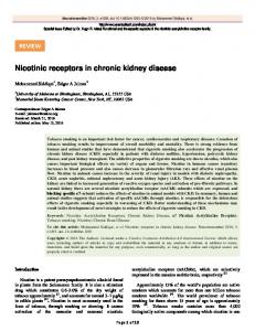
Fig. 1. Indirect immunofluorescent staining with monoclonal antibodies to apo B-100 (A—C) and apo E (D) in patients with IgA nephritis with mild (A) or marked (B and D) mesangial proliferation, and in a patient with focal glomerular sclerosis (C). A. Apo B is visible in the mesangium, and
staining is weak along the glomerular capillary walls (immunofluorescence score 1). B. Mesangial deposition of apo B is prominent (immunofluorescence score 2). C. Intense staining for apo B is seen in an almost globally sclerotic glomerulus (immunofluorescence score 2). D. Positive staining for apo E is prominent in the mesangial area. Original magnification: A—D x400.
nephritis, 15 with mesangial proliferative (non-IgA) glomerulonephritis, 10 with Henoch-Schonlein purpura nephritis (HSPN),
10 with minimal change nephrotic syndrome, 4 with lupus nephritis (WHO Class IV), and 5 with nephrotic syndrome with focal glomerular sclerosis. Laboratory data including urinalysis, 24-hour urinary protein excretion and creatinine clearance
were collected for each patients. Plasma levels of lipids and apoproteins were determined using fasting serum samples obtained from 28 patients with mesangial proliferative glomerulo-
nephritis, including 18 patients with IgA nephritis. Lipids and apoproteins were measured by standard procedures currently used in Japan; total cholesterol and triglyceride were assayed with an autoanalyzer, utilizing enzymatic methods. HDL cholesterol was measured after precipitation of LDL and VLDL cholesterols by a heparin-manganase reagent. Apo A-I, apo B and apo E were immunoturbidometrically determined using the reagent kits (ApoAuto "Daiichi", Daiichi Pure Chemical, Tokyo, Japan) [17].
920
Takemura et al: Apo and LDL receptors in human glomeruli
Table 2. Results of apo B, apo E, LDL receptor and scavenger receptor immunofluorescent staining in glomeruli of patients with various types of glomerular diseases
Disease/Mesangial proliferationa
Total no. of patients
Apo B"
Apo E"
18/30 3
15/30
2+
30 10 14
3+
6
9 6
15
10/15
5 6 4
2 4 4
10 3
3/10
IgA nephritis
1+
Mesangial proliferative (non-IgA) nephritis
1+
2+ 3+ Henoch-SchOnlein purpura nephritis
1+
2+ 3+ Nephrotic syndrome Minimal change Focal glomerulosclerosis Lupus nephritis (WHO Class IV) Total
6
0 2
1
1
10 5
2 7 6 7/15
7/30
Scavenger receptorc 3/15
1
0
3 3 5/15
2 2/7
1
2 2
0 0 2
1/10
1/5
1
0 0
0 0
1
1
1
1
3 3 2/10 0
0/10 4/5
1/10 4/5
Staining in glomeruli LDL receptorb
1
1/10 3/5
1/6
4
3/4
3/4
2/4
2/5 2/4
74
39/74
31/74
19/74
11/42
a Mesangial proliferation was graded as mild (1+), moderate (2+) or marked (3+) b Number
of patients with positive staining/number of patients examined by immunofluorescence Number of patients whose kidney tissues showed increased intensity and distribution of staining compared with normal human kidneys/number of patients examined by immunofluorescence
Three other kidney specimens were obtained; these were lyze the relationship between apoprotein deposition and histofrom patients with renal calculi or renal trauma, and histologi- logic changes, plasma lipid profile and clinical findings, the
cally normal portions were used as normal human kidney patients were divided into three groups: Group 1, negative tissue. For light microscopic observation, the tissues were fixed immunofluorescence (score 0); Group 2, weak immunofluoresin 10% buffered formalin and embedded in paraffin. Three cence (mean score 0< 1.5); and Group 3, strong immunoflu-
sm-thick sections were stained with hematoxylin and eosin,
orescence (mean score 1.5< 2).
periodic acid-Schiff, and methenamine silver. Mesangial prolif-
As controls, kidney sections were incubated with nonimmune
eration was graded as described previously [18]. Cryostat
sera or with unrelated mouse IgG monoclonal antibody, followed by incubation with FITC- or TRIC-labeled goat antimouse IgG, or with secondary antibody alone. These controls were negative. Immunoelectron microscopy was performed using pre-embedding and post-embedding method as described previously [21, 23]. For pre-embedding staining [21], tissues were fixed in paraformaldehyde-lysine-periodate fixative and then embedded in OCT compound. Then they were incubated with the primary antibodies, followed by reaction with the appropriate secondary
sections were stained for neutral lipids with oil red 0.
Antibodies The monoclonal and polyclonal antibodies used in this study are listed in Table 1. Antibody to human scavenger receptor was produced by immunizing rabbits with bovine serum albumin-coupled synthetic peptide (hSRI-2) corresponding to the C-terminus of the collagen-like domain [14]. The following antibodies were purchased from Cappel (Malvern, Pennsylvania, USA): F(ab')2 fragments of goat anti-mouse IgG labeled antibody labeled with peroxidase. The sections were then
with fluorescein-isothiocyanate (FITC) and tetramethyl- treated with diaminobenzidine/hydrogen peroxide solution, rhodamin (TRIC), F(ab')2 fragments of goat anti-rabbit IgG post-fixed with osmic acid, and embedded in Epon 812. Ultralabeled with FITC, and goat anti-mouse IgG labeled with thin sections prepared by microtome were observed under an peroxidase. Goat anti-mouse IgG conjugated with colloidal gold electron microscope. For the post-embedding method [23], (particle size: 15 nm) was obtained from E-Y Laboratories (San specimens were embedded in Lowicryl K4M (Chemische Werke Lowi GmbH, Waldkreiburg, Germany). Ultra-thin secMateo, California, USA). tions, placed on 200-mesh nickel grids, were incubated with Immunocytochemistry primary antibodies and then reacted with secondary antibodies Indirect immunofluorescence and dual staining were per- labeled with gold particles. The sections were then fixed in 0.2% formed as described previously [20—23]. In the dual staining, glutaraldehyde/0.2 M sodium cacodylate buffer, stained with FITC-labeled goat anti-mouse IgG and TRIC-labeled goat anti- uranyl acetate, and viewed under an electron microscope. rabbit IgG were used. The number of glomeruli evaluated Statistical analysis ranged 1 to 10 (mean, 3.4). The intensity and distribution of fluorescence in each glomerulus were scored as negative (score The correlation between the grade of apoprotein immunoflu0), and 1+ positive (score 1) or 2+ positive (score 2) (Fig. 1, A, orescence and that of mesangial proliferation was assessed B and C). The mean score (the sum of scores/the number of using the chi-square test. The relationship between apoprotein glomeruli evaluated) was calculated for each patient. To ana- immunofluorescence and plasma levels of total cholesterol,
921
a-
Takemura et a!: Apo and LDL receptors in human glomeruli
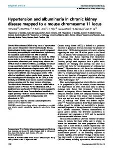
Fig. 2. Ultrastructural observation of apo B
in glomeruli of patients with focal glomerular sclerosis (A) and IgA nephritis (B), stained with monoclonal antibody to apo B-100 and peroxidase-labeled secondary antibody. A. Apo B-positive droplets are seen within the cytoplasm of visceral epithelial cells (arrowheads). B. Granular reaction products (asterisks) accumulated in the mesangial matrix. Original magnification: A x8,000, B x4,000. Abbreviations are: CL, glomerular capillary lumen; EP, glomerular epithelial cell; MC, mesangial cell.
triglyceride, HDL cholesterol, apo A-I, apo B, and apo E, and 24-hour urinary protein excretion and creatinine clearance was evaluated using the Kruskal-Wallis test. A value less than 0.05 was considered statistically significant. Results
Apolipoproteins Apo B and apo E immunofluorescence were not detected in
the glomeruli of normal human kidneys, but were detected focally in the media of arterioles and peritubular capillaries. These apolipoproteins were frequently observed in the glomeruli of patients with IgA nephritis, mesangial proliferative (nonIgA) nephritis, Henoch-SchOnlein purpura nephritis, focal gb-
merular sclerosis and lupus nephritis, but were rarely observed in minimal change nephrotic syndrome (Table 2). Apo B and apo E were localized predominantly in the mesangium (Fig. 1), and the distribution and intensity of their fluorescence conelated significantly with the grade of mesangial proliferation (P < 0.05). Apo B (Fig. 1C) and apo E were also stained in sclerosed areas of glomeruli. Immunoelectron microscopy revealed that apo B- and apo E-positive droplets were present in the cytoplasm of glomerular epithelial cells in focal glomerular sclerosis (Fig. 2A), and in the cytoplasm of mesangial cells in mesangial proliferative types of glomerulonephritis. In addition, apo B (Fig. 2B) and apo E were found as amorphous aggregations in the mesangial area.
922
Takemura et a!: Ape and LDL receptors in human glomeruli
Fig. 3. Immunofluorescent micrographs of ape A-I in normal and nephritic kidneys. A. Apo A-I is stained dominantly in the glomerular epithelial cells of the normal human kidney. B. Ape A-I staining is increased in a glomerulus of minimal change nephritic syndrome. C and D. Dual staining of apo A-I (C) and laminin (D) in a glomerulus of focal glomerular sclerosis. Apo A-I staining is decreased in the sclerosed area (arrows) where laminin staining is intense. Original magnification: A—D x400.
Fig. 4. Dual immunofluorescent staining of LDL receptor (A) and apo B (B) in a glomerulus of a patient with IgA nephritis. Mesangial
co-localization of LDL receptor and ape B is seen only in the sites stained intensely with apo B (arrows).
Monoclonal antibody to apo A-I stained glomerular epithelial cells and weakly stained the cytoplasm of some tubular epithehal cells in normal and nephritic human kidneys. The intensity of the immunofluorescence was increased in the glomeruli of patients with minimal change nephrotic syndrome, focal gbmerular sclerosis, and mesangial proliferative types of gbomer-
(Fig. 3). Post-embedding immunoelectron microscopy revealed that ape A-I was localized in the cytoplasm and foot processes of visceral epithelial cells.
Oil red 0 staining was performed in 20 sections, and the results were compared with the immunofluorescent staining for apo B. The dye was distributed in a scattered fashion, particu-
ulonephritis compared with normal human kidneys, but the larly in the mesangial areas of glomeruli and in the renal immunofluorescence was reduced or lost in scierosed areas interstitium of nephritic kidneys. The distribution of oil red 0
Takemura et at: Apo and LDL receptors in human glomeruli
923
Fig. S. Immunoelectron microscopic
photographs of LDL receptor in a patient with IgA nephritis. A. Colloidal gold staining. Gold particles are distributed on the surface of foot processes of visceral epithelial cells. There are small clusters of these particles (arrows) in some parts (coated pits ?). B. Peroxidase staining. Reaction products are seen on the mesangial cell surface (arrows). Original magnification: A x5,000, B x3,500. Abbreviations are: BM, glomerular basement membrane; FP, foot process of glomerular epithelial cell; MC, mesangial cell.
staining in the mesangial area was similar to that of apo B mesangium and weakly along the glomerular capillary walls. staining, but was weaker in intensity and less diffuse than the Dual staining showed that LDL receptor was occasionally latter.
Lipoprotein receptors In normal human kidneys, LDL receptor immunofluorescence was visible in the cytoplasm of tubular epithelial cells, but was negative in the glomeruli. LDL receptor was positive, however, in the glomeruli of some patients with glomerular
disease (Table 2). It was distributed predominantly in the
co-localized with apo B in the glomeruli, especially in the sites
where apo B staining was intense (Fig. 4). Immunoelectron microscopy showed that LDL receptor was expressed on the membrane surface of visceral epithelial cells, as well as on mesangial cells (Fig. 5). Weak immunofluorescence for scavenger receptor was detected along the glomerular capillary walls and mesangium of normal human kidneys. In some of nephritic kidney tissues, the
Takemura et al. Apo and LDL receptors in human glomeruli
'4,
S
924
Fig. 6. Immunofluorescent photographs of scavenger receptor in patients with IgA nephritis. Dual staining for scavenger receptor (A) and type IV collagen 7S domain (B) in the same tissue section exhibit distribution of the receptor in the mesangium (arrows) and along the glomerular capillary walls. Monocyte/macrophage staining in the serial section containing the same glomerulus was negative. Dual staining for scavenger receptor (C) and monocytes/macrophages (D). The scavenger receptor is intensely expressed by monocytes/macrophages within the glomerulus (arrows), and is weakly expressed in other parts of the glomerulus. Original magnification: A—D x400. Abbreviation is: G, glomerulus.
expression of scavenger receptor was enhanced in these loci glomerular apo B or apo E deposition and plasma levels of total (Table 2, Fig. 6 A and C). Dual immunofluorescent staining with cholesterol, triglyceride, HDL-cholesterol, apo A-I, apo B, or anti-scavenger receptor antibody and anti-type IV collagen (7S apo E. Deposition of apo B and apo E was significantly (P < domain) antibody confirmed mesangial distribution of the re- 0.05) correlated with the patients' 24-hour protein excretion but ceptor (Fig. 6 A and B). The immunofluorescence for scavenger not with their creatinine clearance. Since apo A-I immunofluoreceptor tended to be intense in the glomeruli of patients with rescence was detected in the glomeruli of normal human moderate or marked mesangial proliferation. Immunoelectron kidneys and was reduced or lost in the scierosed glomeruli in microscopic studies further demonstrated that the receptor was nephritis tissues, we did not evaluate the relationship between present on the surface of glomerular epithelial and mesangial apo A-I deposition and patients' laboratory data. cells (Fig. 7 A and B). Dual immunofluorescence for scavenger receptor and monocytes/macrophages revealed strong expres-
sion of the receptor in monocytes/macrophages which are
Discussion
LDL receptors have been demonstrated on most cell types. Culture studies have indicated that rat mesangial cells have nephritis (Fig. 6 C and D). The localization of scavenger specific LDL receptors [7, 9]. Mesangial cells show receptorreceptor was generally discordant with that of apo B. mediated endocytosis of receptor-bound LDL, and high concentrations of LDL are toxic to these cells [15, 16]. Cultured Apolipoprotein deposition and laboratory data human glomerular epithelial cells also have LDL receptors The relationship between apoprotein deposition and labora- which mediate the binding, internalization, and degradation of tory findings in patients with mesangial proliferative types of LDL [8]. Scavenger receptors [13, 141, different from LDL glomerulonephritis, including IgA nephritis and mesangial pro- receptors in both their binding characteristics and protein liferative (non-IgA) glomerulonephritis, was evaluated (Table structure, are expressed in a restricted number of cell types. 3). There was no significant correlation between the grade of Whether glomerular mesangial and epithelial cells possess occasionally infiltrating the glomeruli of lupus nephritis and IgA
925
Takemura et a!: Apo and LDL receptors in human glomeruli
I
I,
us
r
I. BM
t
SS
S
I
Fig. 7. Immunoelectron microscopy of
scavenger receptor in a patient with IgA nephritis. A. Colloidal gold staining for scavenger receptor, displaying labeling of the foot processes of glomerular epithelial cells. B. Peroxidase staining for scavenger receptor, showing labeling of mesangial cells (arrows). Original magnification: A x 12,000, B x6,000 (inset x8,000). Abbreviations are: BM, basement membrane in the paramesangial zone; MES, mesangium; US, urinary space; MC, mesangial cell.
scavenger receptors is unclear, since they apparently did not immunofluorescence study revealed LDL receptor in the gbbind and internalize acetylated LDL [8]. However, recent meruli of some patients with glomerulonephritis, particularly in studies by Coritsidis et al [15] have shown that rat mesangial those with increased mesangial proliferation, although this cells in culture avidly bind and take up oxidized LDL to a receptor was not detected in normal glomeruli. The expression greater extent than native LDL. Our present study immunocytochemically demonstrated the expression of both LDL receptor and scavenger receptor on the glomerular epithelial and mesangial cells of human kidneys. Our
of scavenger receptor was observed in normal glomeruli, and was increased in the glomeruli of nephritic kidneys. The gbmerular expression of LDL receptors was occasionally colocalized with apo B-lOO, a ligand for the LDL receptor. These
926
Takemura et a!: Apo and LDL receptors in human giomeruli
Tab'e 3. Analysis of the relationship between glomerular apoprotein deposition and plasma lipid level or other laboratory findings in patients
with mesangial proliferative type of glomerulonephritis Plasma lipid level mg/dl T-C
Trig
Apoprotein B deposition
Group I (N = 3) Group 2 (N = 8) Group 3 (N = 17)
P
Apoprotein E deposition Group 1 (N = 9) Group 2 (N = 9) Group 3 (N= 10)
P
154
37
148 188
42 48
75 91
NS 157 155 198
NS
41 41 51
7,8
68 27 30 NS
71
77
78
14
96 37 NS
HDL-C 61
67
0.7 12
63 9.2 NS
65
12
Apo A-I 131
120 131
14 23 20
23
63
131
51
NS
2.3
4.7
75
14
3.7
80 22 NS
123 129
NS
25
Apo E
75
NS
65 6.1 11
Apo B
72 72
3.8
17
4.3 4.8
0.9 0.8
10
89 23 NS
4.6 NS
1.6 0.5 1.4
1.8
NS
U0,
g124 hr
0.55 0.85 1.8
1.73 m2
0.21
95
3.1
0.28 1.4
89
12