Indian Journal of Experimental Biology Vol. 45, February 2007, pp. 160-165
Review Article
Application of nanotechnology in biomedicine Rajshri M Navalakhe & Tarala D Nandedkar National Institute for Research in Reproductive Health, Jehangir Merwanji Street, Parel, Mumbai, 400012, India
Nanotechnology is the development of engineered devices at the atomic, molecular and macromolecular level in nanometer range. Nanoparticles have potential application in medical field including diagnostics and therapeutics. Nanotechnology devices are being developed for diagnosis of cancer and infectious diseases which can help in early detection of the disease. Advances in nanotechnology also proved beneficial in therapeutic field such as drug discovery, drug delivery and gene/protein delivery. Nanoparticles can be constructed by various methodology so that effect can be targeted at desired site. In this review, some of the applications of nanoparticles in medicine as diagnostics and therapeutics which can be employed safely at the clinical level have been described. On other hand, as the particles become generally smaller their likehood of causing harm to the lung increases. Therefore, there is a need to study safety of nanoparticles. Keywords: Cancer, Diagnosis, Drug delivery, Nanoparticles, Nanotoxicity, Therapy
“Nanotechnology” is the creation of very small particles, devices and systems; the technology takes place at a very minute level. One nanometer is one billionth of a meter, the width of about 5 atoms. These materials and devices can be designed to interact with cells and tissues at a molecular (i.e. subcellular) level with high degree of functional specificity, thus allowing integration between the device and biological system, not previously attainable1. The emerging field of nanotechnology is not a single scientific discipline but it involves scientists from many different areas including physicists, chemists, engineers and biologists. In this review, the emerging technology being developed for medical application for diagnostic and therapeutic purposes has been reviewed.
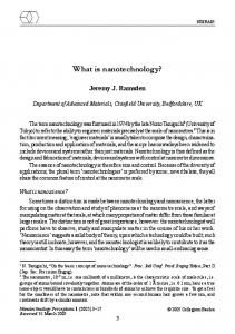
Challenges associated with nanotechnology Today, Nanotechnology is gaining importance in biology due to its small size and targeted effects. Nanoscale devices are 100-10,000 times smaller than the human cell. Because of their small size and larger surface area relative to their volume, nanoscale devices can readily interact with biomolecules (such as enzymes and receptors) on both, the surface of the cell and inside the cell. By gaining access to various areas of the body, nanoparticles have the potential to detect disease at the micro level and deliver treatment. Work is currently being conducted to find ways to safely move these new research tools into clinical practice (Fig.1). Nanoparticles, for example can have multiple functionalities that can provide detailed information on the progression of disease.
A brief overview of nanotechnology and different tools of nanotechnology available for diagnosis and targeting of diseases has been covered in the first section. The second section focuses on application of nanotechnology to medicine, which is the leading issue in today’s world (i.e. cancer, contraception, drug delivery). The third section comprises a description of toxicology of nanomaterials.
Nanoparticles can be made from a vast range of materials, such as metals (gold, silver), metal oxides, [e.g. titanium dioxide (TiO2), Silicon dioxide (SiO2)], inorganic materials (carbon nanotubes, quantum dots), polymeric materials and lipids2. The other new sets of tools is available in nanotechnology are nanocrystals, cantilevers, dendrimers, nanoshells and nanowires. These particles can range from few to several hundreds of nanometers in diameter. Products made from each of these tools can be used for diagnosis (as biomarkers) and therapy.
_____________ *Correspondent author: Phone: 91-22-24192116 Fax: 91-22-24139412 Email:
[email protected]
As reported by Service3 nanotechnology in just 5 years has developed from being a specialty of physicists and chemists to a worldwide scientific and
NAVALAKHE & NANDEDKAR: APPLICATION OF NANOTECHNOLOGY IN BIOMEDICINE
161
Fig. 1⎯ Nanotechnology: multiple applications
industrial enterprise. Currently most of the research work focuses on the use of nanoparticles to treat diseases such as cancer, HIV and diabetes and as carrier for drug delivery (Table 1). Didenko and Baskin4 have described an enzymatic approach for labeling nanotubes with quantum dots. The labeling was performed via enzymatic biotinylation of nanotubes in the tyramide-horseradish peroxidase (HRP) reaction. They achieved both direct and indirect fluorescent labeling of single walled carbon nanotubes (SWNTs) using either biotinyltyramide or fluorescently tagged tyramides. Linking semiconductor nano crystals, quantum dots (Q-dots) on the surface of nanotubes resulted in their fluorescent visualization, whereas conventional fluorophores bound to SWNTs directly or through biotin-streptavidin linkage were completely quenched. Using this approach other organic molecules such as proteins, antibodies or DNA can be conjugated to biotinylated SWNTs, which could be useful for a number of biomedical applications. Nanotechnology and its application to cancer Cancer, a major killer disease is a complex sequence starting from diagnosis till therapy. Currently detection and diagnosis of cancer usually depends on changes in cells and tissues which occur at the nanoscale level inside the cells and are detected either by physical examination or imaging expertise. Scientists would now like to make it possible to detect cancer when the earliest molecular changes occur. Detecting cancer at an early stage before it spreads, completely changes the scenario for treatment of most cancer. Nanotechnology offers a wealth of tools that provide cancer researchers with new and innovative ways to diagnose and treat cancer.
Table 1⎯ Use of nanoparticles in medicine Application
Property
Drug Delivery More effective treatment with existing drug, controlled release of drug Targeted effect Cancer Early Diagnosis, Targeted effects to tumor cells but not healthy cells HIV/AIDS Agent targeted to desired site Evading body’s immune system Diabetes Monitor conditions and acts as an artificial means of regulating and maintaining body’s own hormonal balance Contraception Agent targeted to desired site with minimal side effects
Status Early clinical trials
Clinical trials
Early clinical trials At laboratory level
At laboratory level
National Cancer Institute (NCI) US is working on Nanotechnology in Cancer. NCI has also established the nanotechnology characterization laboratory, which will develop a cascade of assays for further product development and regulatory review. Dr. Gregory Downing, Director of NCI5 has described how nanotechnology can help in product development from ‘bench to bedside’ and improve drug discovery efforts by addressing the complexities of cancer. Nanoscale devices can deliver multiple therapeutic agents to a tumor in order to simultaneously attack multiple points in the pathway involved in cancer. Similarly, nanotechnology generates in vivo biosensors that have the capability of detecting and pointing the location of tumor and metastatic changes that are smaller than those detectable using conventional technologies. Diagnosis Nanodevices such as nanowires and cantilevers can provide rapid and sensitive detection of cancer related
162
INDIAN J EXP BIOL, FEBRUARY 2007
molecules by enabling scientists to detect molecular changes even when they occur only in a small percentage of cells. This would help in early detection of cancer. The attachment of nanomaterials to the molecule of interest can be used as diagnostic markers. The cantilever is one tool with potential aid in cancer diagnosis. Nanoscale cantilevers-tiny bars anchored at one end can be engineered to bind to molecules associated with cancer. When the cancer associated molecule binds to the cantilevers, it changes the surface tension causing the cantilever to bend. By monitoring whether the cantilevers are bent and to what extent, scientists can assess, whether the cancer molecules are present6. One tool Quantum dot can detect early DNA changes in the body. Quantum dots are tiny crystals that glow when they are stimulated by ultraviolet light. The wavelength or color of the light depends on the size of the crystal. Latex beads filled with these crystals can be designed to bind to specific DNA sequences. When the crystals are stimulated by light, the colors they emit act as dyes that light up the sequences of interest. By combining different size quantum dots within a single bead, researchers can create probes that release a spectrum of various colors and intensities of light serving as spectral bar code. To detect cancer, one can design beads containing quantum dots to bind to the sequence of DNA that is associated with cancerous cells. Research is underway to find out innovative ways at the nanoscale level which could be useful to detect early mutagenic changes. Therapy After diagnosis when it is time to treat cancer, nanoscale devices have the potential to improve cancer therapy other than the existing conventional (chemotherapy, radiotherapy) techniques and also to discover new therapeutic agents. It is useful for developing ways to eradicate cancer cells without harming healthy, neighboring cells. Scientists hope to use this technology to create therapeutic agents that can target specific cells and deliver toxins in a controlled, time released manner6. The ultimate goal of researchers is to find out agents of these nanoparticles which can circulate through the body, detect cancer associated molecular changes, assist in imaging, release a therapeutic agent and then monitor the effectiveness of the intervention. It can reduce the unpalatable side-effects that accompany many current cancer therapies.
One such molecule with potential to link treatment with detection and diagnosis is known as dendrimer. A useful feature of dendrimer is their branching shape, which provides a vast surface area so that scientists can attach therapeutic agents or other biological molecules. A single dendrimer can carry a molecule that can recognize cancer cells, a therapeutic agent that kills these cells and a molecule that recognizes the signals of cell death. Majoros et al7. have reported dendrimer based multifunctional cancer therapeutic conjugates, which have been designed and synthesized by them. The functional molecule FITC (an imaging agent), folic acid (FA, targets overexpressed folate receptors on specific cancer cells) and palcitaxel (taxol, a chemotherapeutic drug) were conjugated to the dendrimers. These dendrimer conjugates have been tested in vitro for targeted delivery of chemotherapeutic and imaging agents to specific cancer cells. This experiment has shown that only cells containing the folic acid receptor took up the dendrimer and was highly toxic to the cells. In contrast, the dendrimer construct had no effect on the cells without the folic acid receptor. Dr Hawkins, Chief Medical Officer of American Bioscience, on the basis of clinical trials8, found that Abraxane is safer and more effective than Taxol in treating patients with breast cancer who had failed earlier therapies. He also showed that Abraxane is also effective at treating lung cancer and metastatic melanoma. Nanoshells, another recent invention, are miniscule beads coated with gold. These beads can be designed to absorb specific wavelength of light. The most useful nanoshells are those which can easily penetrate several centimeters of human tissue. The absorption of light by the nanoshells creates an intense heat that is lethal to the cells. Researchers can link nanoshells to antibodies that recognize cancer cells. Metal nanoshells which are intense near-infrared (NIR) absorbers are effective both in vivo and in vitro on human breast carcinoma cells9. Ramsey et al10. focused on liposomes which are being investigated using the lipid based nanotechnology to carry fixed dose anti cancer drug combinations. Liposomes are small spherical systems that are synthesized from cholesterol and non-toxic phospholipids in the body. Liposomes are natural materials considered as attractive, harmless drug delivery carriers that can circulate in the blood stream for a long time11.
NAVALAKHE & NANDEDKAR: APPLICATION OF NANOTECHNOLOGY IN BIOMEDICINE
Application of nanotechnology in drug delivery Currently, the most promising consequence of the application of nanotechnology, with respect to medicine, is of drug delivery. The major problem with most of the new chemical entities is their insolubility. Therefore the first principal aim of nanotechnology is to improve their solubility and bioavailability. The second is to enhance the release rate of the drug. Due to these reasons nanotechnology has focused on targeted drug delivery and controlled drug release. A targeted drug delivery system can convey drugs more effectively, increase patient compliance and extend product life cycle. According to Dubin12, drugs tend to perform more effectively in nanoparticulate form and with fewer side-effects. Further, specific nanosized receptors present on the surface of a cell can recognize the drug and elicit an appropriate response, by delivering and releasing therapy exactly wherever needed. Thus drugs can be loaded via encapsulation, surface attachment or entrapping. The architecture of nanoparticles, material, drug type and targeted location can determine the attachment technique. Encapsulated drugs can be protected from degradation. The drug may be in particles with coating only a few nanometers in thickness. Drugs are normally ingested or implanted and designed to deliver a controlled release of drug, which may last for many months and can be activated at different sites in the body. Nanopores can act as tiny particles for releasing drugs. By making the nanopores only slightly larger than the molecules of drugs, they can control the rate of diffusion of the molecules, keeping it constant, regardless of the amount of drug remaining inside a capsule. Drugs in such a nanocrystalline form can be administered in smaller doses because they can be delivered directly to the tissue and in controlled doses. In this section we summarize different types of nanoparticles which are under investigation and can be useful for drug delivery systems and their prospective therapeutic applications. Due to poor water solubility of drugs, therapeutic drugs can be nanosized in the range of 100-200 nm. Larger particles of drugs cannot enter the tumor pores while nanoparticles in the range of 50-100 nm can easily move into a tumor which would be useful in cancer treatment. Polymers such as polylactide, poly lactide co-glycolide (PLG), poly acrylates etc. can be used to coat nanoparticles which can be useful as a drug carrying device. The use of magnetic
163
nanoparticles in targeted drug delivery systems is under investigation by several research groups13. Therapeutic drug molecules have been immobilized on the surface of magnetic nanoparticles or nanocrystals and directed to specific targeted tissue using a magnetic field gradient. The drug is released by radio frequency (RF) pulse and if magnetic field is applied, nanoparticles become heated, causing destruction of the cancerous cells. Gold coated iron, nickel or cobalt ferromagnetic nanoparticles have been employed in this “tag and drag” method. To improve oral drug delivery one study reports14 the use of two anti-fungal drugs clotrimazole and econazole. Each drug was encapsulated in nanoparticles of a synthetic polymer (PLG) or a natural polymer (alginate stabilized with chitosan). The formulations were orally administered to mice and the drugs were analyzed in plasma by a validated HPLC technique. There was a controlled drug release for 5-6 days with each of the formulations, compared with unencapsulated drugs, which were cleared within 3– 4 hr of oral/intravenous administration. Further, the drugs were detected in tissues (lungs, liver and spleen) until 6-8 days in case of nanoparticles whereas free drugs were cleared by 12 hr. Buxton15 has discussed some of the more promising targets for nanotechnology-based treatment of heart, lung and blood diseases. Yih and Al-fandi11 have described different types of nanoparticles drug delivery systems under investigation and their prospective therapeutic applications. Recently Ferrari’s work on porous nanocontainers has been referred by Service16. These containers can be used to ferry compounds to a site anywhere in the body. Although, he cautions that it could take years to prove safety and efficacy of these containers. Targets of nanotechnology in contraception Between the years 1980 and 2000 total world population increased from 4 billion to above 6 billion and if this trend continues, by 2025 it will reach to 8 billion. Overpopulation is particularly acute in economically developing countries where contraception has become a social necessity. In most of the countries a number of methods for contraception are available over the counter. Oral contraception is popular in western world while IUDs are preferred in many of the developing countries. Injectables and implants are not available in all the countries. Sterilization, vasectomy and tubectomy, after the birth of 2-3 children have been opted by
164
INDIAN J EXP BIOL, FEBRUARY 2007
couples. Yet, barrier methods such as condoms are advocated for birth control as well as prevention of HIV, which is one of the major problems in Southeast Asia and Africa. The inherent link between brain and reproductive function is well recognized. Therefore the contraceptives such as orals, injectables, implants which act through parental route may cause sideeffects than the barrier methods such as condoms, vaginal pessaries and creams. Use of contraceptives should not only prevent unwanted pregnancies but also benefit the individual to maintain good health. This can be achieved by targeting the organ or tissue such as vaginal contraceptives containing spermicides. Nanotechnology may be an ideal device for targeting contraception. A drug which is effective in blocking fertilization can be targeted at oviduct/fallopian tube level while an anti-implantation drug can be targeted at uterine level to prevent pregnancy without interfering with other organs or systems of the body. Studies with a peptide, FSH binding inhibitor (FSHBI) purified from human ovarian follicular fluid have revealed contraceptive effect in mouse17 and monkeys18,19. At present preliminary studies have been initiated in our laboratory to elucidate possible use of nanoparticles on FSHBI. Nanotoxicity Although the area of nanotechnology is exciting, safety of this innovative technology should be tested. Due to the small size and large surface area nanoparticles exhibit greater biological activity per given mass compared to large particles. Nanostructure is so small that the body may clear them rapidly for them to be effective in detection and imaging. On the other hand, large nanoparticles may accumulate in vital organs, thus creating a toxicity problem. Scientists may need to consider factors how nanostructure will behave in human body and how the body will accept it. The increased activity of nanoparticles can be either positive or desirable (e.g. carrier capacity for therapeutics, penetration of cell barriers for drug delivery) or negative and undesirable (e.g. toxicity, induction of oxidative stress or of cellular dysfunction) or a combination of both20. Therefore priority should be given to the safety evaluation of nanoparticles as their applications in medicine are increasing. Research on nanotoxicology will provide data for safety evaluation of nanostructures and devices and it will also help in the
field of nanomedicine by providing information regarding their undesirable effect. The need for nanotoxicity research and funding has been recite rated by Service21. On the international front, Organization for Economic Cooperation and Development (OECD) members are considering setting up a permanent working group on establishing international nanotoxicology priorities. Indian scenario Considering broad scope of nanotechnology worldwide, research efforts need to be accelerated in India. Bhat22 has explored the recent developments and industrial progress in this field. She has also stressed implementation of nanotechnology in our nation. Some of the research groups23,24 in our country are working on liposome as a drug delivery system in different experimental models. It is seen that other countries like United States, Europe are serious about strong knowledge base in this area and spending millions of dollars and euros on nanotechnology. We in India should take appropriate steps to promote research and industries in nanotechnology. Companies such as Yashnanotech, Mumbai25 have initiated global network, to commercialize and explore local technology. Also the research institutes such as Indian Institute of Science, Bangalore and Indian Institute of Technology, Mumbai have conducted workshops and seminars on nanotechnology to encourage scientists and technologists in this upcoming field. Amity Institute of Nanotechnology, Noida, in India has started M.Tech course in Nanotechnology from November 200326. Government of India has invested 100 crores for the next 5 years on nanotechnology research and developments. Considering the importance and developments of this new frontier area, our scientists should be motivated for application of nanotechnology in clinical use which is still at laboratory level. Government should take initiative in starting nanotechnology courses at the undergraduate and graduate levels so that specialist in nanotechnology can emerge. Looking at global scenario; it is high time that Indian government forges a nanotechnology policy in tune with the specific needs of the country and its existing strength. Udate information According to Service27, Rajangam et.al28 have reported novel therapy for wound healing and heart attacks. They prepared new molecules called peptide
NAVALAKHE & NANDEDKAR: APPLICATION OF NANOTECHNOLOGY IN BIOMEDICINE
amphiphiles with heparin binding nanostructure. When the researchers injected a solution containing amphiphiles into the cornea of mice, a standard model for testing new blood vessel growth, the amphiphiles formed fibers, which prompted new blood vessels to grow. Further, to see whether the fibers could help animals to recover from actual injury, they induced heart attacks in mice and those injected with heparin binding amphiphiles nanofibers were found to recover quickly as compared to control animals receiving only growth factors. These novel results offer hope to millions of victims of heart attack and other major injuries. References 1 Silva G A, Introduction to nanotechnology and Its application to medicine, Surg Neuro, 61 (2004) 216. 2 Lynch I, Dawson K & Linse S, Detecting cryptic epitopes created by nanoparticles. scinece’s stke (www.stke.org/cgi/content/full/sigtean); 327(2006)14. 3 Service R F, Nanotechnology grows up, Science 304(2004)1732. 4 Didenko V & Baski D, Horserdish peroxidase-driven fluorescent labeling of nanotubes with quantum dots, Biotech, 40 (2006) 295. 5 http://nano.cancer.gov 6 Nanotechnology and cancer, Cancer mail from the National Cancer Institute http://cancerweb.ncl.ac.uk/cancernet/ 400388.html 7 Majoros I J, Mayc A, Thomas T, Mehta C B & Baker J R, PAMAM dendrimer-based multifunctional conjugates for cancer therapy: synthesis, characterization and functionality, Biomacromol, 7(2006)572. 8 Nanotech News, National Cancer Institute, US http://www.cancer.gov 9 Hirsch L R, Stafford J, Bankson J A, Sershen S R, Rivera B, Price R E, Hazle J D, Halas N J & West I L, Nanoshellmediated near-infrared thermal therapy of tumors under magnetic resonance guidance, Proc Nat Acad Sci, 100 (2003) 13549. 10 Ramsey E C, Dos S N, Dragowsk W H, Laskin J J & Bally M B, The formulation of lipid based nanotechnologies for the delivery of fixed dose anticancer drug combinations, Curr Drug Del, 2 (2005) 341.
165
11 Yih T C & Al-Fandi-M, Engineered nanoparticles as precise drug delivery systems, J.Cell Biochem, 97(2006)1184. 12 Dubin C H, Small steps for mankind: Controlled-release gets smarter thanks to nanotechnology, Drug Del Technol, 4 (2004). 13 http://www.azonano.com 14 Pandey R, Ahmad Z, Sharma S & Khuller G.K, Nanoencapsulation of azole antifungals: Potential applications to improve oral drug delivery, Int J Pharmaceut, 301 (2005)268. 15 Buxton D, The promise of nanotechnology for heart, lung and blood diseases, Expert Opinion Drug Del, 3 (2006) 173. 16 Service R F, New in nanotech: Self folding delivery boxes, Science, 313 (2006),1032. 17 Nandedkar T D, Kadam A L & Moodbidri S B, Control of follicular maturation in mouse by a non steroidal regulator from sheep follicular fluid, Int J Fertil, 32 (1988) 52. 18 Nandedkar T D, Shahid J K, Kholkute S D, Darpe M B & Moodbidri S B, Interference with ovulation and luteal function by human ovarian follicular fluid peptide in bonnet monkeys, Macaca radiate, Contraception, 45 (1992) 379. 19 Wadia P R, Kholkute S D & Nandedkar T D, Antifertility effect of an octapeptide, a fragment of FSH binding inhibitor in the common marmoset (Callithrix jacchus), Contraception, 67 (2003) 161. 20 Oberdorster G, Oberdorster E & Oberdorster J, Nanotoxicology: an emerging discipline evolving from studies of ultrafine particles, Environ Hlth Pers, 113 (2005) 823. 21 Service R F, Calls rise for more research on toxicology of nanomaterials, Science 310 (2005) 1609. 22 Bhat J S, Heralding a new future-Nanotechnology? Curr Sci, 85 (2003)147. 23 Basu M K & Lala S, Macrophage specific drug delivery in experimental leishmaniasis, Curr Mol Med 6, (2004) 681[abstract]. 24 Sinha J, Mukhopadhyay S, Das N & Basu M K, Targeting of liposomal andrographolide to L. donovani-infected macrophage in vivo, Drug Deliv, 4, (2000), 209[abstract]. 25 http://www.yashnanotech.com/index.php 26 http://www.amity.edu/aint 27 Service R F, If I only had a Nano-Heart, Science, 912 (2006) 2. 28 Rajangam K, Behanna H A, Hui M J, Han X, Hulvet J F, Lomasney J W & Stupp S I, Heparin binding nanostructures to promote growth of blood vessels, Nano Lett, 6 (2006) 2086.