TNSRE-2012-00064
1
Arm stiffness during assisted movement after stroke: the influence of visual feedback and training Davide Piovesan, Member,IEEE, Pietro Morasso, Psiche Giannoni, and Maura Casadio, Member,IEEE
Abstract— Spasticity and muscular hypertonus are frequently found in stroke survivors and may have a significant effect on functional impairment. These abnormal neuro-muscular properties, which are quantifiable by the net impedance of the hand, have a direct consequence on arm mechanics and are likely to produce anomalous motor paths. Literature studies quantifying limb impedance in stroke survivors have focused on multijoint static tasks and single joint movements. Despite this research, little is known about the role of sensory motor integration in post-stroke impedance modulation. The present study elucidates this role by integrating an evaluation of arm impedance into a robotically mediated therapy protocol. Our analysis had three specific objectives: 1) obtaining a reliable measure for the mechanical proprieties of the upper limb during robotic therapy; 2) investigating the effects of robot assisted training and visual feedback on arm stiffness and viscosity; 3) determining if the stiffness measure and its relationship with either training or visual feedback depend on arm position, speed, and level of assistance. This work demonstrates that the performance improvements produced by minimally assistive robot training are associated with decreased viscosity and stiffness in stroke survivors’ paretic arm and that these mechanical impedance components are partially modulated by visual feedback. Index Terms— stiffness, arm impedance, stroke, robot therapy I.
INTRODUCTION
S
TROKE is one of the most common diseases in the developed world and its incidence continues to rise [1]. Nearly 67% of all stroke survivors are left with physical disability [2] and approximately 25% lose their independence [1, 3]. Spasticity and muscular hyper-tonus are likely to limit the functional use of the paretic limb [4], thus impairing daily living activities. Moreover, reflex hyper-excitability of Manuscript received March 27th, 2012, accepted October 13th, 2012. This research was supported by NNINDS grant 2R01NS035673 and EU grant HUMOUR (FP7-ICT-231724) and by a grant from the Italian Ministry of Research (PRIN 2009). Davide Piovesan (corresponding author) is with the Dept. of Physical Medicine and Rehabilitation at Northwestern University, Chicago, IL 60611 USA and the Sensory Motor Performance Program at the Rehabilitation Institute of Chicago (SMPP-RIC), Chicago, IL 60611 USA (phone: 312-238-1225; fax: 312-238-2208; e-mail:
[email protected]). P.M. is with the Robotics, Brain and Cognitive Sciences Dept, Italian Institute of Technology, Genoa, ITALY is (e-mail:
[email protected]). P.G. is with ART Rehabilitation Center, genova, Italy. M.C. is with the Dept. of Physiology at Northwestern University, Chicago, IL 60611 USA, and with the Dept. Informatics, Bioengineering, Robotics and Systems Engineering, University of Genoa, Genoa, ITALY (e-mail:
[email protected]).
muscles may lead to secondary complications such as pain, contracture, and reduction of range of motion. From a long term perspective, these complications may also result in structural changes in both connective tissue and muscle fibers [5, 6], although the contribution of these changes to functional deficits is controversial [7, 8]. Spasticity is a complex phenomenon defined as an exaggerated, velocity-dependent resistance to stretch; hyper-tonus, on the other hand, implies an overly increased resistance to the amount of stretch. Although both phenomena are difficult to describe with a simple model, it is generally accepted that a quantitative measurement of the mechanical impedance is at least a first order approximation for evaluating the combined effect of spasticity and hyper-tonus [9, 10]. The efficacy of several rehabilitation methods that specifically aim to reduce spasticity, such as the Bobath approach [11-14], is still controversial [15-18], as controversial is the interaction between assistive forces and spasticity during sessions of robotic therapy. The mechanical impedance of a limb depends on the mechanical properties of muscles and on neuromuscular activation patterns. Impedance is influenced by a number of factors, such as task, loading conditions, sensory feedback, adaptation and learning. In stroke survivors, damaged sensorimotor integration is an additional contributor to motor impairment and may influence arm stiffness modulation, as suggested by a recent work [19] in which subjects with high Modified Ashworth Score (MAS) found it easier to control the arm without visual feedback (VF). The clinical scales used to assess the degree of spasticity following stroke, including the most popular, the MAS [20], require a certain degree of subjective judgment. Their limited resolution is sufficient to categorize the gross impairment level of a subject, but insufficient to track finer modifications induced by treatments. In this framework, a reliable measure of the limb mechanical proprieties would be desirable to plan and understand the effect of rehabilitative (e.g. [11, 21]) and pharmacological (e.g. [22]) interventions aiming to reduce spasticity. Such quantitative measure will also allow the identification of differences in limb mechanics between individuals with similar impairments and the determination of the effects of different exercise conditions, such as visual or haptic feedback, via assistive forces. Limb impedance seems an appropriate quantitative descriptor: it has been used to quantify the relationship between limb mechanics and neural control during postural and movement tasks [23-32] and it seems a natural way to assess the control strategies compromised by stroke [33-36]. While changes in impedance
TNSRE-2012-00064 following stroke have been reported in single joint postural studies [10, 37, 38], extrapolation to the moving multijoint case is arduous, and a comprehensive study has yet to be performed. A first endeavor to quantify multijoint limb mechanics in stroke survivors during robotic rehabilitation [9] was limited to postural, passive conditions. When movements are considered, there are few data in the literature to support a clear theory concerning impedance modulation. Impedance depends on passive joint properties [37, 39], volitional intervention [40], and the reflex pathways responsible for the alteration of muscle activation [10, 41]. Recent studies focused on investigating the effect of stretch reflexes on functionally relevant arm conditions by using electromyographic techniques [42-45]. However, a direct estimation of the arm impedance provides a complete characterization of the mechanical properties of the limb at the point of contact with the environment and thus can reveal something significant about the underlying neural control. In the last decades the growing introduction of robots as aids in the rehabilitation treatment of stroke survivors especially for the upper limb - offers an ideal framework for integrating impedance estimates with the rehabilitation process. Robots guarantee precise dynamic-kinematic measures and allow the therapist to create exercises with different levels of assistance and to manipulate feedback signals. However, the techniques typically used to estimate arm impedance in unimpaired individuals are not suitable for use with stroke survivors. Even in the simpler case of postural conditions (e.g. [27, 29]), numerous perturbations must be delivered in different directions under the hypothesis of repeatability over time. While this assumption is generally accepted for healthy subjects, it is more unlikely with patients, considering their tendency toward hyperactive reflexes. Moreover, the problem is further complicated if impedance needs to be estimated during movements, not in static conditions. Most of the approaches proposed in the literature for such a case use a prediction of the reaching trajectory to apply a servo-displacement to the hand [26, 28, 46-48]. A long series of “identical” movements is necessary to construct either a look-up table or an auto regressive model that can be used to predict the trajectory of the movement. Subsequently, while the subject performs a reaching movement, the trajectory is perturbed by a robotic manipulandum on randomly selected trials to keep the perturbation unpredictable. The robot imposes on the subject’s hand the prediction of the actual movement and a known displacement in a chosen direction. When a sufficient number of perturbations are delivered, impedance is calculated by means of a regression between the variation of hand kinematics and the reaction force to the perturbation. Regression methods are not practical for clinical use since they require many trials for estimating both a repeatable trajectory and the stiffness associated with the modifications of the trajectory induced by perturbations. We adopted an innovative method that is capable of characterizing the impedance of the limb with a drastic reduction in movement repetitions [49]. The method is based on a time-frequency analysis and characterizes the mechanical properties of the limb by monitoring the variation of the limb’s resonant frequencies in response to a single mechanical perturbation.
2 The assumptions of the proposed technique are less restrictive than those of regressive methods because they do not include the repeatability and stationarity of the movement. In this context, the time-frequency technique is a valuable tool for understanding how stroke alters the regulation of limb mechanics. The goal of this paper is to assess to what extent integrating an evaluation of arm stiffness into a robotic rehabilitation protocol for stroke survivors can aid in understanding the main factors that influence functional recovery and how factors such as exercise, sensory feedback, and assistive force influence its modulation. The proposed method was validated using a calibrated rig and was then tested on the data collected from a pilot study [19] whose protocol was ideally suited for the novel measuring method because a single disturbance was part of the robot assistance mechanism. An additional validation of the single-perturbation method was carried out by comparing its estimates with those obtained with the classical multiple-perturbations method. The protocol used in the pilot robot-therapy study [19] included trials with different levels of assistive force with and without visual feedback. The assistive force, modulated based on performance, was switched off as soon as a subject reached the current target. Thus, the protocol implicitly included a step-like force perturbation at the end of each movement, as required by the new time-frequency technique for stiffness estimation [49]. We had three specific goals for this study: 1) to obtain a reliable means of measuring the mechanical proprieties of the upper limb during robotic therapy; 2) to assess the effects of robot assisted training and visual feedback on arm stiffness and viscosity; 3) to determine if the stiffness measure and its relation to training and visual feedback depend on arm position, speed and level of assistance. Our results demonstrate that visual feedback and training have opposite effects for stroke survivors: integrating visual feedback with the motor plan increased arm rigidity while minimally assistive robot training had beneficial effects on spasticity reduction. II. MATERIALS AND METHODS A. Novel, fast method for estimating arm impedance. We modeled the upper limb as a planar double pendulum with two degrees of freedom [29, 31, 49]: M (θ , t ) X&& (t ) + C (θ&, θ , t ) X& (t ) + K (θ&&, θ&, θ , t ) X (t ) = 0
(1)
where X (t ) is the Cartesian space trajectory in response to a perturbation and M , C , and K are the matrices of arm inertia, damping, and stiffness, respectively. The regression method proposed by Zatziosky [50] allowed the determination of the inertia M , from the height and weight of each subject (Table I). This method provides one of the best approximations of the inertia of the human arm [51]. Using a reassigned spectrogram [52, 53], we monitored the output of the system in response to the known force perturbation in order to estimate the instantaneous resonant frequencies ω (t ) and corresponding amplitudes A(t ) of each system’s degrees of freedom (DOF).
TNSRE-2012-00064
3 TABLE I. SUBJECTS ’ DATA
A
DD
FM
FM
before
after
MAS
G
E
W
Ht
PH
S1
72
28
6
8
3
M
I
90
165
L
20
S2
69
25
12
18
1+
F
I
60
155
R
12
S3
57
40
17
21
3
M
I
90
170
L
9
S4
34
24
13
23
1+
F
I
67
178
R
9
S5
30
12
6
9
2
F
I
57
168
L
6
S6
46
26
6
13
2
F
H
50
155
L
8
S7
55
76
36
41
1
F
H
55
165
L
4
S8
59
39
5
8
3
F
I
65
170
R
20
S9
53
39
41
45
1
F
H
58
168
R
5
AF
A=Age: years. DD= duration of disease (months) FM = upper arm FuglMeyer score, max 66/66; before, after the robot therapy sessions, MAS= Modified Ashworth score, G=Gender: M=male, F=female; E= Etiology: I=ischemic, H= Hemorrhagic; PH=paretic hand: L=Left, R=Right; W=Weight [kg]; Ht=Height [cm]; AF=level of assistive force at which we estimated the stiffness [N]
The measured A(t ) and ω (t ) are sufficient to solve the inverse vibrational problem (i.e. estimating the stiffness K and the damping C from the system response) by using classical modal analysis, as explained in details in [49]. The method produces repeatable estimations and does not need numerous stimulus-response patterns; therefore it is ideal for fast estimations during dynamic tasks. We validated the measurement technique during movement using a calibrated mechanical prototype and we further tested the method on healthy individuals under static conditions by comparison with standard regression techniques (Appendix A).
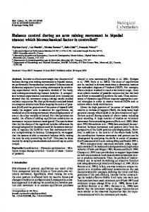
force field during postural tasks a natural strategy for preserving postural stability is to increase muscles’ cocontraction, which affects ellipse size while leaving shape and orientation invariant [55]. In general, when comparing two different conditions, both the determinant and eccentricity will change to a certain extent. To determine if the cause of the stiffness increase is co-contraction or modulation of reflexes, we need to analyze the shape of the ellipse by examining its eccentricity and orientation: a consistent change in eccentricity or orientation of the stiffness ellipses is considered evidence of reflexive modulation [28, 47]. C. The pilot clinical study. Nine chronic stroke survivors with different levels of impairment participated in the experiment; their clinical and anthropometric data are listed in Table I. The task was designed according to the Bobath concept and aimed to facilitate the active execution of outward movements of large amplitude [19]. Subjects had to hit a set of targets arranged on three layers: proximal (A), intermediate (B) and distal (C) with respect to the shoulder joint (see Figure 1, panel A, for details). The exercise was composed of blocks of trials in which subjects had to reach each of the 7 targets on the outward level (C), which were presented in random order, three times via a large, single movement and then return to the A level via B. The subsequent backward movements were divided in two shorter sub-movements (from C to B, from B to A) in order to disrupt the dominating flexion patterns that characterize most stroke survivors. Each block consisted of 21
B. Indicators Stiffness and damping matrices of a two degree of freedom system can be graphically represented as ellipses [29]. Any of these ellipses (let us name A the corresponding matrix) is completely characterized by three parameters: determinant, eccentricity, and orientation [54]: Determinant. The determinant is the product of the two semiaxes (a and b) of the ellipse and is proportional to its area: Area = π ⋅ a ⋅ b = π ⋅ det( A)
(2)
Eccentricity. Can be computed as follows:
(a − tr( A)) + (b − tr( A)) 2
Ecc( A) = 2
tr( A)
2
(3)
where tr ( A) = a + b (trace of A ). Eccentricity varies from 0 to 1: Ecc ( A) = 1 corresponds to a degenerate ellipse in which either axis is null; Ecc ( A) = 0 corresponds to a circle (a = b) . Orientation. The orientation is computed as the angle between the major semi-axis a and the axis x of the reference frame. When a is aligned with x , the orientation is null. These indicators are closely related to physiologically relevant aspects of stiffness and damping. The determinant is directly associated with the metabolic energy required to stabilize the limb. Moreover, when the arm is perturbed by a
Figure1: Experimental setup and protocol of robot-therapy. A) Configuration of subject and robotic device for the experiments. The origin of the workspace is located at the center of the shoulder, which is constrained by safety straps. In the central position of the workspace, shoulder is at 45° and elbow is at 90°. B) Time-course of the assistive force during the basic trial: 1. the visual target and the force field are activated; 2. linear increase of assistive force, starting with visual target activation, with a duration of 1s, up to a preset force value; 3. the preset force is maintained at a constant level up to the time of target contact; 4. Target and assistive force are suddenly turned off; 5. A wait time (1s) follows, with no target and no force.
TNSRE-2012-00064
4
reaches and 42 sub-movements. A planar manipulandum (Braccio di ferro [56] ) provided assistive forces to help stroke survivors accomplish the task. Subjects sat in front of the robot with their shoulders strapped to the chair, holding the end-effector of the robot with their impaired hand. The hand was securely fastened using a custom made cast. Three feedback signals were presented: visual, acoustic, and haptic. A different sound was associated with each target layer. There were two types of blocks of trials: with and without VF. When the VF was provided, subjects saw their arm, the robot, and a computer monitor (1 meter away at eye level) that displayed the end-effector position and the target to reach (Figure 1, panel A). The target and the cursor corresponding to the endeffector position were represented on the monitor as round disks of different colors with a radius of 1 cm. In the trials without VF, the subject’s view of both the arm and the screen was occluded by means of a blindfold. The presentation order of the VF conditions was selected by the therapist and differed between subjects. The haptic rendering of the task was generated according to the equation: ( x − x H ) Br 0 Fm = ρ ( AF ) T + x& H + Fw (x H , x w , K w ) xT − x H 0 Br
(4)
The assistive force Fm provided by the manipulandum includes 3 elements: 1) a force field of constant amplitude AF , which attracts to the target x T the position of the hand/end-effector x H ; 2) a mild viscous field ( B r = 12 Ns/m ), intended to stabilize the arm posture; 3) a stiff wall Fw ( K w = 1000 N/m ) beyond the ‘C’ targets’ level, which represents the boundary of the workspace. Panel B of Figure 1 summarizes the different phases of a basic trial: 1) The visual target and the force field are activated. 2) The assistive force field is aimed at the target ( x − xH ) location using the vector T . The intensity of the force xT − x H grows linearly according to ρ (F ) and within 1s reaches the pre-selected force value. The time raise is independent of the final force intensity. 3) The assistive force is maintained until the time of contact with the target. 4) The visual target and the force field are suddenly switched off. 5) A wait time follows, with no target and no field, before repeating the cycle with another target. It is worth remarking that the assistive force field is not elastic; thus, its intensity does not grow with the distance from the target, but remains constant throughout a trial except for the initial smooth activation phase. The assistive force level, AF , was adapted to the impairment severity of the different subjects and its starting and highest value is listed in the rightmost column of Table I. The empirical criterion of selection was to identify an approximate threshold below which subjects would be unable to complete the task without robotic assistance. In the described experiments the evaluation of the threshold was done manually by a therapist supervising the experiments, although more automatic procedures could be designed in the future. The hand speed at the target depends on several factors. If the arm was a pure inertial load, given the selected
force, the hand would uniformly accelerate in the direction of applied force producing a rather large velocity at the target. On the other hand, the intrinsic stiffness at the joint hinders the movement, reducing that value. Moreover, there is no time constraint for accomplishing the task and, as training proceeds, the movement of the subjects improves [19], creating a combination of the active reaching movement and the trajectory driven by the assistive force [57]. Hence, the velocity upon reaching the target was low at the beginning of training, but it increased substantially as training progressed, thus making the task more similar to “hitting” than to “reaching”. We estimated the stiffness and damping of the arm according to the time frequency technique described above, analyzing the residual vibration of the arm in the wait time period after “hitting” the target when the force dropped to zero. The force drop can be considered in engineering terms as a negative step of force, and it was previously defined as a “hold and release” type of action [58]. If the same technique is applied to a passive linear system, the residual oscillation of the system would be independent of the amplitude of the step perturbation. On the other hand, since the human arm is by no mean passive, it makes sense to verify the influence of hitting velocity and force amplitude on the estimation of stiffness. The protocol included 10 sessions, with a duration that ranged from 45 to 75 minutes. Each session started with the same initial force, selected by the therapist as the minimal force allowing the subject to initiate the movement in the first session. After the first two blocks, the therapist could decide to extend the exercise with additional blocks with lower levels of force in accordance with the subject’s residual ability and fatigue. For each subsequent session, while always starting with the force level selected at the beginning of the first session, subjects experienced a further decrease in the assistive force, with the ultimate goal being – when possible - to reach the target with no assistive force. D. Applying the stiffness measurement technique to the clinical setting Two questions are of particular interest for the considered clinical protocol: 1) Are the performance improvements induced by robot training (such as speed and smoothness of the performed reaching movements) correlated with a reduction in stiffness? 2) Does visual feedback increase or decrease stiffness? These questions are clearly related to evaluating how treatment produces changes in muscle activity. Such evaluation is impossible with the usual clinical scales, like MAS, given their limited resolution. To highlight the reflexes’ contribution to the stiffness, all the estimations and relative statistics were calculated at 200ms after the perturbation onset. Later estimates are influenced by voluntary control, among other factors. Subjects did not experience the same levels of force because of the different degrees of impairment. The selected force for each subject is reported on Table I. For all sessions, with and without VF, the same level of force was used in our calculations. The trials we selected were not part of the first block in order to avoid effects due to the initial acquaintance with the exercise. We only considered the trials corresponding to the forward movements (A to C). The 21 measures at the C layer were averaged independently
TNSRE-2012-00064 and used for the statistical analysis. We performed a repeated measures ANOVA with 3 factors: training (sessions), visual feedback (with/without), and position on the C layer. Significance level was set at p