0013-7227/05/$15.00/0 Printed in U.S.A.
Endocrinology 146(5):2481–2488 Copyright © 2005 by The Endocrine Society doi: 10.1210/en.2004-1606
Genomic Regions that Mediate Placental Cell-Specific and Developmental Regulation of Human Cyp19 (Aromatase) Gene Expression in Transgenic Mice Amrita Kamat, Margaret E. Smith, John M. Shelton, James A. Richardson, and Carole R. Mendelson Departments of Biochemistry (A.K., C.R.M., M.E.S.), Obstetrics-Gynecology (C.R.M.), Internal Medicine (J.M.S.), and Pathology and Molecular Biology (J.A.R.), University of Texas Southwestern Medical Center, Dallas, Texas 75390-9038 The human aromatase (hCYP19) gene is controlled by tissuespecific promoters that lie upstream of tissue-specific first exons. Placenta-specific exon I.1 lies approximately 100,000 bp upstream of exon II. Previously, we observed that genomic sequences within 501 bp upstream of exon I.1 mediate placenta-specific expression. In the present study, transgenic mice were created carrying hCYP19I.1⫺246:hGH/hGX, hCYP19I.1⫺201:hGH, and hCYP19I.1⫺125:hGH fusion genes to further delineate 5ⴕ-flanking sequences within 501 bp of exon I.1 that are required to mediate placenta-specific hCYP19 gene expression. As little as 246 bp of hCYP19 exon I.1 5ⴕflanking sequence was sufficient to direct placenta-specific expression in transgenic mice. By contrast, transgenes containing 201 or 125 bp of exon I.1 5ⴕ-flanking DNA were not expressed in mouse placenta. Furthermore, hCYP19I.1⫺246: hGX transgene expression was developmentally regulated; ex-
G
ROWTH OF THE human placenta is driven by replication of mononuclear cytotrophoblasts, a stem cell population in the placenta (1, 2). Upon maturation, cytotrophoblasts within the floating chorionic villi stop dividing and fuse to form the outer terminally differentiated, multinucleated syncytiotrophoblast layer that is bathed in maternal blood. Differentiation of cytotrophoblasts to syncytiotrophoblast generates a cascade of regulatory signals that result in the production of a diverse array of polypeptide hormones, growth factors, steroids, and steroid-metabolizing enzymes, including aromatase. The human placenta acquires increased capacity for aromatization of C19 steroids synthesized by the fetal adrenals with advancing gestation, so that after the ninth week of gestation, the human placenta becomes the primary source of circulating estrogens (3). The aromatase enzyme complex, comprised of the ubiquitous flavoprotein, NADPH-cytochrome P450 reductase and aromatase P450 (P450arom; product of the hCYP19 gene), catalyzes the final and rate-limiting step in the synthesis of estrogens from androgens (4 – 6). In most vertebrates, CYP19 expression is restricted to the gonads and brain; however, in humans, it is expressed in specific cell First Published Online January 27, 2005 Abbreviations: E, Embryonic day; FBS, fetal bovine serum; Gcm1, glial cells missing transcription factor; Mash-2, mammalian achaete scute homologous protein-2; P450arom, aromatase P450. Endocrinology is published monthly by The Endocrine Society (http:// www.endo-society.org), the foremost professional society serving the endocrine community.
pression was observed as early as embryonic d 7.5 (E7.5) in several cells of the trophoblast ectoderm, on E8.5 in some trophoblast giant cells, and by E9.5 in giant cells and the labyrinthine layer. By contrast, expression of the hCYP19I.1⫺501:hGH transgene was first observed on E10.5 and was restricted to the labyrinthine layer, which is most analogous to the human syncytiotrophoblast. This suggests the presence of regulatory elements between ⴚ501 and ⴚ246 bp that may bind inhibitory transcription factors expressed in giant cells. These findings from transgenic experiments together with deletion mapping studies using transfected human placental cells indicate that the concerted interaction of strong placenta-specific enhancers and silencers within this 501-bp region mediate labyrinthine and syncytiotrophoblastspecific CYP19 gene expression. (Endocrinology 146: 2481–2488, 2005)
populations of a diverse set of tissues, including the syncytiotrophoblast layer of the placenta; granulosa and luteal cells of the ovary; Leydig, Sertoli, and germ cells of the testis; stromal cells of adipose tissue; bone; and discrete nuclei within the brain and in fetal liver (7, 8). In humans, hCYP19 is encoded by a single copy gene, approximately 130 kb in length, that is localized on chromosome 15q21.1 (8). hCYP19 gene expression in various estrogen-producing tissues appears to be driven by tissuespecific promoters upstream of tissue-specific alternative first exons, which encode the 5⬘-untranslated regions of hCYP19 mRNA transcripts. These alternative first exons, which are located from approximately 110 –100,000 bp upstream of the hCYP19 translation initiation site in exon II, are alternatively spliced onto a common site just upstream of the translation start site in exon II so that the protein encoded in each of these tissues is identical. In placenta, the majority of the hCYP19 mRNA transcripts contain sequences encoded by exon I.1, which lies about 100,000 bp upstream of the start site of translation in exon II. In previous studies using primary cultures of human placental cells, we observed that differentiation of cytotrophoblasts to syncytiotrophoblast is oxygen dependent and associated with a marked induction of aromatase activity and hCYP19 gene expression (9, 10). Transfection of placental and nonplacental cells with reporter gene constructs indicated that placenta-specific exon I.1 5⬘-flanking sequences between ⫺501 and ⫺42 bp mediates trophoblast-specific hCYP19 gene expression (9). Studies using transgenic mice also suggested
2481
2482
Endocrinology, May 2005, 146(5):2481–2488
that as little as 501 bp of exon I.1 5⬘-flanking DNA directed reporter gene expression exclusively to the placenta and specifically to the labyrinthine trophoblast layer, which is most analogous to the human syncytiotrophoblast (11). Because mouse placenta does not express aromatase, these findings indicate that placental transcription factors that mediate hCYP19 gene expression are conserved between mouse and human, whereas the genetic response elements that bind these factors are not. Collectively, these findings suggest that the 5⬘-flanking DNA within 501 bp of exon I.1 of the hCYP19 gene contains cis-acting elements, which bind placenta-specific transcription factors that are conserved between humans and mice. In the present study we used transgenic mice and transfected cells to further delineate the 5⬘-flanking sequences upstream of hCYP19 exon I.1 that mediate placental cellspecific and temporal regulation of expression. Our findings indicate that placenta-specific and temporal regulation of hCYP19 expression requires the concerted interaction of strong placenta-specific enhancers within ⫺246 bp and silencers between ⫺246 and ⫺501 bp flanking the 5⬘ end of exon I.1. Materials and Methods Construction of hCYP19I.1:hGH/hGX transgenes The hCYP19I.1⫺501;hGH transgene was constructed as described previously (11). Genomic sequences comprised of 246, 201, and 125 bp of 5⬘-flanking DNA and 103 bp of untranslated exon I.1 (hCYP19I.1) of the hCYP19 gene were subcloned into the HindIII/Sal I and BamHI sites of plasmid pACsk20GH to create recombinant hCYP19I.1⫺501:hGH, hCYP19I.1⫺246:hGH, hCYP19I.1⫺201:hGH, and hCYP19I.1⫺125:hGH plasmids, respectively (Fig. 1). Plasmid pACsk20GH contains the promoterless hGH structural gene (12, 13). The recombinant plasmid, hCYP19I.1⫺246:hGX, was created by subcloning 246 bp of 5⬘-flanking DNA and 103 bp of untranslated hCYP19 exon I.1 into the Sal I and BamHI sites of plasmid pUChGX. Plasmid pUChGX contains a mutated form of hGH (hGX), which does not bind to GH or prolactin receptors (14). The recombinant plasmids were digested with SalI and XbaI to release the fusion genes. The DNA was then isolated on agarose gels and purified for microinjection (11, 15).
Kamat et al. • hCYP19 Gene Expression in Transgenic Mice
Generation and identification of transgenic mice carrying hCYP19I.1:hGH/hGX fusion genes All animal studies were approved by the institutional animal care and research advisory committee of University of Texas Southwestern Medical Center. Transgenic mice were produced by microinjection of several hundred molecules of each hCYP19I.1:hGH or hCYP19I.1:hGX fusion gene DNA, described above, into the male pronucleus of ICR (Institute of Cancer Research) hybrid mouse eggs. The eggs were introduced into the oviducts of pseudopregnant ICR mice and allowed to develop until term. Transgene-positive animals were then identified, and the copy number of fusion genes per genome was ascertained by quantitative Southern dot-blot analysis of tail or fetal tissue DNA using a 32P-labeled hGH or hGX cDNA probe, as described previously (16). Transgenic founders (F0) were bred to establish at least five independent lines.
Northern blot analysis of hGH mRNA in transgenic mouse tissues For Northern blot analysis, various tissues were obtained from adult F1 or F2 or fetal F1 mice carrying hCYP19I.1:hGH/hGX fusion genes. Total RNA was isolated from these tissues as previously described (11). The pelleted RNA was resuspended in water, and approximately 30 g were electrophoresed in formaldehyde-containing agarose gels. The RNA then was transferred to nitrocellulose and probed using a 32P-labeled hGH or hGX cDNA fragment.
In situ hybridization analysis of hGH mRNA in placental tissues of transgenic fetal mice Placental tissues from embryonic d 7.5 (E7.5), E8.5, E9.5, E10.5, E11.5, and E17.5 transgenic fetal mice were fixed for approximately 16 h at 4 C in 4% paraformaldehyde-PBS (pH 7.4), then rinsed and placed in cold saline. The tissues were embedded in paraffin and sectioned. In situ hybridization analysis was performed on these paraffin sections (17) using 35S-labeled sense and antisense hGH cRNA transcripts, as described previously (11, 18).
Isolation and culture of human placental cells and maintenance of cell lines Midtrimester human placental tissues were obtained in accordance with the Donors Anatomical Gift Act of the State of Texas after obtaining consent in writing. In all cases, consent forms and protocols were ap-
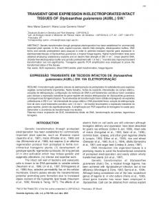
FIG. 1. Schematic representations of the hCYP19 (aromatase) gene and hCYP19I.1:hGH fusion genes introduced into transgenic mice. Exons II–X (䡺), which encode the aromatase protein, and their introns (black lines) comprise a region approximately 34 kb in size. The heme-binding region (HBR) and two polyadenylation signals in the 3⬘-untranslated region (o) are encoded in exon X. Exons IIa, I.4, and I.1 (f) encode the 5⬘-untranslated regions of P450arom mRNAs in gonads, adipose tissue, and placenta, respectively. The region containing these alternative first exons encompasses about 100 kb. The hCYP19I.1:hGH fusion genes are comprised of hCYP19 DNA sequences encoding 501, 246, 201, and 125 bp of DNA flanking the 5⬘ end of exon I.1 (solid line) and the first 103 bp of exon I.1 (u) fused to either the wild-type (hGH) or mutated, biologically inactive form (hGX) of the hGH structural gene, as reporter (䡺). The arrow indicates the position of the transcription initiation site and the direction of transcription for all fusion gene constructs.
Kamat et al. • hCYP19 Gene Expression in Transgenic Mice
Endocrinology, May 2005, 146(5):2481–2488
proved by the institutional review board of University of Texas Southwestern Medical Center. Human cytotrophoblasts were isolated and cultured as described previously (9). Briefly, trophoblast cells were isolated from midterm human placenta by trypsin digestion; cytotrophoblasts were purified using a 5–70% Percoll gradient. The cells that sedimented in the middle of the gradient (density, 1.048 –1.062 g/ml) were suspended in DMEM containing 2% fetal bovine serum (FBS), plated at a density of 2 ⫻ 106 cells/dish on 35-mm dishes, and incubated in a humidified atmosphere of 95% air/5% CO2. A549 lung adenocarcinoma cells (ATCC CCL-185, American Type Culture Collection, Manassas, VA) were maintained in Waymouth’s MB 752/1 medium supplemented with 10% FBS.
Preparation of recombinant adenoviruses carrying hCYP19I.1:hGH fusion genes Recombinant adenoviruses carrying hCYP19I.1:hGH fusion genes were created by in vivo homologous recombination in 293 cells, a permissive human embryonic kidney cell line, as described previously (9), or by using a protocol involving in vivo recombination in bacteria (10, 19). Briefly, the hCYP19I.1:hGH fusion genes were subcloned into the pShuttle vector to generate pShuttle-hCYP19I.1:hGH. The recombinant adenoviral particles containing hCYP19I.1:hGH fusion genes were then generated by cotransformation of pShuttle-hCYP19I.1:hGH and pAdEasy-1 into electrocompetent BJ5183 bacteria. PacI-digested recombinant adenoviral hCYP19I.1 fusion genes were transfected into 293 cells for recombinant adenoviral packing and propagation. Viral DNA was analyzed to confirm the presence of the fusion genes by restriction endonuclease digestion, PCR, and DNA sequencing. The recombinant adenoviruses were then titrated in 293 cells at least three times to determine the number of infectious viral particles (plaque-forming units).
Infection of trophoblasts in primary culture with recombinant adenoviruses Freshly isolated cytotrophoblast cells, plated at a density of 2 ⫻ 106 cells/35-mm dish in DMEM containing 10% FBS or lung A549 cells cultured in Waymouth’s medium containing 10% FBS, were infected with 1 ⫻ 106 recombinant adenoviral particles to achieve a multiplicity of infection of approximately 0.5. After an overnight incubation, media were collected and replaced with DMEM containing 2% FBS. Media from infected cells were then collected at 24-h intervals and replaced daily for the next 96 h. The collected media were assayed for hGH by
2483
RIA, using an Allegro hGH kit (Nichols Institute Diagnostics, San Juan Capistrano, CA).
Results Analysis of placental expression of hCYP19I.1:hGH/hGX fusion genes in transgenic mice
Previously, we observed that hCYP19I.1⫺501:hGH fusion genes were expressed in a placenta-specific manner, exclusively in the labyrinthine trophoblast in transgenic mice (11). To further define genomic sequences within this ⫺501 bp region responsible for placental labyrinth-specific expression, in the present study transgenic mice carrying fusion genes containing 246, 201, or 125 bp of DNA flanking the 5⬘ end and the first 103 bp of exon I.1 linked to hGH, as reporter, were generated and compared with mice expressing the 501bp-containing transgene (Fig. 1). To ensure that hGH produced in tissues expressing the transgene had no effect on tissue- and cell-specific expression, transgenic mice were also generated carrying an hCYP19I.1⫺246:hGX transgene. In this case, the hGH reporter gene was mutated at a unique BglII site in exon 5 to produce a physiologically inactive hGH peptide (14). Transgenic founder mice (F0) identified by dotblot analysis were bred to establish independent lines (F1). Placental tissues from the F1 mice were then analyzed for hGH/hGX mRNA expression. Copy numbers and expression of fusion genes in placentas and other tissues of transgenic mouse lines are presented in Table 1. In three independent lines of transgenic mice, hCYP19I.1⫺501:hGH fusion gene expression was detected in the placenta. In nontransgenic littermates, hGH expression was undetectable in the placenta (data not shown), indicating the absence of crossreacting endogenous mRNA transcripts. These results confirm those previously reported (11) in five other independent transgenic mouse lines carrying the hCYP19I.1⫺501:hGH fusion gene and demonstrate that 501 bp of hCYP19 exon I.1
TABLE 1. Expression of hCYP191.1-501:hGH, hCYP191.1-246:hGH/hGX, hCYP191.1-201:hGH, and hCYP191.1-125:hGH fusion genes in transgenic mice Transgene
No. of linesa
No. transgene copiesb
Placenta
Nonplacental tissues
hCYP191.1-501:hGH
1 2 1 1 5 1 1 1 1 2 2 2 1 6 1 1 1 1
6 1 5 35 1 6 5 1 9 3 2 1 1 1 2 1 3 1
⫹ ⫹ ⫹ ⫹ ⫹ ⫹ ⫹ ⫺ ⫺ ⫺ ⫺ ⫺ ⫺ ⫺ ⫺ ⫺ ⫺ ⫺
⫺ ⫺ ⫺ ⫺ ⫺ mamm (very low) adr, ov, test (very low) ⫺ ⫺ ND ND ⫺ kid (very low) ⫺ ⫺ ov, adr (very low) brain (very low) Liv, kid, ov, mamm,adr (very low)
hCYP191.1-246:hGH hCYP191.1-246:hGX
hCYP191.1-201:hGH
hCYP191.1-125:hGH
adr, Adrenals; mamm, mammary; ov, ovary; liv, liver; kid, kidney; test, testis; ND, not determined. a The number of independent transgenic founder lines analyzed carrying a specific number of genomically integrated copies of each of the five transgenes. Transgenic founders were identified and copy numbers analyzed by quantitative Southern dot blot analysis of tail DNA. b Transgene copy number was estimated by comparing the intensity of hybridization of the 32P-labeled probe (hGH or hGX) to genomic DNA with a known hGH/hGX standard.
2484
Endocrinology, May 2005, 146(5):2481–2488
5⬘-flanking sequence are sufficient for expression in mouse placenta. In one line of transgenic mice carrying a fusion gene containing 246 bp of exon I.1 5⬘-flanking DNA linked to an hGH reporter (hCYP19I.1⫺246:hGH) and in eight of nine lines carrying a 246-bp-containing fusion gene with an hGX reporter (hCYP19I.1⫺246:hGX), high levels of expression also were detected in the placenta (Table 1). In contrast, when the hCYP19 exon I.1 5⬘-flanking sequence was reduced further to 201 bp (hCYP19I.1⫺201:hGH), the eight transgenic mouse lines that were generated failed to manifest any placental expression. Similarly, placental expression was undetectable in nine of the 10 transgenic mouse lines carrying the hCYP19I.1⫺125: hGH fusion gene. One transgenic mouse line carrying the hCYP19I.1⫺125:hGH fusion gene manifested low levels of hGH expression in the placenta; however, hGH expression was also detected in liver and in a number of other tissues. No effect of hGH expression on reproduction or fetal growth was evident in any of the transgenic mice analyzed. Expression of hGH/hGX mRNA in various tissues of transgenic mice
Representative Northern blots of hGH/hGX mRNA in placental and various tissues of lines of adult transgenic mice carrying the hCYP19I.1⫺501:hGH, hCYP19I.1⫺246:hGX/hGH, hCYP19I.1⫺201:hGH, and hCYP19I.1⫺125:hGH fusion genes are shown in Fig. 2. As previously reported (11), the hCYP19I.1⫺501:hGH fusion gene was expressed in a placentaspecific manner in transgenic mice. hGH mRNA was undetectable in adult tissue samples from brain, testes, ovary, adipose, adrenals, kidneys, spleen, liver, lung, and heart (Fig. 2 and Table 1). In six of the nine independent lines carrying the hCYP19I.1⫺246:hGX transgene and in the single mouse line carrying the hCYP19I.1⫺246:hGH transgene, expression was also found to be placenta-specific (Fig. 2). In two other lines carrying the hCYP19I.1⫺246:hGX transgene, in addition to
Kamat et al. • hCYP19 Gene Expression in Transgenic Mice
very high levels of reporter gene expression in the placenta, there were much lower levels of expression in the adrenals, testes, ovary, and mammary tissues (Table 1). These findings indicate that the expression of the 246-bp-containing transgene was placenta-specific regardless of whether the reporter gene used was hGH or the mutated form, hGX. In all mouse lines carrying the hCYP19I.1⫺201:hGH transgene and in nine of the 10 lines carrying the hCYP19I.1⫺125: hGH transgene, expression was undetectable in the placenta, although various levels of expression were observed in several other adult tissues (Table 1 and Fig. 2). However, in the only mouse line that expressed the hCYP19I.1⫺125:hGH transgene in the placenta, roughly equivalent levels of expression were observed in most of the other tissues analyzed (Table 1). This may be due to position effects of transgene integration. Taken together, these findings suggest that sequences within 246 bp upstream of the transcription start site within exon I.1 contain cis-acting elements that mediate selective expression of hCYP19 in placenta. Cellular localization of hCYP19I.1:hGH/hGX fusion gene expression in transgenic mouse placenta
To analyze cellular localization of transgene expression, in situ hybridization analysis was performed, as described in Materials and Methods, using an antisense hGH cRNA probe. As shown in Fig. 3, A and C (dark-field), and Fig. 3E (brightfield) and as reported previously (11), hCYP19I.1⫺501:hGH transgene expression was detected on E11.5 specifically in the labyrinthine trophoblast layer. Reporter gene expression was undetectable in the giant cell or spongiotrophoblast layer. By contrast, the hCYP19I.1⫺246:hGX transgene was expressed at very high levels in the labyrinthine as well as the trophoblast giant cell layer on E11.5 (Fig. 3, B, D, and F). In contrast, hybridization signal was absent in the placenta of a nontransgenic littermate (data not shown), indicating that the radiolabeled hGH cRNA probe does not cross-react with endogenous mouse placental hormones. These results, therefore, suggest that although sequences between ⫹103 and ⫺246 bp flanking the 5⬘ end of exon I.1 mediate expression of hCYP19 promoter I.1 in the labyrinthine trophoblast layer of the mouse placenta, repressors may bind to the region between ⫺501 and ⫺246 bp to suppress expression in the trophoblast giant cells. Developmental regulation of hCYP19I.1:hGH/hGX fusion gene expression in transgenic mouse placenta
FIG. 2. Two hundred and forty-six base pairs of hCYP19 exon I.1 5⬘-flanking sequence are sufficient to mediate placenta-specific expression in transgenic mice. Aliquots of total RNA (30 g) isolated from placentas of an E17.5 F1 transgenic mice or from various tissues of adult F1 male or female mice carrying the hCYP19I.1⫺501:hGH, hCYP19I.1⫺246:hGH, hCYP19I.1⫺201:hGH, or hCYP19I.1⫺125:hGH transgenes were analyzed by Northern blotting using a 32P-labeled hGH cDNA probe, as described in Materials and Methods.
Because we observed that fusion genes containing either 501 or 246 bp of exon I.1 5⬘-flanking sequence directed placenta-specific expression, it was of interest to analyze their expression in the placenta at early embryonic stages to assess expression in trophoblast lineages. In previous studies of transgenic mice carrying the ⫺501-bp-containing fusion gene, we found that transgene expression was detectable on E10.5; however, earlier time points were not analyzed (11). In situ hybridization analyses were, thus, performed on E7.5, E8.5, E9.5, and E10.5 placentas using an antisense hGH probe. As shown in Fig. 4 (left panels), hCYP19I.1⫺501:hGH transgene expression was first detectable on E10.5, specifically in the labyrinthine layer; transgene expression was not
Kamat et al. • hCYP19 Gene Expression in Transgenic Mice
Endocrinology, May 2005, 146(5):2481–2488
2485
FIG. 3. The hCYP19I.1⫺246:hGX transgene is expressed in both the labyrinthine trophoblast layer and giant cells on E11.5, whereas expression of the hCYP19I.1⫺501:hGH transgene is labyrinthine trophoblast-specific. Placentas obtained on E11.5 from transgenic mice carrying either the hCYP19I.1⫺501:hGH (A, C, and E) or the hCYP19I.1⫺246:hGX (B, D, and F) fusion gene were processed for in situ hybridization using an 35S-labeled antisense hGH cRNA probe and were exposed to photographic emulsion for 1–2 wk. Bright- and dark-field microscopies were then performed. A and B, Dark-field micrographs (low magnification) of placental tissue sections from E11.5 transgenic mice carrying the ⫺501- and ⫺246-bp-containing fusion genes, respectively. C and D, Dark-field micrographs of the placental tissue sections (high magnification) shown in A and B, respectively. E and F, Bright-field micrographs of the hematoxylinstained E11.5 placental tissue sections shown in C and D, respectively. gc, Trophoblast giant cell; sp, spongiotrophoblast; lab, labyrinthine trophoblast.
detected at earlier embryonic stages (E7.5, E8.5, or E9.5). In contrast, as shown in Fig. 4 (right panels), hCYP19I.1⫺246:hGX transgene expression was detected as early as E7.5, specifically in a few cells of the trophoblastic ectoderm. On E8.5, hCYP19I.1⫺246:hGX transgene expression was observed in several trophoblast giant cells; by E9.5, expression was readily detected throughout the giant cell layer, although mRNA expression appeared to be stronger in some cells than in others, suggesting some heterogeneity in expression levels within giant cells (Fig. 4, right panels). Additionally, very low levels of hCYP19I.1⫺246:hGX transgene expression were detected at this stage in the primitive labyrinthine layer. However, by E10.5, relatively high levels of hCYP19I.1⫺246:hGX transgene expression were observed in both the well vascularized labyrinthine layer and the trophoblast giant cell layer (Fig. 4, right panels). The findings that the ⫺246-bp-containing transgene was expressed in trophoblast giant cells and in the labyrinthine layer, whereas the ⫺501-bp-containing transgene was labyrinth-specific indicates that sequences between ⫺246 and ⫺501 bp repress hCYP19 promoter I.1 activity in giant cells.
FIG. 4. The hCYP19I.1⫺246:hGX transgene is expressed as early as E8.5 in trophoblast giant cells, whereas hCYP19I.1⫺501:hGH transgene expression is evident only on E10.5, specifically in the labyrinthine trophoblast layer. Placental tissues obtained from E7.5, E8.5, E9.5, and E10.5 fetal mice carrying either the ⫺501-bp (left panel) or ⫺246-bp (right panel)-containing transgene were processed for in situ hybridization using an 35S-labeled antisense hGH cRNA probe and exposed to photographic emulsion for 1–2 wk. Bright- and dark-field microscopies were then performed. Left panels, Dark-field micrographs of placental tissue sections from E7.5, E8.5, E9.5, and E10.5 transgenic mice carrying the hCYP19I.1⫺501:hGH fusion gene. Right panels, Dark-field micrographs of placental tissue sections from E7.5, E8.5, E9.5, and E10.5 transgenic mice carrying the hCYP19I.1⫺246: hGX fusion gene. Bright-field micrograph of hematoxylin-stained E10.5 placental tissue section from mice carrying ⫺501- or ⫺246-bpcontaining transgene are shown above their respective dark-field micrographs. gc, Trophoblast giant cell; sp, spongiotrophoblast; lab, labyrinthine trophoblast.
5⬘-Flanking sequences between ⫺300 and ⫺246 bp repress fusion gene expression in primary cultures of syncytiotrophoblast and in a lung cell line
In previous cell transfection studies we observed that hCYP19I.1⫺246:hGH fusion genes were highly expressed in
2486
Endocrinology, May 2005, 146(5):2481–2488
human syncytiotrophoblast, but were also expressed in lung and kidney cell lines. By contrast, expression of ⫺501-bpcontaining fusion genes was human syncytiotrophoblastspecific (9). To further define the region between ⫺246 and ⫺501 bp upstream of exon I.1 that may be involved in labyrinth/syncytiotrophoblast-specific hCYP19 gene expression, hCYP19I.1:hGH fusion genes comprised of ⫺246, ⫺300, ⫺350, ⫺400, or ⫺501 bp of exon I.1 5⬘-flanking DNA were incorporated into recombinant replication-defective adenoviral viral particles (1 ⫻ 106 plaque-forming units/dish) and introduced into freshly isolated human trophoblast cells (2 ⫻ 106) and A549 lung adenocarcinoma cells by infection. Fusion gene expression was analyzed 3 d after infection (when most primary trophoblast cells had fused to form syncytia) by assay of hGH secreted into the medium over a 24-h period of culture. As observed previously (9), fusion genes containing 246 bp of 5⬘-flanking sequence were highly expressed in the human syncytiotrophoblast cells and A549 cells (Fig. 5). In contrast, expression of the hCYP19I.1⫺300:hGH fusion gene was essentially undetectable, suggesting the presence of transcriptional repressors in both cell types that bind to elements between ⫺246 and ⫺300 bp. In human placental cells, expression levels of fusion genes containing 350, 400, and 501 bp of exon I.1 5⬘-flanking DNA were increased compared with that of the 300-bp construct, whereas in lung A549 cells, the expression of these longer fusion genes was barely detectable (Fig. 5). This suggests that the repression in trophoblast cells was overcome in part by syncytiotrophoblastspecific-activating factors binding to the region between ⫺300 and ⫺501 bp. It is possible that analogous repressors also restrict the expression of the 501-bp transgene to the labyrinthine layer in transgenic mice. Discussion
Our understanding of the transcriptional regulation and cell-specific expression of various genes in placenta is complicated by its structural diversity among species and differences in the endocrine functions of various trophoblast cell types. This is exemplified by comparison of human and rodent hemochorial placentas, which are bathed in maternal blood, but differ in gross anatomy and capacity to express the hCYP19 gene. The trophoblast stem cells of the human placenta are the FIG. 5. Region between ⫺300 and ⫺246 bp upstream of hCYP19 exon I.1 binds transcriptional repressors. Freshly isolated human cytotrophoblasts and lung A549 adenocarcinoma cells in culture were infected with 1 ⫻ 106 recombinant adenoviral particles containing hCYP19I.1⫺501:hGH, hCYP19I.1⫺400:hGH, hCYP19I.1⫺350: hGH, hCYP19I.1⫺300:hGH, or hCYP19I.1⫺246:hGH fusion genes. Culture media were harvested and replaced with fresh media every 24 h over a 4-d period. Shown here are the levels of hGH that accumulated in the culture medium between d 2 and 3 of culture. Values are the mean ⫾ SEM (n ⫽ 3) of data from a representative of three independent experiments.
Kamat et al. • hCYP19 Gene Expression in Transgenic Mice
highly proliferative cytotrophoblasts, which reside in either the anchoring or floating villi of the chorion. The cytotrophoblasts of the anchoring villi physically connect the embryo to the uterine wall and are highly invasive. Cytotrophoblasts of the floating villi, which are bathed by maternal blood, do not contact the uterine wall, but differentiate upon fusion to form the multinucleated syncytiotrophoblast that covers the villi (1, 2). Human cytotrophoblasts do not express the hCYP19 gene, but the highly differentiated syncytiotrophoblast, which mediates gas and nutrient exchange for the developing embryo, manifests very high levels of hCYP19 gene expression (9, 20). In contrast to that in the human, the rodent placenta does not express aromatase (21). During mouse placental development, which begins on E3.5, there is formation of three distinctive layers of trophoblast derivatives that persist throughout gestation (22, 23). These include an outer trophoblast giant cell layer, an intermediate spongiotrophoblast layer, and the inner labyrinthine layer. The giant cell layer, which interfaces with the maternal decidua, is the site of production of various angiogenic factors, enzymes and hormones, including the placental lactogens and prolactin. These cells also facilitate the breakdown and invasion of the decidua and in this respect are similar to the invasive cytotrophoblast cells of human placenta. The underlying spongiotrophoblast, which is formed after E7.5, is morphologically distinct from the giant cell layer; however, it also manifests endocrine activities, including the expression of a subset of members of the prolactin gene family. The innermost labyrinthine layer, formed after E9, is most similar to the human syncytiotrophoblast, because it serves as a site of nutrient and gas exchange between maternal and fetal blood compartments. Additionally, the labyrinth, which is trilaminar, has an inner layer comprised of syncytial cells (22, 23). In previous studies using transgenic mice, we observed that 501 bp of hCYP19 exon I.1 5⬘-flanking DNA was sufficient to mediate placenta-specific expression (8, 11). This provided definitive evidence that mouse placenta contains the necessary transcription factors to activate the hCYP19 gene placenta-specific promoter, but that the mouse genome lacks cis-acting elements required for endogenous CYP19 gene expression. In the present study we observed that expression of a transgene containing as little as 246 bp of exon I.1 5⬘-
Kamat et al. • hCYP19 Gene Expression in Transgenic Mice
flanking DNA was placenta-specific, whereas transgenes containing only ⫺201 or ⫺125 bp of 5⬘-flanking sequence were not expressed in placenta or other tissues. Collectively, these findings suggest that the genomic region between 201 and 246 bp upstream of hCYP19 exon I.1, which lies about 100,000 bp upstream of exon II containing the hCYP19 translation initiation site, contains response elements critical for directing its placenta-specific expression. Although placentaspecific transcription factors that bind to this region have not been identified, we previously found that a G/C box at ⫺233 bp, which binds the transcription factor Sp1 and other unidentified proteins, was required for high levels of CYP19 promoter I.1 activity in transfected human syncytiotrophoblast cells (9). Although Sp1 is ubiquitously expressed, it may act in concert with other factors that confer tissue specificity. In studies of the chorionic gonadotropin -subunit gene, Sp1 was found to act through complex enhancers that also bind activating protein-2 to mediate basal and cAMP-induced expression (24). The binding of activating protein-2 family members to the hCYP19 gene G/C box has not as yet been ascertained. The hCYP19I.1 ⫺246-bp 5⬘-flanking region also contains putative response elements for a member of the GATA family of transcription factors and for the placentaenriched transcription enhancer factor-5 (25). A GATA-like factor was found to act together with transcription enhancer factor-5 to mediate placenta-specific expression of the 3hydroxysteroid dehydrogenase type I gene (26). In studies using in situ hybridization, expression of the ⫺501-bp-containing transgene was first detectable on E10.5 and was restricted to the labyrinthine trophoblast, which is structurally and functionally similar to the human syncytiotrophoblast that expresses aromatase. By contrast, expression of the ⫺246-bp-containing transgene was observed as early as E7.5 in a few cells of the trophoblastic ectoderm and by E8.5 in several trophoblast giant cells residing between the ectoplacental cone and the decidua. On E10.5, very high levels of expression of the 246-bp-containing transgene were observed in giant cells and in the well developed labyrinthine layer. Thus, initiation of labyrinthine expression of the ⫺246 bp transgene coincides with differentiation of this trophoblast layer, which begins after the allantois fuses with the chorion with subsequent invasion of the allantoic mesoderm and blood vessels into the chorionic plate (27). The differences in temporal and spatial expression of the ⫺246- and ⫺501-bp-containing transgenes suggest the presence of silencer elements between ⫺246 and ⫺501 bp that prevent activation of hCYP19 promoter I.1 in giant cells. The transcription factor, glial cells missing (Gcm1), which is expressed as early as E7.5 in clusters of chorionic trophoblast cells and later in regions of chorioallantoic folding (27, 28), is essential for chorioallantoic branching and formation of the labyrinth (27). Interestingly, Gcm1 was reported to bind to a genomic region between ⫺205/⫺184 bp upstream of hCYP19 exon I.1, which is required for expression of hCYP19I.1 promoter activity in transfected JEG-3 cells (29). Our finding that expression of the 246-bp-containing transgene was detectable on E7.5–E8.5 in placental cells known to express Gcm1 suggests that Gcm1 may activate the hCYP19 promoter in giant cells of the trophoblastic ectoderm during early phases of placental development.
Endocrinology, May 2005, 146(5):2481–2488
2487
Both the ⫺501- and ⫺246-bp-containing transgenes were highly expressed in the labyrinthine on E10.5. At this stage, the embryonic vasculature has invaded and branched extensively into the labyrinth to facilitate efficient transport of nutrients and oxygen to the embryo (22, 23). This indicates that the transcription factors required for activation of these transgenes are expressed in the labyrinth and raises the possibility that O2 may play a permissive role in their expression. In studies using human trophoblast cells in culture, we observed that syncytiotrophoblast differentiation and induction of hCYP19 gene expression are prevented when the cells are cultured under hypoxic (2% O2) conditions (10). This suggests the presence of response elements within the 501-bp region that bind hypoxia/O2-regulated transcription factors, which, in turn, control hCYP19I.1 promoter activity. We observed that increased expression of the basic-helix-loop-helix transcription factor mammalian achaete scute homologous protein-2 (Mash-2) under hypoxic conditions prevented induction of hCYP19 gene expression in cultured human trophoblast cells (10). The inhibitory effect of Mash-2 appears to be mediated directly by increased binding of upstream stimulatory factor-1 and -2 as heterodimers to E boxes within the 5⬘-flanking region (⫺58 bp) and first exon (⫹26 bp) of the hCYP19 gene (30). In mouse placenta, Mash-2 is expressed in the spongiotrophoblast and labyrinth (22, 31). Interestingly, Mash-2 expression decreases on E10.5 (32), a time that coincides with increased vascularization of the labyrinth and induced expression of the 501p- and 246-bp-containing transgenes. Therefore, elevated Mash-2 under the hypoxic conditions extant in murine placenta before E10.5 could inhibit hCYP19I.1⫺501:hGH transgene expression by preventing binding of stimulatory transcription factor(s). In previous cell transfection studies, we observed that fusion genes containing 501 bp of hCYP19I.1 5⬘-flanking DNA were expressed in cultured human syncytiotrophoblast, but not in other cell lines (9). In contrast, the ⫺246bp-containing fusion gene was not only highly expressed in human syncytiotrophoblast, but also in mammalian cell lines derived from lung and kidney (9). This is of interest in light of our present finding that the ⫺501-bp-containing transgene was expressed exclusively in labyrinth, whereas the ⫺246 bp transgene was expressed in both labyrinth and giant cells. Thus, sequences between ⫺246 and ⫺501 bp may bind inhibitory transcription factors that block expression in other cell types. To further define the genomic region between ⫺501 and ⫺246 bp involved in syncytiotrophoblast/labyrinth-specific expression, human trophoblast cells in primary culture and lung adenocarcinoma cells were transfected with hCYP19I.1: hGH fusion genes containing 246, 300, 350, 400, and 501 bp of exon I.1 5⬘-flanking DNA. As reported previously (9), the ⫺246-bp-containing fusion gene manifested very high levels of expression in the placental cells. Although the ⫺501-, ⫺400-, and ⫺350-bp-containing fusion genes were expressed in the primary cell cultures, albeit at lower levels than the ⫺246-bp-containing fusion gene, expression of the ⫺300 bp construct was essentially undetectable. This suggests that the region between ⫺300 and ⫺246 bp contains a strong silencer element(s) that binds inhibitory transcription factors expressed in placental cells. Although the ⫺246 bp fusion gene
2488
Endocrinology, May 2005, 146(5):2481–2488
was also highly expressed in the lung cell line, expression of the ⫺300, ⫺350, ⫺400, and ⫺501 bp fusion genes was barely detectable. Thus, although inhibitory transcription factors are expressed in syncytiotrophoblast cultures and in nonplacental cells, the syncytiotrophoblast also expresses placenta-specific activating transcription factors, which bind to sequences between ⫺300 and ⫺501 bp and override the effects of the downstream silencer(s). In contrast to our findings using transfected cells, in transgenic mice the ⫺246-bp-containing fusion gene was expressed in a placenta-specific manner. Because the ⫺201-bpcontaining transgene was not expressed in placenta, these findings collectively indicate that the genomic region between ⫺201 and ⫺246 bp contains response elements that bind both general and placenta-specific activating factors. Although in transiently transfected cells, the general activators can bind to enhancers within the ⫺246 bp region, resulting in increased expression in placental as well as other cell types, in transgenic mice, expression is probably restricted to placenta because of constraints imposed by the tissue-specific chromatin structure. Studies are in progress to identify response elements, transcription factors, and chromatin modifications that mediate placental cell-specific expression of aromatase. It is anticipated that our findings will provide important insight into the mechanisms involved in trophoblast differentiation and the temporal and spatial regulation of hCYP19 gene expression.
Kamat et al. • hCYP19 Gene Expression in Transgenic Mice
10.
11.
12. 13. 14. 15.
16.
17. 18. 19. 20.
Acknowledgments 21.
Received December 13, 2004. Accepted January 20, 2005. Address all correspondence and requests for reprints to: Dr. Carole R. Mendelson, Department of Biochemistry, University of Texas Southwestern Medical Center, 5323 Harry Hines Boulevard, Dallas, Texas 75390-9038. E-mail:
[email protected]. This work was supported by National Institutes of Health Grant R01-DK-31206 (to C.R.M.). Current address for A.K.: Department of Medicine (Geriatrics), University of Texas Health Science Center San Antonio, 7703 Floyd Curl Drive, San Antonio, Texas 78229.
22. 23. 24.
25.
References 1. Kaufmann P 1982 Development and differentiation of the human placental villous tree. Bibl Anat 22:29 –39 2. Ringler GE, Strauss III JF 1990 In vitro systems for the study of human placental endocrine function. Endocr Rev 11:105–123 3. Everett RB, MacDonald PC 1979 Endocrinology of the placenta. Annu Rev Med 30:473– 488 4. Mendelson CR, Wright EE, Evans CT, Porter JC, Simpson ER 1985 Preparation and characterization of polyclonal and monoclonal antibodies against human aromatase cytochrome P-450 (P-450AROM), and their use in its purification. Arch Biochem Biophys 243:480 – 491 5. Nakajin S, Shinoda M, Hall PF 1986 Purification to homogeneity of aromatase from human placenta. Biochem Biophys Res Commun 134:704 –710 6. Kellis Jr JT, Vickery LE 1987 Purification and characterization of human placental aromatase cytochrome P-450. J Biol Chem 262:4413– 4420 7. Simpson ER, Zhao Y, Agarwal VR, Michael MD, Bulun SE, Hinshelwood MM, Graham-Lorence S, Sun T, Fisher CR, Qin K, Mendelson CR 1997 Aromatase expression in health and disease. Recent Prog Horm Res 52:185–213 8. Kamat A, Hinshelwood MM, Murry BA, Mendelson CR 2002 Mechanisms in tissue-specific regulation of estrogen biosynthesis in humans. Trends Endocrinol Metab 13:122–128 9. Kamat A, Alcorn JL, Kunczt C, Mendelson CR 1998 Characterization of the
26.
27. 28. 29. 30. 31. 32.
regulatory regions of the human aromatase (P450arom) gene involved in placenta-specific expression. Mol Endocrinol 12:1764 –1777 Jiang B, Kamat A, Mendelson CR 2000 Hypoxia prevents induction of aromatase expression in human trophoblast cells in culture: potential inhibitory role of the hypoxia-inducible transcription factor Mash-2 (mammalian achaetescute homologous protein-2). Mol Endocrinol 14:1661–1673 Kamat A, Graves KH, Smith ME, Richardson JA, Mendelson CR 1999 A 500-bp region, approximately 40 kb upstream of the human CYP19 (aromatase) gene, mediates placenta-specific expression in transgenic mice. Proc Natl Acad Sci USA 96:4575– 4580 Alcorn JL, Gao E, Chen Q, Smith ME, Gerard RD, Mendelson CR 1993 Genomic elements involved in transcriptional regulation of the rabbit surfactant protein-A gene. Mol Endocrinol 7:1072–1085 Selden RF, Howie KB, Rowe ME, Goodman HM, Moore DD 1986 Human growth hormone as a reporter gene in regulation studies employing transient gene expression. Mol Cell Biol 6:3173–3179 Idzerda RL, Behringer RR, Theisen M, Huggenvik JI, McKnight GS, Brinster RL 1989 Expression from the transferrin gene promoter in transgenic mice. Mol Cell Biol 9:5154 –5162 Alcorn JL, Hammer RE, Graves KR, Smith ME, Maika SD, Michael LF, Gao E, Wang Y, Mendelson CR 1999 Analysis of genomic regions involved in regulation of the rabbit surfactant protein A gene in transgenic mice. Am J Physiol 277:L349 –L361 Hammer RE, Swift GH, Ornitz DM, Quaife CJ, Palmiter RD, Brinster RL, MacDonald RJ 1987 The rat elastase I regulatory element is an enhancer that directs correct cell specificity and developmental onset of expression in transgenic mice. Mol Cell Biol 7:2956 –2967 Benjamin IJ, Shelton J, Garry DJ, Richardson JA 1997 Temporospatial expression of the small HSP/␣ B-crystallin in cardiac and skeletal muscle during mouse development. Dev Dyn 208:75– 84 Shelton JM, Lee MH, Richardson JA, Patel SB 2000 Microsomal triglyceride transfer protein expression during mouse development. J Lipid Res 41:532–537 He TC, Zhou S, da Costa LT, Yu J, Kinzler KW, Vogelstein B 1998 A simplified system for generating recombinant adenoviruses. Proc Natl Acad Sci USA 95:2509 –2514 Fournet-Dulguerov N, MacLusky NJ, Leranth CZ, Todd R, Mendelson CR, Simpson ER, Naftolin F 1987 Immunohistochemical localization of aromatase cytochrome P-450 and estradiol dehydrogenase in the syncytiotrophoblast of the human placenta. J Clin Endocrinol Metab 65:757–764 Terashima M, Toda K, Kawamoto T, Kuribayashi I, Ogawa Y, Maeda T, Shizuta Y 1991 Isolation of a full-length cDNA encoding mouse aromatase P450. Arch Biochem Biophys 285:231–237 Hemberger M, Cross JC 2001 Genes governing placental development. Trends Endocrinol Metab 12:162–168 Rossant J, Cross JC 2001 Placental development: lessons from mouse mutants. Nat Rev Genet 2:538 –548 Johnson W, Jameson JL 1999 AP-2 (activating protein 2) and Sp1 (selective promoter factor 1) regulatory elements play distinct roles in the control of basal activity and cyclic adenosine 3⬘,5⬘-monophosphate responsiveness of the human chorionic gonadotropin- promoter. Mol Endocrinol 13:1963–1975 Jacquemin P, Sapin V, Alsat E, Evain-Brion D, Dolle P, Davidson I 1998 Differential expression of the TEF family of transcription factors in the murine placenta and during differentiation of primary human trophoblasts in vitro. Dev Dyn 212:423– 436 Peng L, Huang Y, Jin F, Jiang SW, Payne AH 2004 Transcription enhancer factor-5 and a GATA-like protein determine placental-specific expression of the type I human 3-hydroxysteroid dehydrogenase gene, HSD3B1. Mol Endocrinol 18:2049 –2060 Anson-Cartwright L, Dawson K, Holmyard D, Fisher SJ, Lazzarini RA, Cross JC 2000 The glial cells missing-1 protein is essential for branching morphogenesis in the chorioallantoic placenta. Nat Genet 25:311–314 Basyuk E, Cross JC, Corbin J, Nakayama H, Hunter P, Nait-Oumesmar B, Lazzarini RA 1999 Murine Gcm1 gene is expressed in a subset of placental trophoblast cells. Dev Dyn 214:303–311 Yamada K, Ogawa H, Honda S, Harada N, Okazaki T 1999 A GCM motif protein is involved in placenta-specific expression of human aromatase gene. J Biol Chem 274:32279 –32286 Jiang B, Mendelson CR 2003 USF1 and USF2 mediate inhibition of human trophoblast differentiation and CYP19 gene expression by Mash-2 and hypoxia. Mol Cell Biol 23:6117– 6128 Guillemot F, Nagy A, Auerbach A, Rossant J, Joyner AL 1994 Essential role of Mash-2 in extraembryonic development. Nature 371:333–336 Nakayama H, Liu Y, Stifani S, Cross JC 1997 Developmental restriction of Mash-2 expression in trophoblast correlates with potential activation of the notch-2 pathway. Dev Genet 21:21–30
Endocrinology is published monthly by The Endocrine Society (http://www.endo-society.org), the foremost professional society serving the endocrine community.