Taiwanese Journal of Obstetrics & Gynecology 54 (2015) 455e458
Contents lists available at ScienceDirect
Taiwanese Journal of Obstetrics & Gynecology journal homepage: www.tjog-online.com
Research Letter
Array comparative genomic hybridization characterization of multiple interstitial deletions involving 7p22.1, 7q11.23, 7q21.3-q22.1, 19p13.3p12, and 19q13.11-q13.43 in a fetus associated with split handefoot malformation. Role of EPS15L1 in pathogenesis Ya-Lien Hsueh a, Yi-Ning Su b, Hsin-Yu Lin c, Chien-Nan Lee c, Jin-Chung Shih c, * a b c
Department of Obstetrics and Gynecology, Cardinal Tien Hospital Yunghe Branch, New Taipei City, Taiwan Department of Obstetrics and Gynecology, School of Medicine, College of Medicine, Taipei Medical University, Taipei, Taiwan Department of Obstetrics and Gynecology, National Taiwan University Hospital and National Taiwan University College of Medicine, Taipei, Taiwan
a r t i c l e i n f o Article history: Accepted 11 December 2014
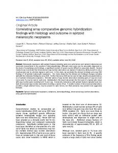
A 39-year-old woman, gravida 4, para 1, abortus 2, was referred at 20 weeks' gestation due to limb abnormality disclosed by a routine second-trimester anomaly scan. No relevant family history was traced. Our Level II exam revealed a male singleton complicated with ectrodactyly manifested by deep median cleft of both hands and feet, as a result of the absence of the central digital rays. Syndactyly and hypoplasia of phalanges, metacarpals and metatarsals were found in all four limbs as well (Figure 1). No additional fetal anomaly was disclosed at the same time. Isolated split-hand/ split-foot malformation (SHFM) was diagnosed after a thorough anatomical survey. She opted to keep the pregnancy after counseling for the condition and its prognosis. Array comparative genomic hybridization (aCGH) was advised since ectrodactyly may sometimes associate with certain genetic defects, such as ectrodactylyeectodermal dysplasiaecleft lip/palate syndrome (EEC) [1] or Cornelia de Lange syndrome [2]. Amniocentesis was done in the same week. aCGH (GRCh36/ hg18) revealed five distinct deletions involving mostly chromosomes 7 and 19. These were one 1.9-Mb microdeletion at chromosome 7p22.1p22.1(4,583,819-6,498,129), one 3.9-Mb micro deletion at chromosome 7q11.23q11.23(72,119,820-75,977,247), one 4.1-Mb microdeletion at chromosome 7q21.3q22.1(97,723,732101,812,625), one 24.0-Mb deletion at chromosome 19p13.3p12 (210,424-24,170,303), and one 26.2-Mb deletion at chromosome
* Corresponding author. Department of Obstetrics and Gynecology, National Taiwan University Hospital and National Taiwan University College of Medicine, Number 8, Chung Shan South Road, Zhongzheng District, Taipei City 10041, Taiwan. E-mail address:
[email protected] (J.-C. Shih).
19q13.11q13.43(37,601,047-63,787,200) on aCGH (CytoChip Oligo; BlueGnome, Cambridge, UK; 100-kb resolution; genome reference: International Standard Cytogenomic Array Consortium and Database of Genomic Variants) using the uncultured amniocytes (Figure 2). The patient and her family were counseled about the likely outcome of these deletions. Since SHFM is sometimes associated with EEC, sensorineural deafness or mental retardation, the patient chose an elective termination at 23 weeks' gestation. The gross features of the fetus were identical to those that were depicted by antenatal ultrasound (Figure 3). SHFM occurs in one of 18,000 live births, and accounts for 8e17% of all limb malformations [3]. SHFM is a diverse congenital limb abnormality. The diverse severities of digital defects and varied limb numbers of involvement have been described in the SHFM. In our patient with isolated SHFM, all four limbs were involved, with different presentations of both partial and complete absence of central rays. It may be an isolated abnormal finding or syndromic presentation such as EEC, acro-dermato-ungual-lacrimal-tooth syndrome, lacrimo-auriculo-dento-digital syndrome, CHARGE sequence (coloboma of the eye heart defects, atresia of the nasal choanae, retardation of growth and/or development, genital and/or urinary abnormalities, and ear abnormalities and deafness), VACTERL association (vertebral anomalies, anal atresia, cardiovascular anomalies, trachea-esophageal fistula, renal and/or radial anomalies, limb defects), mental retardation and sensorineural deafness [1]. The formation and development of limb buds are mediated by the signal produced by these three cell groups: apical ectodermal ridge (AER), the progress zone and the zone of polarizing activity. Among them, aberrations of maintaining AER result in the formation of SHFM manifestation [4]. So far, seven chromosomal loci associated with SHFM have been described, including SHFM1e6 and SHFM/SHFLD. Translocations, inversions, and duplications were reported in these involved regions, but in most instances they were
http://dx.doi.org/10.1016/j.tjog.2014.12.009 1028-4559/Copyright © 2015, Taiwan Association of Obstetrics & Gynecology. Published by Elsevier Taiwan LLC. All rights reserved.
456
Y.-L. Hsueh et al. / Taiwanese Journal of Obstetrics & Gynecology 54 (2015) 455e458
Figure 1. (AeC) 2D and 3D US of right hand. (D) 2D US of fetal left hand; (EeG) 2D and 3D US of fetal right leg. (F) 2D US of the fetal left leg. (AeH) Show the absence of the central digital rays of limbs. 2D ¼ two-dimensional; 3D ¼ three dimensional; US ¼ ultrasound.
deletions [1]. Furthermore, TP63, WNT10B, and DLX5 are also known as candidate genes for SHFM4 (3q28, OMIM 605289), SHFM6 (12q13, OMIM 601906), and SHFM1 (7q21, OMIM 183600) [1], respectively. In our patient, the 4.1-Mb microdeletion located on 7q21.3q22.1 (97,723,732-101,812,625) was initially suspected to contain the SHFM1 locus at the same region of chromosome 7. However, after detailed matching of the sequence, the deletions did not include DLX5 (hg18 chr7:96487638-96492079) and DLX6 (hg18 chr7:96473226-96478288) gene sequences, which are responsible for SHFM1 phenotypes (7q21, OMIM 183600) [5]. DLX5 and DLX6 were hypothesized to be the repressors of downstream target genes that reduce cell proliferation in AER, which is crucial for limb development [6]. In fact, SHFM1 phenotypes were also found in patients with interrupted genes adjacent to SHFM1 region, without direct missing the region of DLX5 and DLX6 [7,8]. The authors hypothesized that downregulation of these genes by disruption of a control element could be a cause of the syndrome. Further study is needed to investigate our region related to this phenotype. In fact, all the five distinct deletions of our patient did not contain SHFM 1e6 or SHFM/SHFLD that were well-established connections of specific phenotypes of SHFM. We then ran a detailed search in OMIM for the possible link between these deletions and the phenotypes of this affected fetus. Surprisingly, another 24.0-Mb deletion at 19p13.3p12 (210,424-24,170,303), which contains EPS15L1 gene sequence located at 19p13.11 region (hg 18 chr19:16,327,059-16,443,762), was identified in this fetus. EPS15L1 gene was mapped at the 19p13.11 region (hg 18 chr19:16,327,059-16,443,762), and is assumed to be a candidate
gene for SHFM [9]. EPS15L1ecoded protein acts as a substrate for the tyrosine kinase activity of the epidermal growth factor receptor, which is associated with limb morphogenesis [9]. Aten et al [10] first described the presentations of SHFM, tetralogy of Fallot, and clinical phenotypes similar to Angleman syndrome in a patient with a de novo 1-Mb microdeletion in 19p13.11. Bens et al [11] described a patient with a de novo microdeletion at 19p13.11, which was almost identical with the sequence reported by Aten, except that EPS15L1 and CALR3 genes are retained. The two patients shared similar phenotypes (e.g., deep-set eyes, strabismus, and developmental delay) except for SHFM features and heart defects that were not seen in the patient of Bens et al [11]. Therefore, the contribution of EPS15L1 deletion to SHFM manifestation was hypothesized. Our case supports this notion. Nevertheless, more solid evidence is required to prove the genotypeephenotype correspondence. In addition, the aCGH of this fetus also contained one 3.9-Mb microdeletion at 7q11.23 and one 26.2-Mb deletion at 19q13.11q13.43. The microdeletion of 7q11.23 locus (OMIM 194050) is responsible for WilliamseBeuren syndrome, which is characterized as an atypical facial profile, cardiovascular malformations (mostly supravalvular aortic stenosis and/or pulmonary artery stenosis), and moderate mental retardation [12]. 19q13.11 microdeletion syndrome (OMIM 613026) is characterized by lowset ears, retrognathia/micrognathia, broad nasal root, and thin lips, cutis aplasia, and pre- and postnatal developmental delay [13]. Nonetheless, the contributions of these two microdeletions in this fetus were not ascertained. Neither abnormal facial profile nor cardiovascular malformations were observed in this fetus at 23
Y.-L. Hsueh et al. / Taiwanese Journal of Obstetrics & Gynecology 54 (2015) 455e458
457
Figure 2. Five different sizes of deletions involving chromosomes 7 and 19 are illustrated in the array comparative genomic hybridization using uncultured amniocytes.
Figure 3. Postnatal images. (A) Gross features including facial appearance. (BeE) Highlights of right hand, left hand, right foot and left foot respectively.
weeks gestation. We were not able to locate the role of these deletions and the clinical manifestations in this fetus. Conflicts of interest The authors have no conflicts of interest relevant to this article. References [1] Anna SS, Magdalena S, Aleksander J. Split-hand/foot malformation e molecular cause and implications in genetic counseling. J Appl Genet 2014;55: 105e15.
[2] Dempsey MA, Knight Johnson AE, Swope BS, Moldenhauer JS, Sroka H, Chong K, et al. Molecular confirmation of nine cases of Cornelia de Lange syndrome diagnosed prenatally. Prenat Diagn 2014;34:163e7. [3] Czeizel AE, Vitez M, Kodaj I, Lenz W. An epidermiological study of isolated split hand/foot in Hungary, 1975e1984. J Med Genet 1993;30:593e6. [4] Duijf PH, van Bokhoven H, Brunner HG. Pathogenesis of split-hand/split-foot malformation. Hum Mol Genet 2003;12:R51e60. [5] Crackower MA, Scherer SW, Rommens JM, Hui CC, Poorkaj P, Soder S, et al. Characterization of the split hand/split foot malformation locus SHFM1 at 7q21.3-q22.1 and analysis of a candidate gene for its expression during limb development. Hum Mol Genet 1996;5:571e9. [6] Robledo RF, Fajan L, Li X, Lufkin T. The Dlx5 and Dlx6 homeobox genes are essential for craniofacial, axial, and appendicular skeletal development. Genes Dev 2002;16:1089e101.
458
Y.-L. Hsueh et al. / Taiwanese Journal of Obstetrics & Gynecology 54 (2015) 455e458
[7] van Silfhout AT, van den Akker PC, Dijkhuizen T, Verheij JB, OlderodeBerends MJ, Kok KS, et al. Split hand/foot malformation due to chromosome 7q aberrations(SHFM1): additional support for functional haploinsufficiency as the causative mechanism. Eur J Hum Genet 2009;17:1432e8. [8] Bernardini L, Palka C, Ceccarini C, Capalbo A, Bottillo I, Mingarelli R, et al. Complex rearrangement of chromosomes 7q21.13-q22.1 confirms the ectrodactyly-deafness locus and suggests new candidate genes. Am J Med Genet A 2008;146:238e44. [9] Wong WT, Schumacher C, Salcini AE, Romano A, Castagnino P, Pelicci PG, et al. A protein-binding domain, EH, identified in the receptor tyrosine kinase substrate Eps15 and conserved in evolution. Proc Natl Acad Sci USA 1995;92: 9530e4. [10] Aten E, den Hollander N, Ruivenkamp C, Knijnenburg J, van Bokhoven H, den Dunnen J. Split hand-foot malformation, tetralogy of Fallot, mental
retardation and a 1 Mb 19p deletion e evidence for further heterogenecity? Am J Med Genet A 2008;152:2053e6. [11] Bens S, Haake A, Tonnies H, Varter I, Stephani U, Hoterhus PM, et al. A de novo 1.1 Mb microdeletion of chromosome 19p13.11 provides indirect evidence for EPS15L1 to be a strong candidate for split hand split foot malformation. Eur J Med Genet 2011;54:e501e4. [12] Lacroix A, Pezet M, Capel A, Bonnet D, Hennequin M, Jacob MP, et al. WilliamseBuren syndrome: a multidisciplinary approach. Arch Pediatr 2009;16: 273e82. [13] Kulharya AS, Michaelis RC, Norris KS, Taylor HA, Garcia-Heras J. Constitutional del(19)(q12q13.1) in a three-year-old girl with severe phenotypic abnormalities affecting multiple organ systems. Am J Med Genet 1998;77:391e4.