Avian Pathology (1999) 28, 545±557
Arthropathic and amyloidogenic Enterococcus faecalis infections in brown layers: a study on infection routes W. J. M. Landman1*, D. R. Mekkes1, R. Chamanza 2,3, P. Doornenbal1 and E. Gruys3 1
Animal Health Service, Poultry Health Centre, P.O. Box 9, 7400 AA Deventer, The Netherlands, Department of Poultry Health, Faculty of Veterinary Medicine, Harare University, Zimbabwe, and 3 Department of Veterinary Pathology, Faculty of Veterinary Medicine, Utrecht University, The Netherlands 2
Intravenous, intra-articular and intraperitoneal inoculation of 6-week-old brown-layer pullets with an arthropathic and amyloidogenic strain of Enterococcus faecalis resulted in amyloid arthropathy, while intramuscular, oral and intratracheal inoculation did not. Oral inoculation of 1-day-old chickens did not cause any pathology. However, intramuscular inoculation with 106 colony forming units resulted in severe growth retardation and arthritis in 60% of the birds, and amyloid arthropathy in approximately 40%. In egg transmission studies, neither egg dipping nor inoculation of the air chamber with E. faecalis reproduced the condition, although a few chicks became septicaemic. Yolk sac inoculation of 6-day-old embryos caused embryonic death within 2 days. In contrast, egg albumen inoculation with E. faecalis led to arthritis in one of six of the progeny, indicating the possibility that vertical transmission of E. faecalis by the oviductal route could lead to arthritis. The presence of antibodies to E. faecalis was con® rmed by enzyme-linked immunosorbent assay in 14/15 of experimental birds that had developed arthritis.
Introduction Amyloid arthropathy was found mainly in heavy brown rearing layers with growth retardation and generalized amyloidosis, and the joints as the major deposition site (Landman et al., 1994). The main joints involved were the hip, knee and hock. Amyloid was found to a lesser extent in wing joints and internal organs. Articular amyloid has also been found in guinea fowl (Numida meleagris galeata) (Maestrini & Pascucci, 1970) and in turkeys (Meleagris gallopavo gallopavo) (Shivaprasad et al., 1991). Recently, amyloid arthropathy has been described in an Indian peafowl (Pavo cristatus) (Landman & Gruys, 1998) and broiler breeders (Landman et al., 1998), indicating that in Galliformes, the joint is a major target site for amyloid deposition. In ® eld cases of amyloid arthropathy, Enterococcus faecalis was isolated from 10 to 12% of the
amyloid positive joints (Landman et al., 1994, 1998). Some of the isolates were used to develop an animal model for amyloid arthropathy in which joint amyloidosis developed in all birds following a single intravenous or intra-articular injection of high doses (108 to 109 colony forming units (cfu)) of E. faecalis (Landman et al., 1997). Further assessment of ® eld cases and the evaluation of various agents as a potential cause for amyloid arthropathy established the importance of E. faecalis in the aetiology of joint amyloidosis in brown layers. Moreover, pulsed-® eld gel electrophoresis of various E. faecalis isolates, collected over a 4-year period from several European countries, showed similar chromosomal DNA restriction endonuclease digestion patterns for all amyloidogenic and amyloid associated isolates analyzed so far, suggesting clonal spread (Landman et al., 1998). Biochemical characterization of joint amyloid
* To whom correspondence should be addressed. Fax: 1 31 570 660175. E-mail:
[email protected] Received 2 September 1998. Accepted 1 July 1999. ISSN 0307-9457 (print)/ISSN 1465-3338 (online)/99/060545-13
Ó 1999 Houghton Trust Ltd
546 W. J. M. Landman et al.
protein showed it was of AA type, with an amino acid sequence quite similar to duck AA (Landman et al., 1996). Its precursor protein, serum amyloid A (SAA), is encoded by a single gene. SAA mRNAs in brown and white layers give rise to primary translation products with identical amino acid sequence (OvelgoÈnne et al., 1998), indicating that reported differences in breed susceptibility are related to mechanisms other than the SAA gene coding sequences. The histopathological and ultrastructural features of chicken amyloid arthropathy have been extensively described (Peperkamp et al., 1997). E. faecalis has been described as a cause of omphalitis, yolk sac in¯ ammation, endocarditis, meningitis, ® brinous arthritis and/or tenosynovitis (Gross & Domermuth, 1962). However, the existence of isolates with strong amyloidogenic potential has not been described by authors outside our group. Our E. faecalis induction model for amyloid arthropathy was used as a positive control to further study the pathogenesis of arthropathic and amyloidogenic E. faecalis infections in brownlayer pullets. We focused on various ages and different infectious routes that might lead to haematogeneous spread of E. faecalis, enabling chronic (poly)arthritis and subsequent amyloid arthropathy to occur. Also, to assess the presence of antibodies to E. faecalis in experimental birds, a whole-cell antigen enzyme-linked immunosorbent assay (ELISA) was performed. Material and Methods Experimental animals All experiments were performed using brown replacement pullets and brown replacement pullet hatching eggs. The chickens were housed in separated ¯ oor pens or isolators (Beyer & Eggelaar, Utrecht, The Netherlands). In each animal experiment, a negative and positive control group were included. Only high-quality commercial hatching eggs were used, i.e. free from salmonella and mycoplasma, and not soiled (not ¯ oor eggs and not washed). Birds were weighed at the start and end of all experiments. In all experiments, the chickens were fed ad libitum with a commercial layer rearing pellet containing 10.9 megajoules (MJ)/kg of metabolic energy. A lighting scheme consisting of 16 h of light and 8 h of dark was applied. Birds were housed, handled and treated following approval by the Institutional Animal Experimental Committee in accordance with the Dutch regulations on experimental animals.
Inocula For all experiments, E. faecalis (isolate 6085.94) isolated from a spontaneous case of amyloid arthropathy (Landman et al., 1994) and known to induce amyloid arthropathy (Landman et al., 1997) was used. Frozen beads containing the isolate ( 2 70°C) were rolled on a sheep-blood agar plate. After overnight incubation at 37°C, colonies were scraped off and a suspension of 109 cfu was prepared in peptone saline (International Standard Organisation 6887, 1993). Control of the bacterial concentrations was performed by means of bacterial counting according to international standards (International Standard Organisation 7402, 1985).
Experimental design Experiment 1: different routes of administration. Five-week-old brown pullets were obtained from a commercial farm. The birds had been routinely vaccinated against Marek’ s disease, infectious bronchitis, Newcastle disease and Gumboro disease. After 1 week of acclimatization, ® ve chickens per group were injected either intravenously (vena ulnaris), intraperitoneally, intra-articularly (left knee), intramuscularly (musculus pectoralis), orally or intratracheally with 109 cfu of E. faecalis prepared as already described. As negative controls, ® ve chickens per group were inoculated with nutrient broth. During the experimental period, the birds were housed in two isolated litter ¯ oor pens. The birds inoculated parenterally were in one pen, and those infected orally and intratracheally were in the other. The birds were necropsied 10 weeks after start of the experiment. Experiment 2: inoculation of 1-day-old chicks with E. faecalis. Hatching eggs, which had been incubated for 18 days, were purchased from a commercial hatchery and further incubated after disinfection with formaldehyde gas (20 g potassium permanganate and 30 ml formalin 40% per m3 ). At hatch, eight chickens were inoculated orally with E. faecalis (108 cfu) inoculum. Four control chicks were inoculated with peptone saline (PS). Different doses of E. faecalis, as detailed in Table 1, were injected intramuscularly, with 16 chicks per dose (in the musculus gastrocnemius, following Marek’ s disease vaccination). As negative controls, eight chickens were injected with PS. All groups were housed separately in isolators. The birds were necropsied 7 weeks after the start of the experiment. Experiment 3: transmission studies in eggs. Preliminary studies. Before starting the experiment, the maximum PS volume uptake in time, by means of temperature gradient, was established in an extra set of hatching eggs. Groups of 10 eggs were dipped for eight different time periods (5, 10, 15, 20, 30, 40, 50 and 60 min). PS uptake of eggs was induced using a temperature gradient: PS at 6.3°C and eggs at 25.6°C. An E. faecalis dilution series was used to examine differences in the degree of egg contamination. Groups of 30 eggs were dipped in four different concentrations of E. faecalis: 102 , 104 , 106 or 108 cfu/ml. A control group of 29 eggs was dipped in PS. The eggs were at a temperature of 25.5°C, while the suspensions were cooled at 6.4°C. Eggs were then incubated for 18 days and then examined for bacteriological contamination. Effect of egg air-chamber inoculation and egg dipping (Flock 1). Brown replacement pullet hatching eggs were purchased from a commercial hatchery and used to study the effect of egg dipping (batch A, 50 eggs) and egg air-chamber inoculation at the ninth day of incubation (batch B, 50 eggs), with E. faecalis. As negative controls, eggs were either dipped (25 eggs) or air-chamber inoculated (25 eggs) with PS. Inoculated eggs were given 0.25 ml E. faecalis suspension (108 cfu/ml) in the air chamber, after eggshell disinfection with a 1% iodine (J2, Merck 4761) solution (in 70% ethanol). Eggs were sealed with a droplet of molten paraf® n wax and fumigated with formaldehyde gas before further incubation. Dipped eggs (26°C) were submersed in E. faecalis broth with 108 cfu/ml at 6°C. On the 19th day of incubation, candled eggs (i.e. infertile eggs and eggs containing dead embryos), in addition to randomly selected eggs up to a maximum of 10 eggs per treatment group and ® ve eggs per control group, were collected for bacteriological examination of the yolk sac. The birds were housed in separated ¯ oor pens, while the controls were placed in isolators. At 11 weeks of age, one-half of the ¯ ock which hatched was necropsied and the remaining birds were debeaked. The latter group was necropsied at 16 weeks of age. Effect of yolk-sac inoculation (Flock 2). Yolk-sac inoculation of 132 brown replacement pullet hatching eggs with a dilution series (20, 102 , 104 , 106 and 108 cfu) of E. faecalis (0.75 ml/egg) was performed on
b
12.8 6
16 5 cfu 0/16 0/16 0/16 0/16 0.4B
16 165 cfu 2/16 0/14b 2/16 1/13 (25.2%) 13.8 6 0.5C
E. faecalis
Intramuscular
0.18);
16 105 cfu 1/16 0/16 1/16 3/15 (21.1 to 46.3%) 13.9 6 0.4D
0.50); A±E, P , 0.001; B±C, not signi® cant (P 5
4 1 ml 0/4 0/4 0/4 1/4 (19.2%) 14.4 6 0.6G
PSa
PS, Peptone saline; s.e., standard error of the mean. Missing birds died early due to E. faecalis septicaemia. c Range percentage positivity (PP) above 18.6%. d Comparison between treatment groups: A±B, P , 0.05; A±C, not signi® cant (P 5 0.39); A±D, not signi® cant (P 5 B±D, not signi® cant (P 5 0.09); B±E, P 5 , 0.001; C±D, not signi® cant (P 5 0.80); C±E, P , 0.001; D±E, P , 0.001; F±G, not signi® cant (P 5 0.97).
a
8 108 cfu 0/8 0/8 0/8 4/8 (19.3 to 26.1%) 14.5 6 0.3F
n Dose Arthritis Congo red positive joint smears or histology Re-isolation from joint Positive E. faecalis ELISA (PP)c Average daily growth at necropsy (g 6 s.e.) d
Oral
E. faecalis
Inoculation route Inoculum
16 106 cfu 9/16 6/16 6/16 5/13 (58.8 to 88.4%) 9.3 6 0.5E
14.5 6
8 1 ml 0/8 0/8 0/8 0/7
PS
0.7A
Table 1. Effect of the administration route of arthropathic and amyloidogenic E. faecalis on the occurrence of amyloid arthropathy after inoculation in 1-day-old brown replacement pullets (Experiment 2)
Pathogenesis of E. faecalis infections in chickens 547
548 W. J. M. Landman et al. the sixth day of incubation. As a control, 22 PS inoculated eggs were used. Eggs were candled at the 19th incubation day. All eggs were examined bacteriologically. Effect of egg albumen inoculation (Flock 3). Egg albumen inoculation of 125 brown replacement pullet breeding eggs with a dilution series (30, 103 , 105 and 107 cfu) of E. faecalis (0.75 ml/egg) was performed in embryonated eggs incubated for 6 days. As controls, 25 PS inoculated eggs were used. Eggs were candled at the 19th day of incubation. All non-hatching eggs were examined bacteriologically. The birds were housed in separated ¯ oor pens. The birds were necropsied at 7 weeks of age. Assessment of embryo mortality. To accurately assess the embryo mortality pattern, 300 additional eggs were either yolk-sac or albumen inoculated with 10, 102, 103, 105 and 107 cfu of E. faecalis (25 eggs per group). Control eggs were inoculated with PS. Eggs were candled at the 19th incubation day. All non-hatching eggs and hatched chicks were examined bacteriologically.
SPF sera. Serum from 80 5-week-old SPF White Leghorn birds, kept under ® ltered air positive pressure conditions, were tested as already described and used to determine the cut-off value. The PP values were calculated for each serum individually, relative to the positive control. The positive-negative cut off PP for the ELISA was determined as the mean PP plus three times the standard deviation calculated from this set of 80 SPF sera (Wright et al., 1993).
Statistical analysis Differences in average daily weight gain between treatment groups were tested with analysis of covariance (SAS Institute Inc., 1991). The weight of pullets at the start of the experiment was entered in the statistical model as a covariate to account for unequal starting weights between treatment groups.
Results Postmortem and further procedures Birds were killed, using an electric current (10 s at 340 V). Jugular vein blood was obtained for E. faecalis serology by means of ELISA. Bacteriological analysis of specimens was performed as previously described (Landman et al., 1994). Whole joints and 1 cm3 samples of liver, spleen, kidney, jejunum, pancreas and proventriculus were ® xed in 4% formalin, and paraf® n embedded. Sections were stained with haematoxylin and eosin, and Congo red (Stokes, 1976). Smears of synovial ¯ uid from the knee joint and, if unaffected, the hock joint were ® xed in 4% buffered formaldehyde for alkaline (Stokes, 1976) and aqueous (Romha nyi, 1971) Congo red staining.
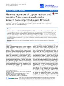
Experiment 1: different routes of administration As illustrated by the results in Table 2, both the intravenous and intra-articular inoculation of E. faecalis resulted in amyloid arthropathy (Figure 1) in all birds. After intraperitoneal application of E. faecalis, 40% of the birds developed joint amyloidosis. Intratracheal, oral and intramuscular inoc-
Detection of antibodies to E. faecalis (ELISA) An ELISA was performed by modifying the method described by Arduino et al. (1994). The antigen was prepared from arthropathic and amyloidogenic E. faecalis isolate 6085.94 (Landman et al., 1997) grown overnight on sheep-blood agar plates. Colonies were swabbed off into phosphate-buffered saline (PBS) (pH 7.2) and subsequently washed three times in PBS. The antigen was diluted 1:1000 in 0.1 M sodium carbonate buffer (pH 9.6) and coated at 100 ml per well in rows A, C, E and G of 96-well polystyrene plates. Plates were kept overnight at 4°C. Sodium carbonate buffer (pH 9.6) without E. faecalis antigen served as control antigen and was coated in rows B, D, F and H. Following coating, wells were blocked with 1% bovine serum albumin in 0.1 M sodium carbonate buffer (pH 9.6) for 1 h at room temperature. Wells were washed three times with PBS containing 0.05% Tween 20 (PBST). All sera were assayed against E. faecalis and control antigen simultaneously, using a serum dilution of 1:500 in PBST. A speci® c pathogen free (SPF) serum pool from 10 individual birds was used as a negative control, and a blank (PBST) and positive control were included on each plate. The positive control consisted of pooled sera from ® ve birds infected with E. faecalis by intravenous injection (109 cfu/bird). Plates were incubated for 1 h at room temperature and subsequently washed three times with PBST. To each well, 100 ml rabbit anti-chicken immunoglobulin G (IgG) horseradish peroxidase conjugate was added at a 1:20 000 dilution in PBST. Following incubation for 1 h at room temperature, wells were washed three times with PBST. To each well, 100 ml substrate solution containing 3,39 ,5,59 -tetramethylbenzidine (at 0.1 g/l) and H2 O2 (1.3 mmol/l) in 0.1 M sodium acetate buffer (pH 6.0) was added. Plates were incubated for 15 min at room temperature and the reaction was stopped by adding 0.12% hydrogen ¯ uoride. Optical densities (ODs) were read at 650 nm and corrected ODs were calculated for all tested samples and the respective controls using the formula: corrected OD 5 (ODantigen ± ODcontrol antigen). For each sample, results were calculated from corrected OD values and expressed as percentage positivity (PP) related to the positive control used: PP 5 (corrected OD ± corrected ODnegative control)/(corrected ODpositive control ± corrected ODnegative control) (Wright et al., 1993).
Figure 1. Medial view of a femoro-tibial and tibio-metatarsal joint with amyloid arthropathy after intravenous inoculation of E. faecalis. Note the enlargement of both joints due to amyloid deposition, particularly in the synovial recesses.
Pathogenesis of E. faecalis infections in chickens 549
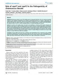
Figure 2. Two brown replacement pullets (4 weeks of age) from the same hatch; the left bird is a healthy control, the right bird was inoculated intramuscularly at hatch with 106 cfu of arthropathic and amyloidogenic E. faecalis (Experiment 2).
ulations of E. faecalis did not cause amyloid arthropathy. Re-isolation of E. faecalis was only achieved from birds inoculated by the intravenous (3/3) or intra-articular routes (3/4). These groups also had the lowest average daily growth, and this was signi® cantly different (P , 0.001 and P , 0.05, respectively) from the average daily growth of controls (Table 2). Experiment 2: inoculation of 1-day-old chicks with E. faecalis Oral inoculation of 1-day-old chicks with high doses of E. faecalis did not cause amyloid arthropathy or any other pathology (Table 1). Birds looked healthy and developed well. Lower doses of E. faecalis injected intramuscularly resulted in similar joint pathology to the higher doses, although only a few birds were affected and no amyloid was found in the joints. In the group injected with 165 cfu, 2/16 chickens developed arthritis, while in the group receiving 105 cfu, only one bird was affected (Table 1); E. faecalis was re-isolated from these affected joints. For the group receiving 106 cfu/bird, E. faecalis was re-isolated from 6/16, while 9/16 had severe arthritis. Amyloid arthropathy was present in six of the 16 birds with arthritis. Birds with joint involvement showed severe growth retardation (Figure 2). On histopathological examination, joint amyloid deposits were found in lesioned synovial membrane, on the surface of articular cartilage and around the nutritional vessels of the articular carti-
lage (Figure 3A,B). The arthritis was evidenced by proliferation of synoviocytes, with purulent exudate in the articular recesses, and in® ltration of the synovial membrane by lymphocytes, plasma cells and heterophils (Figure 3C,D). The synovial membrane presented either irregular extensions or villous hypertrophy. Half of the birds had arthritis deformans. Birds infected intramuscularly with 106 cfu showed the lowest daily average growth, and were signi® cantly lighter than the controls and birds receiving other doses (P , 0.001). The average daily growth of the other groups did not differ from each other nor from the controls, except for the group inoculated with 5 cfu. This group was signi® cantly lighter than the controls (P , 0.05) (Table 1). The orally inoculated group did not differ in average daily growth from the control group (Table 1). Experiment 3: transmission studies in eggs Preliminary studies. Determination of the PS volume uptake with time during egg dipping. PS volume absorbtion reached a plateau after a dipping time of 20 min, when the average volume was 0.29 6 0.026 ml (mean 6 standard deviation). Longer dipping time did not signi® cantly increase the volume uptake (Figure 4). Effect of E. faecalis concentration during egg dipping on egg infection rate. The average volume
550 W. J. M. Landman et al.
Figure 3. Congo red stain of chicken knee joint after intramuscular inoculation with 106 cfu of arthropathic and amyloidogenic E. faecalis at hatch viewed in full (A) and in polarized light (B). Note the darker stained areas (arrows) in A and the corresponding birefringent areas (bright) in B representing amyloid. Other bright areas represent collagen (white arrows in B). Note also the central area with cartilage deformation where the articular surface has lost its convexity (area between arrow heads). Bar 5 200 m . C, haematoxylin and eosin stain of articular recesses showing extensive cellular in® ltration. Bar 5 200 m . At higher magni® cation (3D), heterophil (arrow heads), lymphocytic and plasmacellular in® ltrations (arrows) are visible. Bar 5 50 m .
Pathogenesis of E. faecalis infections in chickens 551
uptake at 20 min was $ 0.29 6 0.100 ml for all groups, except for the group treated with the highest dose showing an average uptake of 0.22 6 0.100 ml. No E. faecalis organisms were found in the control eggs. The numbers of E. faecalis-infected
eggs found by bacteriological examination were 22/30, 16/30, 30/30 and 27/30, respectively, for doses 102, 104, 106 and 108 cfu/ml. In the subsequent studies, a dipping time of 20 min was used and, in ¯ ock 1, the highest E. faecalis concentration (108 cfu).
0/5
0/5
16.0 6
3/3*
3/3
3/3 (70.1 to 96.7%)
5.2 6
0.8B
Comparing the effect of each treatment with controls: A±B, P , 0.001; C±D, P , 0.05; E±F, P 5 0.29; G±H, P 5 0.28; I±J, P 5 0.095; K±L, P 5
**
Missing birds died due to E. faecalis septicaemia. Missing birds died due to cannibalism. *** Range percentage positivity (PP) above 18.6%. s.e.: standard error of the mean.
*
0/5
5 109 cfu
n Dose Congo red positive joint smears Reisolation of E. faecalis from joint Positive E. faecalis ELISA (PP)*** Average daily growth in g 6 s.e.
0.9A
5 1 ml
E. faecalis
Inoculum
Nutrient broth
Intravenous
Inoculation route
1.5C
15.3 6 1.2D
0/5
0/5
0/5
5 1 ml
Nutrient broth
0.33 (not signi® cant).
7.9 6
4/4 (21.4 to 80.9%)
3/4
4/4**
5 109 cfu
E. faecalis
Intra-articular
0/4
0/4**
5 1 ml
Nutrient broth
13.4 6 1.0E
15.0 6 1.1F
2/5 0/4 (86.1 to 94.6%)
0/5
2/5
5 109 cfu
E. faecalis
Intraperitoneal
15.6 6 0.4G
0/5
0/5
0/5
5 109 cfu
E. faecalis
Intramuscular
16.2 6 0.4H
1/5 (22.5%)
0/5
0/5
5 1 ml
Nutrient broth
15.9 6 0.4I
4/5 (32.3 to 82.3%)
0/5
0/5
5 109 cfu
E. faecalis
Intratracheal
17.1 6 0.4J
1/5 (30.8%)
0/5
0/5
5 1 ml
Nutrient broth
17.1 6
0/5
0/5
0/5
0.4K
5 109 cfu
E. faecalis
Oral
16.4 6 0.4L
0/5
0/5
0/5
5 1 ml
Nutrient broth
Table 2. Effect of the route of administration on the occurrence of amyloid arthropathy after inoculation with arthropathic and amyloidogenic E. faecalis in 6-week-old brown replacement pullets (Experiment 1)
552 W. J. M. Landman et al.
Pathogenesis of E. faecalis infections in chickens 553
seventh and eighth day of incubation. There were no deaths in the PS-inoculated controls. E. faecalis was re-isolated from all eggs except the controls. Effect of egg albumen inoculation (Flock 3).
Figure 4. Volume uptake of eggs with time during egg dipping in peptone saline (box and whisker plot; Experiment 3).
Effect of egg air chamber inoculation and egg dipping (Flock 1). Egg dipping resulted in a volume uptake of 0.20 6 0.147 ml for the E. faecalis group and 0.20 6 0.062 ml for the controls. The numbers of E. faecalis-positive eggs sampled on the 19th day of incubation were: 8/10 (3/8 positive eggs had live embryos, the others were dead) for those injected into the air chamber with E. faecalis; 0/5 for the injected controls; 9/10 for those dipped in E. faecalis (5/6 live embryos); and 0/5 for the dipped controls. The hatch results for the remaining eggs were: 77.2% for the injected controls, 64% for the eggs injected with E. faecalis, 91% for the dipped controls and 78% for the eggs dipped in E. faecalis suspension. Three birds died during the ® rst week of life due to E. faecalis septicaemia. Two of these chickens hatched from injected eggs and one from the dipped group, indicating low transmissions of 4 and 2%, respectively. No E. faecalis was re-isolated from joints of any treatment group at necropsy, nor were any amyloid positive birds found at necropsy. Joint smears were also negative for amyloid. Regarding growth (data not shown), dipping with E. faecalis did not in¯ uence growth (P 5 0.43), in contrast to air chamber injection (P , 0.05) in birds necropsied at 11 weeks of age. In the remainder, which were necropsied at 16 weeks, there was no signi® cant difference in weights.
Egg albumen inoculation of E. faecalis also resulted in high embryonic death rates when high doses of E. faecalis were used ( $ 96% for 103, and 100% for 105 and 107 cfu). However, embryonic death rate was lower (76%) at lower doses (30 cfu). In the latter group, transmission of E. faecalis with associated arthritis (re-isolation from joint) on a small scale was achieved, i.e. 1/6 hatched chicks (1/25 inoculated eggs). The affected bird showed severe growth depression, weighing 233 g (controls, 647 6 83.7 g; mean 6 s.d.), ruf¯ ed feathers and the characteristic stiff gait observed also in amyloid arthropathic birds in the ® eld. The only chick that hatched from the 103 cfu dose group, died during the ® rst week due to E. faecalis septicaemia. The hatching percentages were: 88% for the controls, 24% for the 30 cfu and 4% for the 103 cfu group. E. faecalis was re-isolated from all non-hatching eggs except the controls. Assessment of embryo mortality. All embryos from yolk-sac inoculated eggs died between day 7 and 8 of incubation, except for one that died at day 17. The hatching percentage of controls was 56%. Embryonic death occurred later in time for some lower doses of albumen-inoculated eggs, allowing a small number of chicks to hatch and con® rming the results of the previous experiment (Flock 3). The hatching percentages of the albumen-inoculated eggs were; 84% for the controls, 12% the 102 cfu, and 4% the 103 cfu. E. faecalis was re-isolated from the non-hatched eggs and the yolk sac of hatched chicks, except for the controls. Detection of antibodies to E. faecalis (ELISA) SPF sera. The cut-off value of the E. faecalis ELISA, calculated from the 80 SPF sera as mean plus three times the standard deviation and expressed as PP, was 18.6%. Therefore, in all subsequent experiments, sera with PP values $ 18.6% were considered positive.
Effect of yolk-sac inoculation (Flock 2). Yolk-sac inoculated eggs were candled at day 19. At this time, all embryos of the E. faecalis dilution series were dead. The embryos died between the
Sera from experimental birds. The ELISA detected antibodies in all birds with amyloid arthropathy (9/9) of Experiment 1. Anti-
554 W. J. M. Landman et al.
bodies were also detected in 4/5 intratracheally inoculated birds and two controls, the latter showing low (22.5 to 30.8%) PP values (Table 2). Low titres (19.2 to 26.1%) were found in 4/8 orally inoculated birds and one control (Table 1). Oral infection did not result in measurable antibody levels in Experiment 1, but did in Experiment 2. When the ELISA results from the control groups from Experiments 1 and 2 were combined, 3/49 birds reacted positive, resulting in a speci® city of 93.9% (95% con® dence interval of 87.9 to 97.0%) based on these experiments. Sensitivity was calculated from all E. faecalis-infected groups in both experiments, in which one or more birds developed joint pathology. Sensitivity was found to be 25.0% (95% con® dence interval of 18.0 to 33.3%). In total, the ELISA successfully detected antibodies in 14 out of 15 birds with arthritis, resulting in a relative sensitivity of 93.3%, again based on these experiments. Discussion In human patients, several types of (peri)articular amyloid deposits have been described associated with joint disease. In haemodialysis patients, amyloid deposits consisting of b 2 microglobulin precipitate have been found within the joints (Athanasou et al., 1991; Okada et al., 1993), while in patients with multiple myeloma, periarticular AL (immunoglobulin light chain) deposits occur (Donnelly et al., 1993). In old age, ATTR (transthyretin amyloid) deposits have been found in dystrophic joints (Gof® n et al., 1985). In other mammals, articular amyloid deposits (AA-like) have only been reported in two dogs with signs of rheumatoid arthritis (Colbatzky et al., 1991), while in senescence-accelerated mice, apoAII amyloid deposits in intervertebral discs have been described (Higuchi et al., 1991). In Galliformes, articular amyloid deposits appear to be more common (Maestrini & Pascucci, 1970; Shivaprasad et al., 1991; Landman & Gruys, 1998). Although human articular amyloidosis is of clinical signi® cance, chicken amyloid arthropathy (Landman et al., 1997) is the only animal model which has been described to study the pathogenesis of this condition and the tissue factors involved in localized amyloid deposition. E. faecalis arthritis has not so far been described in association with amyloid arthropathy in humans or other species apart from the chicken. Although E. faecalis causes 80 to 90% of human enterococcal infections (Murray, 1990), human E. faecalis arthritis is rare (Raymond et al., 1995) as opposed to bacteraemia and endocarditis (Maki & Agger, 1988; Gullberg et al., 1989; Jett et al., 1994; Centers of Disease Control and Prevention, 1998), suggesting that in the human, enterococci have a lower af® nity for joint tissue than for
cardiac valves. The use of synovial biopsies may be necessary to establish the diagnosis of E. faecalis arthritis in man, while treatment of this condition generally has a poor outcome (Mitchell et al., 1989). In rabbits, induction of chronic arthritis after inoculation with heat-killed E. faecalis has been reported (Yamamoto & Komatsuzaki, 1987). The lesions resembled those of rheumatoid arthritis, and it was suggested that an Arthus reaction of both immediate hypersensitivity and delayed-type hypersensitivity are necessary to the development of this arthritis. In rats, where induction of arthritis with antigens of streptococci of groups A, B and D was performed, it was postulated that differences in antigen processing by phagocytes may determine the outcome of the arthritis induced: insensitive to phagocyte processing (group A), no or chronic arthritis; intermediate sensitivity to phagocyte processing (group B), chronic arthritis; and sensitive (group D), transient arthritis (Spitznagel et al., 1983). Whether similar processes play a role in the outcome of the E. faecalis arthritis induced in brown layers is currently unknown. The results of Experiment 1 are in accordance with those of the animal model for amyloid arthropathy (Landman et al., 1997), where high doses (108 to 109 cfu) of E. faecalis isolates from ® eld outbreaks induced amyloid arthropathy in 6-week-old brown pullets after intravenous or intra-articular inoculation. Intraperitoneal injection also resulted in joint amyloidosis, although at a lower rate, while oral (also in 1-day-old chicks), intratracheal and intramuscular administration did not. This, suggests that intensive contact with the bloodstream is necessary for E. faecalis to access the joints, enabling (poly)arthritis to develop, with subsequent amyloid arthropathy. Considering the pathogenesis of bacterial arthritis in general, septicaemia is a risk factor for joint infection, as has been noted in humans, where 1.2% of bacteraemic individuals develop arthritis (Espersen et al., 1991), the knee joint being most frequently affected (Peters et al., 1992). Although infection from contiguous infected tissues or penetrating wounds is theoretically possible, it seems irrelevant in outbreaks of E. faecalis-related amyloid arthropathy in brown layers, as some affected ¯ ocks were reared on litter where penetrating wounds were not likely to occur. One-day-old chicks, which like most neonatal animals (Hauser et al., 1986; Wilson, 1986) will not be fully immunocompetent, were susceptible to intramuscular inoculation of E. faecalis, with approximately 60% developing arthritis and 40% amyloid arthropathy after treatment with 106 cfu. In contrast, 6-week-old brown pullets did not develop any pathology after intramuscular inoculation of 109 cfu, re¯ ecting the occurrence of age resistance to this route. This is in accordance with the research ® ndings that demonstrated increased
Pathogenesis of E. faecalis infections in chickens 555
susceptibility to infection with other pathogens, such as salmonellae in chickens (Ziprin et al., 1989) and Salmonella enteritidis in turkeys (Lowry et al., 1997) during the ® rst week posthatch, which was attributed to inef® cient T-cell response, heterophil phagocytosis and killing activities at this age. Direct yolk-sac inoculation with different dilutions of E. faecalis at day 6 of incubation caused massive and rapid embryonic death in all cases. This ® nding suggests that transovarian transmission is unlikely. In contrast, re-isolations from the yolk sac of live embryos from eggs that were dipped or inoculated into the air chamber at day 9 indicate that later infection of the yolk sac may not have such detrimental consequences as early inoculation or contamination. However, in birds that subsequently hatched from eggs in these groups, no joint pathology was found. Direct inoculation of egg albumen also resulted in large numbers of embryonic death for the highest doses. Mortality was lower after inoculation with 30 cfu, while in small numbers of survivors, E. faecalis arthritis (4%) was achieved, suggesting the oviduct as a possible infection route. This was similar to the small-scale egg transmission of E. faecalis by egg dipping (2%) and air-chamber inoculation (4%) to offspring, which died from septicaemia. Our ELISA showed a high speci® city for E. faecalis. The sensitivity was much lower, which was probably caused by the fact that not all inoculated birds developed an (systemic) infection and an antibody response. The fact that several intratracheally and orally inoculated chickens did respond serologically suggests that invasion and immunostimulation is achievable for this isolate through natural infection routes, although it did not progress into (poly)arthritis or amyloid arthropathy. The control birds found positive in the ELISA may have resulted from an aerogenic infection as the experimental birds were housed in separated ¯ oor pens where air transmission would be possible. Alternatively, the cut-off level of our ELISA test may have been set too low. Furthermore, an ELISA system based on speci® c E. faecalis surface antigens that are fully expressed after growth in serum and induce IgG in humans with endocarditis (Shorrock et al., 1990), was not found suitable to distinguish those patients from normal controls by another group of scientists (Arduino et al., 1994). The latter used a whole-cell ELISA, which gave satisfactory results. The latter was modi® ed to perform our ELISA. The fact that 1-day-old chicks, as opposed to older brown-layer pullets, are susceptible to induction of amyloid arthropathy with E. faecalis after intramuscular inoculation, raises the possibility of inducing E. faecalis bacteraemia, leading to amyloid complicated (poly)arthritis through Marek’ s disease vaccination at the hatchery. Epidemiological studies at the hatchery assessing the occurrence
of arthropathic and amyloidogenic E. faecalis in its environment are planned. In the present study, natural infection routes for arthropathic and amyloidogenic E. faecalis were not found. However, additional studies of natural infection routes are in progress. Acknowledgements We acknowledge P. L. de Leur and M. H. C. H. Arts for preparing the inocula and performing the bacteriological analysis, J. L. Nieuwenhuisen for preparing the sections for histology, Dr A. L. J. Gielkens and Dr J. H. H. van Eck for critically reading the manuscript, and Dr. E. Goren for his valuable suggestions. References Arduino, R.C., Murray, B.E. & Rakita, R.M. (1994). Roles of antibodies and complement in phagocytic killing of enteroccoci. Infection and Immunity, 26, 987±993. Athanasou, N.A., Ayers, D., Rainey, A.J., Oliver, D.O. & Duthie, R.B. (1991). Joint and systemic distribution of dialysis amyloid. Quarterly Journal of Medicine, 78, 205±214. Centers of Disease Control and Prevention. National Nosocomial Infection Surveillance (NNIS) System report, data summary from October 1986±April 1998, issued June 1998. American Journal of Infection Control, 26, 522±533. Colbatzky, F., Brunnberg, L., Linke, R.P., Giesel, O. & Hermans, W. (1991). AA-like amyloid deposits con® ned to arthritic joints in two dogs with rheumatoid arthritis. Journal of Comparative Pathology, 105, 331±343. Donnelly, S., Bourne, J.T., Levison, D.A., Doyle, D.V. & Hammond, A. (1993). Amyloid arthritis associated with IgM kappa lymphoplasmocytoid lymphoma. British Journal of Rheumatology, 32, 1004±1007. Espersen, F., Frimodt-Moller, N., Thamdrup Rosdahl, V., Skinhoj, P. & Bentzon, M.W. (1991). Changing pattern of bone and joint infections due to Staphylococcus aureus: study of cases of bacteraemia in Denmark, 1959±1988. Review of Infectious Diseases, 13, 347±358. Gof® n, Y.A., McCrickard, E.L., Ameryckx, J.P., Malmendier, C.L., Hiden, M. & Cornwell III, G.G. (1985). Amyloidosis of the joints: evidence that human hip capsules have a unique predisposition for amyloid of the senile systemic type. Applied Pathology, 3, 88±95. Gross, W.B. & Domermuth, C.H. (1962). Bacterial endocarditis of poultry. American Journal of Veterinary Research, 23, 320±329. Gullberg, R.M., Homann, S.R. & Phair, J.P. (1989). Enterococcal bacteraemia: analysis of 75 episodes. Reviews of Infectious Diseases, 2, 74±85. Hauser, M.A., Koob, M.D. & Roth, J.A. (1986). Variation of neutrophil function with age in calves. American Journal of Veterinary Research, 47, 152±153. Higuchi, K., Naiki, H., Kitagawa, K., Hosokawa, M. & Takeda, T. (1991). Mouse senile amyloidosis. Asam amyloidosis in mice presents universally as a systemic age-associated amyloidosis. Virchows Archives on B Cell Pathology including Molecular Pathology, 60, 231±238. International Standard Organisation 6887 (1983). MicrobiologyÐGeneral Guidance For The Preparation Of Dilutions For Microbiological Examination, 1st edn. Geneva: International Standard Organisation. International Standard Organisation 7402 (1985). MicrobiologyÐGeneral guidance for the enumeration of Enterobacteriaceae without resuscitationÐ MPN technique and colony count technique, 1st edn. Geneva: International Standard Organisation. Jett, B.D., Huycke, M.M. & Gilmore, M.S. (1994). Virulence of Enterococci. Clinical Microbiology Reviews, 7, 462±478.
556 W. J. M. Landman et al. Landman, W.J.M. & Gruys, E. (1998). Amyloid arthropathy in an Indian peafowl. Veterinary Record, 142, 90±91. Landman, W.J.M., Gruys, E. & Dwars, R.M. (1994). A syndrome associated with growth depression and amyloid arthropathy in layers: a preliminary report. Avian Pathology, 23, 461±470. Landman, W.J.M., Sletten, K., Koch, C.A.M., Tooten, P.C.J. & Gruys, E. (1996). Chicken joint amyloid protein is of the AA type. I. Characterization of the amyloid protein. Scandinavian Journal of Immunology, 43, 210±218. Landman, W.J.M., Peperkamp, N.H.M.T., Koch, C.A.M., Tooten P.C.J., Crauwels, P.A.P. & Gruys, E. (1997). Induction of amyloid arthropathy in chickens. Amyloid: The International Journal of Experimental and Clinical Investigation, 4, 87±97. Landman, W.J.M., Van den Bogaard, A.E.J.M., Doornenbal, P., Tooten, P.C.J., Elbers, A.R.W. & Gruys, E. (1998). The role of various agents in chicken amyloid arthropathy. Amyloid: The International Journal of Experimental and Clinical Investigation, 5, 266±278. Lowry, V.K., Genovese, K.J., Bowen, L.L. & Kogut, M.H. (1997). Ontogeny of the phagocytic and bactericidal activities of turkey heterophils and their potentiation by Salmonella enteritidis-immune lymphokines. FEMS on Immunology, Medicine and Microbiology, 19, 95±100. Maestrini, N. & Pascucci, S. (1970). Amyloidosis in guinea fowl. Atti della SocietaÁ Italiana delle Scienze Veterinarie, 24, 485±486. Maki, D.G. & Agger, W.A. (1988). Enterococcal bacteraemia: clinical features, the risk of endocarditis, and management. Medicine (Baltimore), 67, 248±269. Mitchell, D., Duncan, I., Brook, A. & Collignon, P. (1989). Streptococcus faecalis arthritis. Journal of Rheumatology, 16, 138±139. Murray, B. (1990). The life and times of the enterococcus. Clinical Microbiology Reviews, 3, 46±65. Okada, M., Miyazaki, S. & Hirasawa, Y. (1993). Increases in plasma concentration of ubiquitin in dialysis patients: possible involvement of in b 2 microglobulin amyloidosis. Clinica Chimica Acta, 220, 135±144. OvelgoÈnne, J.H., Landman, W.J.M., Gielkens, A.L.J., Peeters, B. & Gruys, E. (1998). Two breeds of chickens with strikking difference in susceptibility to develop AA-amyloidosis appear to have identical SAAs. Proceedings of the VIII International Symposium on amyloidosis (p. 157). Rochester, MN. Peperkamp, N.H.M.T., Landman, W.J.M., Tooten, P.C.J., Ultee, A., Voorhout, W.F. & Gruys E. (1997). Light microscopic, immunohistochemical and electron microscopic features of amyloid arthropathy in chickens. Veterinary Pathology, 34, 271±278. Peters, M.B., Rasker, J.J., Jacobs, J.W., Prevo, R.L. & Karthaus, R.P. (1992). Bacterial arthritis in a district hospital. Clinical Rheumatology, 11, 351±355. Raymond, N.J., Henry, J. & Workowski, K.A. (1995). Enterococcal arthritis: case report and review. Clinical Infectious Diseases, 21, 516±522. Romha nyi, R. (1971). Selective differentiation between amyloid and connective tissue structures based on the collagen speci® c topo-optical staining reaction with Congo red. Virchows Archive A Pathological Anatomy and Histopathology, 354, 209±222. SAS Institute Inc. (1991). SAS/STAT Useº s Guide, Release 6.04 Edition. Cary, NC: SAS Institute. Shivaprasad, H.L., Meteyer, C.U. & Jeffrey, J.S. (1991). Amyloidosis in turkeys. Proceedings of the 1991 Annual Meeting of the American Association of Veterinary Laboratory Diagnosticians (pp. 34, 57). San Diego, CA. Shorrock, P.J., Lambert, P.A., Aitchison, E.J., Smith, E.G., Farrell, I.D. & Gutschik, E. (1990). Serological response in Enterococcus faecalis endocarditis determined by enzyme-linked immunosorbent assay. Journal of Clinical Microbiology, 28, 195±200. Spitznagel, J.K., Goodrum, K.J. & Warejcka, D.J. (1983). Rat arthritis due to whole group B streptococci. Clinical and histopathologic features compared with groups A and D. American Journal of Pathology, 112, 37±47. Stokes, G. (1976). An improved Congo red method for amyloid. Medicine and Laboratory Science, 33, 79±80. Wilson, C.B. (1986). Immunologic basis for enhanced susceptibility of the neonate to infection. Journal of Pediatrics, 108, 1±3.
Wright, P.F., Nilsson, E., Van Rooij, E.M.A., Lelenta, M. & Jeggo, M.H. (1993). Standardisation and validation of enzyme-linked immunosorbent assay techniques for the detection of antibody in infectious disease diagnosis. Scienti® c and Technical Review, 12, 435±450 (Paris, Of® ce International des E pizooties). Yamamoto, H. & Komatsuzaki, T. (1987). Pathological study of Streptococcus faecalis antigen-induced arthritis in New Zealand white rabbits. Jikken Dobutsu, 36, 17±25. Ziprin, R.L., Corrier, D.E. & Elissalde, M.H. (1989). Maturation of resistance to salmonellosis in newly hatched chicks: inhibition by cyclosporin. Poultry Science, 68, 1637±1642. RE SUME Etude des voies d’infection d’Enterococcus faecalis entraõ à nant une arthropathie et une amyloõ È dose chez les pondeuses L’ inoculation par voies intraveineuse, intra-articulaire et intrape ritone ale de futures pondeuses aà ge es de 6 semaines avec une souche de E. faecalis induisant d’ une arthropathie et une amyloõ È dose a bien entraõ à neÂune arthropathie amyloõ È dienne, alors que l’ inoculation par voies intramusculaire, orale et intratrache ale ne l’ a pas fait. L’ inoculation orale de poussins d’ un jour n’ a entraõ à neÂaucune pathologie. Cependant, l’ inoculation intramusculaire de 106 ufc a induit un retard de croissance important et une arthrite chez 60% des oiseaux ainsi qu’ une arthropathie amyloõ È dienne chez approximativement 40% d’ entre eux. Ni les essais de transmission par l’ ú uf, ni ceux de trempage des ú ufs, ni l’ inoculation d’ E. faecalis dans la chambre aÁ air n’ ont pu reproduire la maladie naturelles, bien que quelques poussins e taient septice miques. L’ inoculation dans le vitellus d’ ú ufs embryonne s de 6 jours a entraõ à neÂla mort des embryons en deux jours. Par contre, l’ inoculation dans l’ albumen de l’ ú uf d’ E. faecalis a entraõ à neÂune arthrite chez la descendance dans une proportion de 1/6. Ceci indique la possibiliteÂd’ une transmission verticale d’ E. faecalis par l’ oviducte entraõ à nant une arthrite. La pre sence d’ anticorps anti E. faecalis a e teÂcon® rme e par ELISA chez 14/15 des oiseaux qui ont de veloppeÂune arthrite. ZUSAMMENFASSUNG Arthropathische und amyloidogene Enterococcus faecalis-Infektionen bei braunen LegehuÈhnern: eine Untersuchung der Infektionswege Die intravenoÈse, intraartikulaÈre und intraperitoneale Inokulation von 6 Wochen alten braunen Lege-Junghennen mit einem arthropathischen und amyloidogenen Stamm von E. faecalis fuÈhrte zu amyloider Arthropathie, wogegen intramuskulaÈre, orale und intratracheale Inokulationen nicht diesen Effekt hatten. Die orale Inokulation von EintagskuÈken verursachte uÈberhaupt keinen pathologischen Befund. Die intramuskulaÈre Inokulation mit 106 KBE fuÈhrte jedoch bei 60% der Tiere zu starker WachstumsverzoÈ gerung und Arthritis und bei etwa 40% zu amyloider Arthropathie. Bei EiuÈbertragungsversuchen konnte die Erkrankung weder durch Tauchen der Eier noch durch Inokulation der Luftkammer mit E. faecalis reproduziert werden, obgleich einige KuÈken septikaÈmisch wurden. Die Inokulation 6-taÈgiger Embryos in den Dottersack bewirkte ein Absterben der Embryos innerhalb von 2 Tagen. Im Gegensatz dazu fuÈhrte die Inokulation mit E. faecalis in das Eiklar bei 1/6 der KuÈken zu Arthritis, was auf die MoÈglichkeit hindeutet, dass die vertikale UÈbertragung von E. faecalis uÈber den Eileiter zu Arthritis fuÈhren koÈnnte. Das Vorhandensein von AntikoÈrpern gegen E. faecalis wurde mittels ELISA bei 14/15 der VersuchskuÈ ken nachgewiesen, die Arthritis bekommen hatten. RESUMEN Infecciones por Enterococus faecalis artropaÂtico y amiloidogeÂnico en ponedoras marrones: estudio de las võ as de infeccion La inoculacio n intravenosa, intraarticular e intraperitoneal de pollitas de ponedora marro n de 6 semanas de edad con una cepa artropa tica
Pathogenesis of E. faecalis infections in chickens 557 y amiloidoge nica dio lugar a artropatõ  a amiloide; mientras que la inoculacio n intramuscular, oral e intratraqueal, no. La inoculacio n oral de pollitos de 1 dõ  a, no dio lugar a patologõ  a alguna. Sin embargo la inoculacio n intramuscular con 106 ufc dio lugar a un marcado retraso en el crecimiento y a artritis en el 60% de las aves y artropatõ  a amiloide en el 40% aproximadamente. En estudios de transmisio n por el huevo, ni la inmersio n del huevo, ni la inoculacio n de la ca mara de aire del huero con E. faecalis reprodujo el cuadro, aunque unos pocos pollos presentaron septicemia.
La inoculacio n de embriones de 6 dõ  as, en saco vitelino, dio lugar a muerte embrionaria en 2 dõ  as. Por otra parte la inoculacio n del albumen con E. faecalis dio lugar a artritis en 1/6 de la progenie, sugiriendo la posibilidad de que la transmisio n vertical de E. faecalis por võ  a oviductal podõ  a dar lugar a artritis. Se con® rmoÂla presencia de anticuerpos frente a E. faecalis mediante te cnicas de ELISA en 14/15 de las aves de experimentacio n que habõ  an presentado artritis.