Journal of the American Association for Laboratory Animal Science Copyright 2006 by the American Association for Laboratory Animal Science
Vol 45, No 3 May 2006 Pages 8–11
Reports
Assessment of the Physiologic Stress Response by Quantification of Fecal Corticosteroids Marie-Odile M Chelini,1,* Nívea L Souza,2 Silvia RG Cortopassi,3 Érika CG Felippe,1 and Cláudio A Oliveira1 Noninvasive techniques to monitor reproductive or stress hormones are now widely used in captive and free-ranging wildlife. These methods offer great advantages and deserve to be used also in laboratory rodents. However, we remain naïve about factors that may influence the accuracy of these techniques. The aim of this study was to evaluate the adequacy of measuring the concentration of cortisol fecal metabolites to assess the physiologic stress response. Ten adult female Syrian hamsters were ovariectomized, and all feces voided were collected daily for 4 d before and 5 d after surgery. Cortisol fecal metabolites were extracted and quantified by radioimmunoassay. We determined per-gram fecal cortisol metabolite concentrations, total 24-h fecal output and total 24-h fecal cortisol metabolite production. Surgery considerably affected fecal output, and using per-gram versus total cortisol metabolites led to different conclusions: whereas concentrations increased significantly just after ovariectomy and decreased on subsequent days, the total excreted cortisol metabolites varied in a symmetrical pattern. Therefore, the relative per-gram measure of hormones may not reflect the total amount of circulating hormones, because these measures are comparable only if the volume of the material in which the hormone is contained is the same in the 2 groups.
Monitoring and assessing physiologic response to stress is a basic tool for comprehending and improving animal health and welfare.28 Among laboratory rodents, Syrian hamsters (Mesocricetus auratus) are widely used for behavioral and stress-related studies.4,7,24,30 In this species, as in most mammals, environmental stimuli activate the hypothalamic–pituitary–adrenal cortex axis, increasing the plasma levels of glucocorticoids, mainly cortisol in the hamster.24 Measuring the activity of this system can be useful for attempting to assess how an animal copes with stressful events.17 A conventional means of measuring stress hormones is the analysis of blood. However, although relatively small, the volume of blood required to quantify serum hormones by radioimmunoassay makes it difficult to perform longitudinal long-term assessment of hormone levels in very small animals such as the Syrian hamster. Generally in small animals, to guarantee statistical precision, samples are collected from many animals by invasive and highly stressful procedures, such as cardiac puncture, or by decapitation. Developing alternative methods to monitor hormone fluctuations in these animals in conformity with ethical and humanitarian principles of animal experimentation21 is, therefore, of great interest. The quantification of fecal steroid metabolites, widely used with free-ranging and intractable animals,3,9,13,16,23,25 appears to be an attractive, noninvasive, and inexpensive alternative tool. This technique avoids the stress effects related to blood sampling, which can seriously compromise the experimental results. It provides quite precise results by enabling frequent sampling of the same specimen (and thus longitudinal studies), and individuals can be used as their own controls. Moreover, no animal material is easier to collect than feces, and sampling can be done without disturbing the animals. Although these characteristics justify the use of this Received: 30 June 2005. Revision requested: 30 Sept 2005. Accepted: 30 Sept 2005. 1Hormone Quantification Laboratory, Department of Animal Reproduction, 2Department of Pathology, 3Department of Surgery, Faculty of Veterinary Medicine, University of São Paulo, São Paulo, Brazil. *Corresponding author. Email:
[email protected]
8
technique with laboratory and domestic animals, studies applying this method in small rodents are rare and are limited to studies measuring sex steroids in fecal samples of house mice,2,18 rats,22 and hamsters,6 and a few publications on fecal glucocorticoid measurements in laboratory mice,27 voles and deer mice,10-12 and rats.1,5 The aim of our study was to evaluate the validity of measuring cortisol metabolites in the feces of female Syrian hamsters as a tool for monitoring adrenal response to a stressor stimulus, such as a surgical procedure.
Materials and Methods
Animals and housing conditions. Ten heterogenic, adult (age, 10 wk), sexually mature female Syrian hamsters were supplied by the Laboratory Animal Facility of the Department of Pathology, Faculty of Veterinary Medicine, University of São Paulo, Brazil, whose Ethics Committee approved the experimental design. The animals were housed in standard individual propylene cages in the same animal facility under conventional conditions (12:12-h light:dark, lights on at 0600 h; room temperature, 22 ± 2 °C; 20 changes of air per hour; air pouch filters) according to guidelines from the Institute for Laboratory Animal Research.14 Specific pelleted food (Nuvilab CR1, Nuvital, Curitiba, Brazil) and filtered bottled tap water were supplied ad libitum. In order to facilitate feces collection, absorbent paper pads were substituted for the traditional wood-shavings bedding and were replaced daily. To habituate the hamsters to this paper bedding and to the sampling procedure, we placed them in this housing system for 8 d before the surgery. Prior to beginning the trial, we monitored at least 2 consecutive 4-d estrous cycles per female, to confirm sexual maturity. Surgical procedure. Surgical procedures were carried out under aseptic conditions according to the Principles of Laboratory Animal Care formulated by the National Society for Medical Research and the Guide for the Care and the Use of Laboratory Animals.19 Hamsters were sedated with intraperitoneal ketamine hydrochloride (66 mg/kg) and midazolam (5 mg/kg). Anesthesia was main-
Fecal corticosteroid measurement
tained by mask inhalation of isoflurane. Ovariectomy was through a dorsal incision. During its recovery from anesthesia, each animal received 2 mg/kg morphine intramuscularly. Sample collection. All feces voided by each animal were collected daily at 1300 h during the 4 d before (days 1 through 4) and 5 d after (days 6 through 10) surgery. The entire sample was weighed, immediately identified and stored in a freezer at –20 °C to avoid possible influence of environmental temperature and moisture on the steroid concentrations.29 By using regularly scheduled daily samples, we avoided possible error from circadian variation in corticosteroid levels. Fecal extraction. Fecal steroids were extracted using the ethanol-based procedure described by Thompson and Monfort.26 An aliquot of 0.800 g feces from each sample was weighted and extracted. Because hamster feces are very dry, lyophilization was not necessary. Because they are very compact, the feces–90% ethanol mixture was homogenized in a 10-ml glass tube with a mixer (Polytron, Kinematica, Berne, Switzerland) and then boiled for 20 min. All tubes were centrifuged at 500 × g for 10 min. The supernatant was decanted into a new tube, and the pellet was resuspended in 5 ml 90% ethanol, vortexed for 1 min, and recentrifuged. Both ethanol supernatants were combined, dried completely, and then redissolved in 1 ml methanol, vortexed for 1 min, and kept frozen until assayed. Extraction efficiency was checked by adding of 3H-cortisol to 8 separate fecal samples. These samples were treated as just described. An aliquot (100 ml) from each methanol-dissolved extract was transferred to a scintillation vial, and after addition of 3 ml scintillation fluid, radioactivity was counted using a liquid scintillation counter (Packard, Boston, MA). The counts were compared to those added to determine percentage of recovered radioactivity. The recovery of added 3H-cortisol was 73% ± 0.76%. Radioimmunoassay. Cortisol fecal metabolites were quantified using a commercial solid-phase radioimmunoassay with antibody-coated tubes previously shown to produce reliable results in hamster blood15 by following the manufacturer’s protocol (CoatA-Count Cortisol, Diagnostic Products, Los Angeles, CA). The accuracy of the antibodies for hamster feces was checked, demonstrating parallelism between dilutions of pooled fecal extracts and the standard curve. The correlation coefficient was r = 0.999 (adjusted r2 = 0.998, y = 0.64 + 1.211x). Serial dilutions of a sample were assayed to define the best dilution for running the samples; the 1:4 dilution was proven to be the most effective. All samples were assayed in duplicate. Assay quality control. Parameters used to check assay quality control were sensitivity (lowest detected concentration), specificity (percentage cross-reactivity), and inter- and intra-assay variations. Mean sensitivity of the assay was 0.0429 mg/dl. The inter- and intra-assay coefficients of variation were all less than 10%. The cortisol antiserum was highly specific, cross-reacting at less than 1% with other naturally occurring steroids. Respective cross-reactivity coefficients are provided by the manufacturer.8 Statistical analysis of results. We calculated the concentration (mg/g feces) of cortisol metabolites in every sample, the 24-h fecal output, and the 24-h cortisol fecal metabolite production of every animal. The daily individual values, which were normally distributed, were averaged. We analyzed and compared how these mean values changed throughout the trial period. Basal mean level was determined as the mean of the data measured during the 4 d prior to surgery. We considered measures higher than the basal mean + 2 standard deviations to be peak values. Pearson correlation analyses were performed to evaluate the relationships between cortisol fecal concentration and 24-h fecal output, cortisol
fecal concentration and 24-h cortisol production, and 24-h fecal output and 24-h cortisol production. All statistical analysis was performed using GraphPad InStat (version 3.01 for Windows 95, GraphPad Software, San Diego, CA).
Results
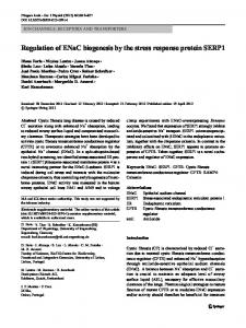
Mean fecal cortisol concentration. Fecal cortisol metabolite concentration varied individually as well as temporally within a wide range (36.27 to 1562.43 mg/g feces). Prior to surgery, daily values (mean ± 1 standard deviation) ranged from 232.46 ± 98.45 mg/g feces to 346.05 ± 169.28 mg/g feces, and the basal level was 302.98 ± 51.12 mg/g feces. Concentrations increased just after ovariectomy, reaching peak values on days 6 and 7 (543.32 ± 410.12 and 408.80 ± 248.50 mg/g feces, respectively) and decreased again on subsequent days. The repeated-measures analysis of variance (Tukey–Kramer test) showed that the difference between the daily measures of concentration of fecal metabolites was significant (P = 0.02) (Figure 1). 24-h fecal output. All feces voided by each animal were collected and weighed daily throughout the trial period. Tukey–Kramer multiple comparisons tests showed that the mean fecal output on day 6, the day after ovariectomy, was significantly (q > 4.603, P < 0.05) lower than that of each of the other days (Figure 1). 24-h production of fecal cortisol metabolites. The 24-h production of fecal cortisol metabolites was calculated by multiplying the per-gram fecal cortisol metabolite concentration by the weight of the total sample for every individual on each day. Basal “normal” production (743.38 ± 76.98 mg) was determined as the average of mean values from day 1 through day 4. Cortisol fecal production decreased sharply on day 6, just after surgery (400.69 ± 218.00 mg) but reached peak values on day 7 (920.67 ± 674.78 mg) and day 9 (942.29 ± 614.60 mg). The repeated measures ANOVA analysis did not confirm the significance of these peaks (Tukey-Kramer test, P = 0.1068) (Figure 1). Intraindividual stability of fecal corticosteroid levels. Repeated measures analysis of variance (Tukey–Kramer test) conducted with the repeated measures of both concentration and production of fecal metabolites by using the identification numbers of the animals as the between-group factor confirmed (P < 0.0001 in both cases) that levels within an individual were stable. Correlation between fecal output, fecal cortisol metabolite concentration, and fecal cortisol metabolite production. As expected, there was a significant (r = –0.9001, r2 = 0.8109, P = 0.0004) negative correlation between fecal cortisol metabolite concentration and 24-h fecal output. Daily fecal cortisol metabolite production and fecal output were also significantly but positively (r = 0.6457, r2 = 0.4169, P = 0.0438) correlated. There was no significant relationship (r = 0.3510, r2 = 0.1232, P = 0.3199) between fecal concentration and production of fecal cortisol metabolites.
Discussion
The hypothalamic–pituitary–adrenal axis response to a stressful event is usually rapid. In rats, maximal glucocorticoid concentrations in the blood are seen about 20 min after the event, but they do not persist for more than 90 min.1 Repeated blood sampling, with all its associated drawbacks, is necessary to assess these changes. Moreover, blood samples only provide point measurements that reflect momentary biologic events, and they are affected by the normal cyclic diurnal variation in corticosteroid levels in the circulation. In contrast, fecal metabolites integrate circulating corticosteroid concentrations over a longer period of time. Therefore, quantifying them allows monitoring short- as well as long-term hormonal reaction to a specific stressor.28 9
Vol 45, No 3 Journal of the American Association for Laboratory Animal Science May 2006
Figure 1. Temporal variation of mean fecal concentration of cortisol metabolites (circles), mean 24-h production of fecal cortisol metabolites (diamonds), and mean 24-h fecal output (triangles) of 10 female Syrian hamsters before (days 1 through 4) and after (days 6 through 10) ovariectomy. Feces were not collected on day 5 (arrow), the day surgery was performed.*, significant (P < 0.05) peak in mean fecal concentration; #, significant (P < 0.05) reduction in 24-h fecal output.
The fecal sampling schedule must be determined according to the specific cortisol metabolism and the kind of event to be studied. After injecting of 3H-corticosterone into male rats, for example, Bamberg and colleagues1 detected peaks of fecal radioactivity 8 to 24 h postinjection. The same treatment in mice of both genders resulted in fecal radioactivity peaks about 10 h postinjection.28 Other variables also affect metabolism and excretion: Touma28 showed that the route and delay of excretion differ widely between male and female mice. In both of these previous studies,1,28 the time of day of hormone administration affected the excretion delay, because passage through the gastrointestinal tract is influenced by the animal’s activity pattern. Because information about cortisol metabolism and excretion in the hamster is scarce, we collected all fecal material at the same time daily. This procedure enabled us to determine daily mean concentrations of cortisol fecal metabolites as well as the total amount of these metabolites that was excreted in each 24-h period, avoiding possible error from circadian variation in corticosteroid levels. Our 1st finding in regard to mean concentration was the enormous interindividual and temporal variation, with the maximum 40-fold higher than the minimum. This result confirms the importance of longitudinal studies, using each animal as its own control, which is impossible in small rodents if blood is sampled. Fecal corticosteroid metabolite concentration reached peak values on days 6 and 7, just after ovariectomy, and decreased again to basal levels. These changes suggest sharp increases in cortisol secretion related to the surgery itself, followed by a rapid return (less than 24 h) to basal level. However, concentration measures can be compared only if the volume of material in which the hormone is contained is the same between the 2 groups.12 One assumes that this condition is fulfilled when working with plasma or serum concentrations, because animals of the same species, age, and gender have approximately the same volume of blood. The situation is otherwise when working with feces. Some events, and especially stressful ones, modify defecation rates. In our study, an important reduction of 24-h fecal output, probably related to fasting, pain, anesthetic drugs, and morphine effects, occurred on the day after surgery. The fecal mass collected on day 6 was only 1/3 of the ‘basal’ amount collected daily on days 1 through 4. As expected, the Pearson correlation coefficient between mean concentrations and fecal 24-h output (r = –0.900) 10
confirmed the linear dependency between these parameters: concentration and fecal output varied together in an almost perfect inverse proportion. In such conditions, the relative per-gram measure of cortisol metabolites may not reflect the total amount of circulating hormone. Because we measured both concentration and fecal output, we were able to calculate 24-h production of cortisol metabolites and analyze its temporal variation. Whereas the changes in concentration values suggested a rapid return to normality after the surgery, the high production of fecal corticosteroid metabolites during the same period indicated a situation of protracted stress, with peak values of fecal metabolite production on days 7 and 9 after surgery, possibly related to pain. However, statistical analysis did not confirm the significance of these peaks. Therefore, it is not clear whether the increase in concentration on day 6 reflects a peak in blood level or is simply related to the dramatic decrease in fecal output, which may also explain the decrease in total fecal corticoid levels on the day after surgery. Conversely, it is possible that the production peaks on the days after surgery resulted from the normalization of fecal output and postponed excretion. We in fact are unable to affirm that surgery itself, performed under favorable conditions of anesthesia and analgesia, with minimal animal manipulation prior to sedation significantly increased cortisol release. In contrast, it is obvious that surgery reduced food input and fecal output and if circulating cortisol concentrations were higher on the day of surgery, it is possible that the supplementary amount of hormone was excreted only 2 d later, when both fecal output and 24-h fecal cortisol metabolites were higher than the basal ones. The relevance of measuring fecal corticosteroid metabolites to monitor adrenocortical activity has already been demonstrated for many species.20 Most studies only determine and compare per-gram measures of corticosteroid metabolite concentrations. However, our results showed that changes in fecal output, either stress-related or not, affect the concentration of fecal metabolites. In that case, the concentration was not significantly correlated with the total amount of excreted hormone, which may be more likely to reflect the level of circulating hormone. Further studies are necessary to determine whether one mechanism for reporting results is more appropriate than the other.
Acknowledgments
We thank Janet W Reid (Virginia Museum of Natural History, Martinsville, VA) for helpful comments on drafts of the manuscript, and Priscila Viau for excellent technical assistance in the laboratory.
References
1. Bamberg E, Palme R, Meingassner JG. 2001. Excretion of corticosteroid metabolites in urine and faeces of rats. Lab Anim 35:307–314. 2. Billiti JE, Lasley BL, Wilson BW. 1998. Development and validation of a fecal testosterone biomarker in Mus musculus and Peromyscus maniculatus. Biol Reprod 59:1023–1028. 3. Brown JL, Wasser SK, Wildt DE, Graham LH. 1994. Comparative aspects of steroid hormone metabolism and ovarian activity in felids, measured non-invasively in feces. Biol Reprod 51:776–786. 4. Cain SW, Karatsoreos I, Gautam N, Konar Y, Funk D, McDonald RJ, Ralph MR. 2004. Blunted cortisol rhythm is associated with learning impairment in aged hamster. Physiol Behav 82:339–344. 5. Cavigelli SA, Monfort SL, Whitney TK, Mechref YS, Novotny M, McClintock MK. 2005. Frequent serial fecal corticoid measures from rats reflect circadian and ovarian corticosterone rhythms. J Endocrinol 184:153–163. 6. Chelini MOM, Souza NL, Rocha AM, Felippe ECG, Oliveira CA. 2005. Quantification of fecal estradiol and progesterone metabolites in Syrian hamster (Mesocricetus auratus). Braz J Med Biol Res 38:1711–1717.
Fecal corticosteroid measurement
7. Cordner AP, Herwood MB, Helmreich DL, Parfitt DB. 2004. Antidepressants blunt the effects of inescapable stress on male mating behaviour and decrease corticotropin-releasing hormone mRNA expression in the hypothalamic paraventricular nucleus of the Syrian hamster (Mesocricetus auratus). J Neuroendocrinol 16:628–636. 8. Diagnostic Products Corporation [Internet]. Radioimmunoassay—international package inserts [cited 20 Aug 2005]. Available at http://www.dpcweb.com/package_inserts/ria/ria_int.html. 9. Graham L, Schwarzenberger F, Mostl E, Galama W, Savage A. 2001. A versatile enzyme immunoassay for the determination of progestagens in feces and serum. Zoo Biol 20:227–236. 10. Harper JM, Austad SN. 2000. Fecal glucocorticoids: a noninvasive method of measuring adrenal activity in wild and captive rodents. Physiol Biochem Zool 73:12–22. 11. Harper JM, Austad SN. 2001. Effect of capture and season of fecal glucocorticoid levels in deer mice (Petomyscus maniculatus) and red-backed voles (Clethrionomys gapperi). Gen Comp Endocrinol 123:337–344. 12. Hayssen V, Harper JM, Defina R. 2002. Fecal corticosteroids in agouti and nonagouti deer mice (Peromyscus maniculatus). Comp Biochem Physiol 132:439–446. 13. Heistermann L, Mohle U, Vervaecke H, van Elsacker L, Hodges JK. 1996. Application of urinary and fecal steroid measurements for monitoring ovarian function and pregnancy in the bonobo (Pan paniscus) and evaluation of perineal swelling patterns in relation to endocrine events. Biol Reprod 55:844–853. 14. Institute of Laboratory Animal Resources (ILAR). 1975. Longterm holding of laboratory rodents. Washington (DC): National Academy Press. 25 p. 15. Jasnow AM, Drazen DL, Huhman KL, Nelson RJ, Demass GE. 2001. Acute and chronic social defeat suppresses humoral immunity of male Syrian hamsters (Mesocricetus auratus). Horm Behav 40:428–433. 16. Morais RN, Mucciolo RG, Gomes MFL, Lacerda O, Moraes W, Moreira N, Graham LH, Swanson WF, Brown JL. 2002. Seasonal analysis of semen characteristics, serum testosterone and fecal androgens in the ocelot (Leopardus pardalis), margay (L. wiedii) and tigrina (L. tigrinus). Theriogenology 57:2027–2041. 17. Mostl E, Palme R. 2002. Hormones as indicators of stress. Domest Anim Endocrinol 23:67–74.
18. Muir C, Spironello-Vella E, Pisani N, DeCatanzaro E. 2001. Enzyme immunoassay of 17 beta-estradiol, estrone conjugates, and testosterone in urinary and fecal samples from male and female mice. Horm Metab Res 33:653–658. 19. National Research Council. 1996. Guide for the care and use of laboratory animals. Washington (DC): National Academy Press. 20. Queyras A, Corosi M. 2004. Non-invasive techniques for analysing hormonal indicators of stress. Ann Ist Super Sanitá 40:211–221. 21. Russel WMS, Burch RL. 1992. [Internet]. The principles of humane experimental technique. [cited 05 August 2005]. Available at http:// altweb.jhsph.edu/publications/humane_exp/het-toc.htm. 22. Schwarz A, Felippe ECG, Bernardi MM, Spinosa HS. 2005. Impaired female sexual behavior of rat offspring exposed to Solanum lycocarpum unripe fruits during gestation and lactation: lack of hormonal and fertility alterations. Pharmacol Biochem Behav. Forthcoming. 23. Schwarzenberger F, Francke R, Goltenboth R. 1993. Concentration of feacal immunoreactive progestogen metabolites during the oestrous cycle and pregnancy in the black rhinoceros (Diceros bicornis michaeli). J Reprod Fertil 98:285–291. 24. Taravosh-Lahn K, Delville Y. 2004. Aggressive behavior in female golden hamsters: development and the effect of repeated social stress. Horm Behav 46:428–435. 25. Thompson KV, Mashburn KL, Montfort SL. 1998. Characterization of estrous cyclicity in the sable antelope (Hippotragus niger) through fecal progestagen monitoring. Gen Comp Endocrinol 112:129–137. 26. Thompson KV, Montfort SL. 1999. Synchronization of oestrous cycles in sable antelope. Anim Reprod Sci 57:185–197. 27. Touma C, Palme R, Sachser N. 2004. Analysing corticosterone metabolites in fecal samples of mice: a noninvasive technique to monitor stress hormones. Horm Behav 45:10–22. 28. Touma C, Sachser N, Mostl E, Palme R. 2003. Effects of sex and time of day on metabolism and excretion of corticosterone in urine and feces of mice. Gen Comp Endocrinol 130:267–278. 29. Washburn BE, Millspaugh JJ. 2002. Effects of simulated environmental conditions on glucocorticoid metabolite measurements in white-tailed deer feces. Gen Comp Endocrinol 127:217–222. 30. Wommack JC, Salinas A, Melloni RH Jr, Delville Y. 2004. Behavioural and neuroendocrine adaptations to repeated stress during puberty in male golden hamsters. J Neuroendocrinol 16:767–775.
11