www.nature.com/scientificreports
OPEN
received: 17 December 2014 accepted: 21 April 2015 Published: 18 June 2015
Assisted reproduction causes placental maldevelopment and dysfunction linked to reduced fetal weight in mice Shuqiang Chen1,2,*, Fang-zhen Sun2,*, Xiuying Huang2, Xiaohong Wang1, Na Tang3, Baoyi Zhu1 & Bo Li1 Compelling evidence indicates that stress in utero, as manifested by low birth weight (LBW), increases the risk of metabolic syndrome in adulthood. Singletons conceived by assisted reproductive technology (ART) display a significant increase in LBW risk and ART offspring have a different metabolic profile starting at birth. Here, used mouse as a model, we found that ART resulted in reduced fetal weight and placental overgrowth at embryonic day 18.5 (E18.5). The ART placentae exhibited histomorphological alterations with defects in placental layer segregation and glycogen cells migration at E18.5. Further, ART treatments resulted in downregulation of a majority of placental nutrient transporters and reduction in placental efficiency. Moreover, the ART placentae were associated with increased methylation levels at imprinting control regions of H19, KvDMR1 and disrupted expression of a majority of imprinted genes important for placental development and function at E18.5. Our results from the mouse model show the first piece of evidence that ART treatment could affect fetal growth by disrupting placental development and function, suggests that perturbation of genomic imprinting resulted from embryo manipulation may contribute to these problems.
Epidemiological studies showing stress in utero, as manifested by low birth weight (LBW) is associated with chronic disease in adulthood. These associations have led to the “fetal programming of adult disease hypothesis” that nutrition and other influences during critical times of fetal life set the tissues and organs for life1–3. Singletons conceived by assisted reproductive technology (ART) have a 2.6-fold greater risk of LBW compared with those conceived naturally, and ART offspring have cardiovascular and metabolic risk factors even in childhood4–6. The causes of LBW are still unclear, as it is difficult to determine whether these problems are related to the ART procedure or to parental factors7,8. Over 5 million ART babies have been delivered worldwide since the breakthrough of in vitro fertilization (IVF) technology in 1978, and the demand for ART is continually increasing. Therefore, it is utmost importance to understand the possible causes of LBW, particularly those that may be associated with ART procedures, from the viewpoint of improving the technology and minimizing the health risks of babies conceived by ART. The placenta plays a critical role in controlling maternal-fetal resource allocation and mediating programming of the fetus for future disease9,10. Studies have showed that variations in gross placental morphology at birth can predict a wide range of disorders in later life10,11. It has been observed that human pregnancies conceived by ART show an increased incidence of anomalous placentation12–14. Further, 1
Department of Obstetrics and Gynecology, Tangdu Hospital, the Fourth Military Medical University, Xi’an 710038, China. 2Laboratory of Molecular and Developmental Biology, Institute of Genetics and Developmental Biology, Chinese Academy of Sciences, Beijing 100080, People’s Republic of China. 3Shaanxi Institute for Food and Drug Control, Xi’an 710038, People’s Republic of China. *These authors contributed equally to this work. Correspondence and requests for materials should be addressed to B.L. (email:
[email protected]) Scientific Reports | 5:10596 | DOI: 10.1038/srep10596
1
www.nature.com/scientificreports/
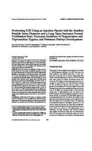
Figure 1. IVF resulted in higher abortion rate at E10.5. (A) Implantation rate of IVC, IVF and control groups at E10.5. (B) Abortion rate of IVC, IVF and control groups at E10.5. Data are presented as Means ± SD, groups with different superscripts differ significantly (p