Cognition, Universite du Quebec a Montreal, CP 8888 Succ A, Mon treal, Quebec .... requiring a press of the index finger on the space bar of a computer ...
Brain Topography, Volume ll, Number 3, 1 999
223
Topographical Analysis of Homotopic Interhemispheric "Relay" Asymmetries in Visual Evoked Potentials Claude M.J. Braun* and Lolc Villeneuve*
Summary: Brown and colleagues (1994) found that most evoked potential studies provide evidence of faster interhemispheric relay from the right to the left hemisphere, as determined from N160 latencies derived from parietal and occipital electrode sites. The experiment reported here was designed to complement those findings by 1) analyzing more electrode sites (several frontal, temporal, and central sites in addition to the previously investigated sites) and more waveforms (P1 and P2 waves); 3) introducing analysis of amplitude data; 4) carrying out site-specific and experiment-wise inference tests of putative interhemispheric relay asymmetry. We found that all of the conclusions of Brown and colleagues, regarding latency effects, could be extended to the ensemble of electrode sites and waveforms. However, amplitude effects were significantly compatible with stronger (though slower) relay from the left to the right hemisphere. Key words: Interhemispheric transfer; Relay; Evoked potential; Poffenberger; Simple reaction time; Asymmetry.
Introduction
Poffenberger (1912) reasoned that since the visual and motor projections are contralateral, the difference in reaction time (RT) between a contralateral and ipsilateral stimulated-field/responding-hand relation ought to be an accurate estimate of interhemispheric relay time (IHRT). Many experiments have reported a small lag of 1 to 5 ms attributable to IHRT (reviewed by Braun 1992; Hoptman and Davidson 1994). Callosal agenesics and callosotomized patients have greatly prolonged IHRT (Clarke and Zaidel 1989; Iacoboni et al. 1994; Jeeves 1969). Attempts to use evoked potentials as indices of the effects of interhemispheric relay (reviewed by Brown et al. 1994) have dealt systematically with latency effects (the IHRT inference), and less systematically with ampli tude effects. Evoked potentials have typically yielded reliable (e.g., consistently large) estimates of IHRT, espe-
* Laboratoire de Neurosciences de Ia Cognition, and Departement de Psychologie, Universite du Quebec a Montreal. Accepted for publication: December 4, 1998. This research was supported by grants from the National Science and Engineering Research Council of Canada and the Fonds Concerte et d'Action des Chercheurs of the government of Quebec to the first author, and from an NSERC doctoral bursary to the second author. Correspondence and reprint requests should be addressed to Pro fessor Claude M.J. Braun, PhD, Laboratoire de Neurosciences de Ia Cognition, Universite du Quebec a Montreal, CP 8888 Succ A, Mon treal, Quebec, Canada, H3C 3P8. Copyright© 1999 Human Sciences Press, Inc.
dally at posterior electrode sites and especially in the mid-latency waveforms (N160). Decrements in ampli tude over the hemisphere ipsilateral to the visual stimu lus have also typically been observed, suggesting that there is a decrease in the signal strength associated with interhemispheric relay. These effects are considerably greater in cases of callosal agenesis (Rugg et al. 1985) and of callosotomy (Kutas et al. 1990). Finally, source analy sis of evoked potentials is concordant with the findings mentioned above. Electrical (Baseler et al. 1994; Gomez Gonzalez et al. 1994) and magnetic (Degg et al. 1992) evoked response studies have consistently shown that simple detection of eccentric localized stimuli yields scalp topographies and source estimates all compatible with the known anatomical contralaterality of the visual projection. However, the opposite pattern occurs when checkerboard full half-field stimuli are used, supposedly due to midline primary striate cortex emplacement of dominant dipole(s) generating signal maxima over the opposed hemiscalp, i.e., ipsilateral to the stimulus (Clem ent et al. 1985; Harding et al. 1991). In this situation, the scalp topography incorrectly suggests a source ipsilateral to the stimulated field, while source derivation does not get fooled. Likewise, electrical (Botzel et al. 1993; Pfurt scheller et al. 1994) and magnetic (Nagamine et al. 1994) source analyses of simple fast motor acts carried out by the index finger (button presses) have consistently shown scalp topography and source estimation which are com patible with the known contralaterality of the motor pro jection. However, the opposite pattern occurs when the
224
required response is a foot or toe movement, presumably again because the dominant dipole is in medial motor cortex generating a current dipole oriented toward the opposite hemisphere (Brunia et al. 1984, and see Vaughan et al. 1968, for a trend). In such situations, the scalp topography incorrectly suggests a source ipsilateral to the responding member, but source analysis is able to provide the required correction. There are nevertheless numerous problems with the use of evoked potentials to investigate interhemispheric dynamics. Some electrode sites yield unreliable or even null or negative IHRT indices. The more reliable IHRT indices from evoked potentials do not correlate with RT indices of IHRT (Saron and Davidson 1989; Potvin et al. 1995). The IHRT indices which are reliable (consistantly positive) (11-18 ms) are not commensurable with IHRT indices obtained in RT (1-4 ms). Responding-hand con figurations have not been controlled or analyzed. Am plitude effects have not been analyzed systematically in the investigation of interhemispheric relay - an issue which is not trivial since amplitude indices of relay could presumably reflect signal strength which could in princi ple dissociate from signal velocity. For example, such a dissociation could reflect a speed accuracy trade-off. This seems all the more likely considering that omission errors yield significantly negative indices of interhemi spheric relay accuracy (Braun et al. 1996). Another indi cation to the effect that such a dissociation is possible was provided by Hoptman and colleagues (1996). They found that IHRT did not change from middle adulthood to senescence in evoked potentials latencies, but that there was indeed a significant decrement in the ampli tudes of so-called "post-relay" components. In the context of meta-analysis of RT research on interhemispheric relay, Braun (1992) proposed a model stating that, depending on experimental conditions, fast and slow channels of interhemispheric relay are selected by the hemispheres: a fast channel being selected by the hemisphere most apt to discriminate the critical stimulus, and a slow channel being selected by the hemisphere least apt to discriminate the stimulus. Marzi and colleagues (1991) completed a meta-analysis of the RT research on interhemispheric relay suggesting faster right-to-left in terhemispheric relay. A follow up experiment provided an ingenious demonstration to the effect that the RT relay asymmetry is likely to be exactly that, rather than an artifact of field or hand effects (Bisiacchi et al. 1994). Brown et al. (1994) reported a meta-analysis as well as their own original data in the EP domain. The results generally supported Marzi's and colleague's position to the effect that relay occurs more rapidly from the right to the left hemisphere than in the other direction. However, they came to this conclusion using very few sites, and a restricted index of brain electrical activity (N160) occur-
Braun and Villeneuve
ing at a disquietingly late latency after stimulation. They ignored waveform amplitude differences across hemi spheres, as did most predecessors. They did not carry out an experiment-wise inference test of the IR asymmetry observed in their own experiment. They also failed to take advantage of an opportunity to bypass the possibil ity of so-called relay asymmetries consisting of none other than a hemisphere effect. Indeed, one way to have more confidence in the idea that one is dealing with a true interhemispheric relay asymmetry, is to obtain the asym metry in conditions where the direct routes (right field left hemiscalp, left field-right hemiscalp) are balanced, i.e., have the same latencies or amplitudes at a homotopic electrode pair. Finally, they directly transposed to the EP domain Braun's postulate (Braun et al. 1992), explained above, concerning RT field effects being linked to asym metries of relay-time estimates. It seems that the more logical test of Braun's model, in the EP domain, would be to determine whether, in the appropriate electrical stimu lus-related potential domain (latency or amplitude), field advantages are concordant with estimates of relay-time asymmetries. Note that the EP estimates of directional IR time are obtained as a difference in latency between the two hemispheres for the same visual field, while for RT the estimates are differences between the visual fields using a given hand. In the EP domain, the directional relay asymmetry is equivalent to a difference between the hemispheres summed over the two visual fields. The association of level of IR asymmetry with level of visual field asymmetry is thus appropriately tested in EPs be cause they are not mathematically dependent. The present report of an evoked potential experi ment comprises an attempt to overcome the above-men tioned problems by: 1) analyzing more electrode sites (several frontal, temporal, and central sites in addition to the previously investigated sites) and more waveforms than covered by Brown et al. 's meta-analysis (particularly the Pl and P2 waves); 3) introducing analysis of ampli tude data; 4) carrying out site-specific and experiment wise inference tests of putative IR asymmetry; 5) analyzing EP indicators of faster or more efficient hemi spheric processing of the stimulus (rather than looking only at RT indicators); and 6) paying special attention to the logical exigency of balanced direct route (the two contralateral field-hemiscalp) conditions as a prereq uisite for any firm conclusion of relay asymmetry, dis tinct from a simple hemisphere (hemiscalp) effect. Method
Subjects Five female and five male undergraduate university students were paid $20.00 CAN to participate as subjects.
225
Interhemispheric Relay
The women's and men's mean ages were 22.4 and 23.2 years respectively, and their mean educational levels were 13.8 and 14.8 years respectively. Right handedness was assured by means of a 16 item questionnaire comprising non-redundant items applying to a single hand. The items were drawn from the Oldfield, Harris and Annett hand edness questionnaires (see Fennell 1986 for details). Sub jects had to be 100% right handed on this questionnaire, and to have reported absence of left handers among first degree relatives, to be admitted as subjects. Behavioral procedure and task The behavioral task consisted of a simple visual task requiring a press of the index finger on the space bar of a computer (PC-486) keyboard upon detection of a laterally eccentric stimulus on a computer screen (Mitsubishi Dia mond Pro14 Color Monitor). Throughout the experi ment, the testing chamber was illuminated by the screen monitor only, so as to minimize reflections on the screen surface. The screen displayed a dark blue background (108 cd/square em) as well as a central fixation point consisting of pale white cross hairs of a length of .5 em. Yellow targets (147 cd/square em) of a dimension of 1 square em appeared at 10 arc degrees (internal border) to the right or left of the central fixation point, and were of 100 ms duration. Subjects' eyes were 50 em from the computer screen. The sequence of stimulations as a func tion of field was random, except for the constraint that no more than three stimulations could occur in a given field consecutively. The subjects' response triggered a timing algorithm for the next stimulus, so that, in a sense, the task was self paced. Intervals between the response and the next stimu lus consisted of random intervals logarithmically distrib uted from 150 to 2,000 ms. If the subject responded during the first 100 msec after stimulus-onset, the response was labelled an "anticipation" error - and the response-to stimulus interval (RSI) timing algorithm was put back into action. If the subject did not respond within the 800 ms following stimulus onset, the trial received the label "omis sion error"- and the RSI algorithm was put back into action. Each subject completed 8 blocks of 200 correct trials each, with one hand in an ABBA hand sequence. Starting hand was counterbalanced across subjects. Missing trials (an ticipation errors and omission errors) were automatically replaced (without disturbing the RSI algorithm) at the end of each block, so that when the entire experiment was complete, each subject had yielded 400 correct right fin gered responses to left-field stimuli, 400 correct right fin gered responses to right-field stimuli, and likewise for the left fingered responses, for a grand total of 1600 correct RTs per subject. The subjects's responding index finger was at 25 em in front of the solar plexus on a table.
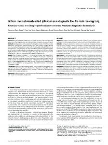
Electrophysiological montage and procedure Eighteen Ag/AgCl electrodes were placed using the 10-20 system. These included three symmetrical frontal sites (FP1-FP2, F7-F8, F3-F4), two temporal (T3-T4, T5T6), one central (C3-C4), one parietal (P3-P4) and one occipital (01-02). The montage was unipolar, linked ear lobes serving as the reference lead. Acquisition of EEG data was done using the Neuroscan software and Sy namps amplifiers. Digitization rate was 200 Hz. High and low pass filters were set at .1 and 39 Hz, with -12dB/octave slopes. Criteria for discarding BEG seg ments, on a trial-by-trial basis, included gross eye move ments detected by the Neuroscan software, or a response occuring within the "anticipation" window (see above) or the "omission" window (see above). The database con sisted of 500 ms of stimulus-synchronized activity, i.e., of activity preceding and 500 ms following the stimulus. It appeared to us that late stimulus-related activity could, in principle, be slightly distorted by emerging response related activity. Averaging of the trials included special conditioning of the data using an iterative procedure related to Woldorff's (1993) "adjacent response filter", decontaminating the potentials related to each event (stimulus or response) from that of the other event in each stimulus-response pair. Our conditioned averaging yielded averaged signals quite appropriate for further waveshape quantification, and smoothing was not re quired (see figure 1). We did not implement a baseline amplitude nor malization procedure, but rotated the electrodes from subject to subject instead. Indeed, an expectancy effect can be seen in the period preceding stimulation (figure 2). This expectancy effect was very variable however. With analysis of variance, we tested the hypothesis to the effect that «indirect» and «direct» conditions would differ at stimulus onset (time zero). The differences never reached significance at any electrode site pair, nor did they do so experiment-wise. We could easily distinguish three waves, respec tively positive, negative and positive, which we termed P1, N1 and P2. The criteria for extraction of the ampli tude and latency values of each of these waves were the following: 1) absence of right-left polarity reversal, occur ing occasionally at anterior temporal and frontal leads, due to horizontal eye movement, 2) the first post-stimu lus positive peak for P1, 3) the next negative peak was determined to be N1, and the next positive peak was P2 - under one constraint however: the component had to be visible at both hemiscalp locations; if the latter condi tion was not met, the next bilaterally identifiable peak was selected. Absence of bilaterality of a wave was very rare (approximately 1 %). Only homotopic electrode sites (any pair of elec-
226
Braun and Villeneuve
LEFT VISUAL FIELD
STIMULUS-RELATED POTENTIALS
·
y 1
, ..... ,...
-• • •• • •
Direct Indirect
,
·
· ...... ...·- ... -------- ---------..
RIGHT VISUAL FIELD
Milliseconds
N1
Figure 2.
500
Tracings from a single subject of the visual
evoked potentials in conditions of direct and indirect stimulation of the hemisphere.
SP1
In the direct condition
(continuous lines), the stimulus is contralateral to the
hemiscalp recorded from, whereas in the indirect condi
tion (dashed lines) the stimulus is ipsilateral to the hemis calp recorded from. The visual field in which the stimulus
was presented is indicated in the figure. The overall left hemisphere latency adavantage observed in this subject
is typical of the entire cohort, and confounds estimation
of directional relay effects Post-relay synchronization oc
curs before pre-relay synchronization In the right-to-left
hemisphere relay conditions (left side of the figure). Post
relay amplitude decrements are more variable and dis
SN1 0
100
200
play less asymmetry in this case, as in the entire cohort.
300
400
Milliseconds Figure 1. An evoked potential of a single subject resulting
from stimulus-onset synchronized averaging prior to the
«adjacent filter>> correction (solid line) and after correction (dashed line). The components identified here are the first
positive one (SP1 ), the first negative one (SN 1) and the
second positive one (SP2). These tracings represent the
electrical activity of one subject at the F4, P4 and 02
electrode sites, upon right visual field stimulation and right hand responding.
trades in symmetric position relative to the sagittal midline) were used for extracting estimates of interhemi-
spheric relay time (IHRT) and interhemispheric relay signal (amplitude) decrement (IHRSD). This does not mean that we assume that interhemispheric relay is ex clusively homotopic. However, this simple approach is sufficient to address the basic question of whether there is a general asymmetry of directional relay. Global IHRT estimates from stimulus-related waveforms consisted of the subtraction of peak latencies in the putative pre-relay condition (sites contralateral to the stimulus) from peak latencies in the putative post-relay condition (sites ipsi lateral to the stimulus). Thus, a positive valence of the IHRT suggests a time cost associated with interhemi spheric relay. Estimates of interhemispheric relay strength decrement (IHRSD) consisted of the subtraction
227
Interhemispheric Relay
of peak amplitudes in the putative pre-relay condition (sites contralateral to the stimulus) from peak amplitudes in the putative post-relay condition (sites ipsilateral to the stimulus). Thus operationalized, an IHRSD of positive valence suggests a gain in signal strength associated with interhemispheric relay for positive waves, and a decre ment of signal strength for negative waves. Estimates of directional relay in stimulus-related po tentials simply consisted of limiting the above calcula tions to one field (data from the left field yields an estimate of relay from the right to the left hemisphere, data from the right field yields an estimate of relay from the left to the right hemisphere). Though we opted to test interhemispheric relay effects with multiple inference tests (c.f. Brown et al. 1994), we do not assume that events occurring at any two electrode sites are independent from events occurring at any other pair of electrode sites. Furthermore, to avoid drawing distorted conclusions from a large set of interdependent electrical events, we planned experiment-wise inference tests. Results
The mean RT was 251 ms from stimulus onset. The mean latencies of the three wave peaks were P1=125.1 (SD = 26 ms), N1=170.3 (SD = 19 ms), and P2=222.9 ms (SD = 26.3 ms) from stimulus onset, and their amplitudes were 1.74 Uv (SD = 4.5 Uv), -2.23 Uv (SD = 3.3 Uv), and 2.49 Uv (SD = 3.5 Uv) respectively. The "post-relay" or "crossed" condition refers to the contralaterality of stimu lated field and responding hand, while the "pre-relay" or "uncrossed" condition consists of an ipsilateral relation. IHRT is the subtraction of the uncrossed RTs from the crossed Rts (crossed-uncrossed difference or CUD). In the present experiment, the mean RT-CUD was 2.1 ms (SO= 2.08) (F(1, 9) = 10.2, p = .011). The mean omission error CUD was l.Oerror (SD = 1.8) (F(1,9) = 3.10, p = .112). In the evoked potential data the mean global estimate of IHRT (all sites and all waves combined, was 7.76 ms (SO = 5.68) (F(1, 9) = 18.67, p = .002). Each global relay effect, each effect of relay asymmetry, and each field or hand effect was subjected to statistical test. The tests having surpassed the alpha criterion of p = .05 are identified with asterisks in the following tables (tables I and II). One problem with the IHRT and IHRSD assump tions is that what is taken to consist of a relay asymmetry may in reality consist of an artifact of differential latencies or amplitudes of the so-called direct (pre-relay) route. If the two conditions corresponding to direct stimulation of the hemisphere (right field stimulation and left scalp electrode sites, or left field stimulation and right scalp electrode sites) are asymmetric, then extraction of IHRT or IHRSD indices is ambiguous, and their significance is more tenuous. An estimate of directional relay can con-
sist of nothing other than a hemiscalp effect (hemisphere advantage). To counter this problem, we carried out a special additional analysis on our own data. We identi fied all the data pertaining to waves and sites and data types (latencies versus amplitudes) c omprising symmetry of the two direct routes. We found 11 balanced latencies in the homotopic direct routes, but none in the amplitudes. Balanced routes are identified in tables I and II by means of the character string SDR - meaning SYM METRICAL DIRECT ROUTE. The question then was whether a pattern of directional relay asymmetry would occur in the corresponding so-called indirect (post relay) routes, i.e., at those homotopic electrode pair sites com prising balanced direct routes. The latency of the earliest component (P1) was com patible with faster right-to-left interhemispheric relay at six of the eight electrode pairs. EP-latency field effects were not systematically concordant with the directional relay asymmetries across sites. The second component (N1) was compatible with faster right-to-left relay at seven of the eight electrode sites. EP-latency field effects were not systematically concordant with the directional relay asymmetries. The third component (P2) yielded estimates compatible with faster right-to-left interhemi spheric relay at seven of the eight electrode sites, and EP-latency field effects were not systematically concor dant with the directional relay asymmetries. See table I. Amplitude estimates of directional relay effects were rarely statistically significant. The P1 wave presented patterns compatible with more efficient relay in the right to-left direction at seven of the eight electrode pairs. EP-amplitude visual field effects were not systematically concordant with the directional relay asymmetries across electrode sites. The N1 wave yielded eight of eight cases compatible with more efficient relay from the left to the right hemisphere. Again, there was no meaningful asso ciation of EP-amplitude field effects with the directional relay asymmetries. The P2 wave yielded directional relay estimates all favoring the right-to-left direction, except at the F3-F4 electrode site. EP-amplitude field effects were not systematically concordant with the estimates of direc tional relay. See table II. Analysis of the ensemble Was there an overall significant asymmetry of indi ces of directional relay? We prepared the data for re peated measures analyses o f variance of relay asymmetries per se. Data had to be recoded for ampli tudes because the meanings of negative deflections (negative amplitude values for the N1 wave) and of a positive deflection (positive amplitude values of the P1 and P2 waves) clash in such a context. Once the data were properly formatted, we were able to test the inde-
228
Braun and Villeneuve
Table I. Visual evoked potential estimates of directional latency interhemispheric relay effects. Electrode site
R to L
L to R
IHRT
IHRT
IHRT latency
Global
asymmetry (ms) IHRT (ms)
(ms)
(ms)
FP1-FP2 __EH8 (Sllill
-3.8
-5.35
1.55
-4.2
10.2.1')
-14.4.1')
1.m
F3-F4 (SDR)
-12.35
6.2
-18.55
-3.08
**
*
21.15
-23
T3-T4 (SDR)
-1.85
-4JiR
Relay direction
EP latency UFA per
Latency UFA all
advantaged
electrode pair
electrodes combined
LR
RiP"ht
Rl
Ri!!ht
RL
Right
* 9.65
RL
15.65
12.05
3.6
P3-P4 (SDR) 01-02 N1
12.3
28.85
*
**
20.4
25.35
*
*
8.95
20.7
**
**
13.85
LR
Right
RL
Right
RL
Left
RL
Right
**
* T5-T6
Left *
* C3-C4
-16.55
20.58 **
-4.95
22.88 **
-11.75
14.83 **
S8
-3.7
-2.1
-4.7S
Rl
Ri!!ht
F7-F8 (SDR)
-14.75
-7.8
-6.95
-11.28
RL
Left
F3-F4
-10.55
0.3
-10.85
-5.13
RL
nil
T3-T4(SDR)
-12
8.45
-20.45
-1.78
RL
Left
C3-C4 (SDR)
9.9
9.7
-0.2
9.8
LR
Left
-20.8
16.85
RL
Right
RL
Left
RL
Right
FP1-FP2
Rilrht
LPft
*
**
T5-T6
*
*
6.45
27.25
** **
** P3-P4 (SDR) 01-02 P?.
18.65
25.35
*
**
8.4
16.55
**
**
-6.7
22 **
-8.15
12.48 ***
-O.fiS
�.O"i
. -3.7
1.?.
RL
Ri2:ht
F7-F8
6
11.85
-5.85
8.93
RL
Left
F3-F4
2.4
10.7
-8.3
6.55
RL
Left
-10.2
12.65
RL
Left
..EI'1-FP2
**
Left
**
* T3-T4 (SDR)
7.55
17.75 **
C3-C4 (SDR)
12.25
T5-T6
-1.55
*
*
3.5
8.75
7.88
LR
Left
19.9
-21.45
9.18
RL
Right
* * ...E&P4f�DR) 01-02
14.fil)
1fi
-1.�1)
l"i."i�
Rl
nil
4.3
14.05
-9.75
9.18
RL
Right
*
**
Legend: P1=first detectable positive wave; N1 =first detectable negative wave; P2=second detectable positive wave; VFA= visual field advantage across both electrodes; ms=millisecond; EP = evoked potential; IHRT= interhemispheric relay time estimate (operationalized as subtraction of the latencies of the scalp contralateral to the stimulus from the latencies of the scalp ipsilatral to the stimulus), IHRT asymmetry= subtraction of left-to-right relay from right-to-left relay; Global IHRT=mean of right-to-left relay and left-to-right relay; LR=left-to-right hemispheric relay advantage; RL=right-to-left hemispheric relay advantage (NB: A relay advantage is considered here to consist of a briefer interval from pre-to post-relay conditions, indicative of faster relav): SDR=svmmetrical direct routes·* =o