B-ENT, 2013, 9, Suppl. 21, 65-79
Auditory neuropathy: a challenge for diagnosis and treatment F. Declau1, A. Boudewyns1, J. Van den Ende2 and P. van de Heyning1 Department of Otorhinolaryngology, Head and Neck Surgery, and Communication Disorders; 2Department of Medical Genetics, University of Antwerp, Antwerp, Belgium 1
Key-words. Hearing loss; neuropathy; genetics; electrophysiology; hearing aids; cochlear implants Abstract. Auditory neuropathy: a challenge for diagnosis and treatment. In current terminology, auditory neuropathy spectrum disorder (ANSD) is a disease involving the disruption of the temporal coding of acoustic signals in auditory nerve fibres, resulting in the impairment of auditory perceptions that rely on temporal cues. There is debate about almost every aspect of the disorder, including aetiology, lesion sites, and the terminology used to describe it. ANSD is a heterogeneous disease despite similar audiological findings. The absence of an auditory brainstem response (ABR) and the presence of otoacoustic emissions (OAE) suggest an ANSD profile. However, to determine the exact anatomical site of the disorder, more in-depth audiological and electrophysiological tests must be combined with imaging, genetics and neurological examinations. Greater diagnostic specificity is therefore needed to provide these patients with more adequate treatment.
Introduction: historical and theoretical issues The first audiological report of ANSD was probably by Hinchcliffe et al.1 (1972), well before OAEs2 were reported describing degenerative neuropathy in Nigerians due to chronic cyanide intoxication. In 1996, Starr et al.3 introduced the term ‘neuropathy’ after studying ten patients with a unique set of auditory problems. Eight patients subsequently developed concurrent peripheral neuropathies. In brief: auditory neuropathy was thought to be a hearing impairment in which outer hair cell function was normal but afferent neural conduction in the auditory nerve was disordered.3 The disorder has also been referred to as type I afferent neuron dysfunction, and neural hearing loss.4,5 In 2001, Berlin et al.6 introduced the term auditory neuropathy/auditory dyssynchrony (AN/AD) to include those cases where no true neuropathy was apparent when the constellation of routine test results did not provide sufficient evidence to differentiate between synaptic dysfunction and “true neuropathy” of the cochlear nerve. Recently, the term auditory neuropathy was extended to auditory neuropathy spectrum disorder (ANSD) to acknowledge the heterogeneous and multifaceted nature of this condition.7 In current terminology, ANSD is a disorder c haracterised by the disruption of the temporal coding
10-declau-.indd 65
of acoustic signals in auditory nerve fibres, resulting in the impairment of auditory perceptions that rely on temporal cues. Abnormal discharge results from lesions involving the auditory nerve (postsynaptic AN), inner hair cells and/or the synapses with auditory nerve terminals (presynaptic AN). Starr et al.8 suggested breaking down auditory neuropathy into types such as type I (presynaptic) or type II (postsynaptic). In 2008, the same authors6 proposed refining the terminology on the basis of the site of the disorder. Rapin and Gravel9 also adopted an anatomical point of view, suggesting that the term auditory neuropathy should be limited to cases in which the pathology is located at the spiral ganglion cells and their axons of the 8th cranial nerve. In recent audiological literature, the term “auditory neuropathy” has also been loosely applied to infants who satisfy the first two audiological criteria only (absent ABR and present OAE) without any further audiological or neurological corroboration. In these cases, the term “ANSD profile” is considered to be more appropriate. However, Rapin admitted that strictly localised pathology was in fact exceptional in cases with functional and anatomical verification and that, in the majority of cases, there was pathology at multiple levels in the auditory pathway.5 One of these strictly localised pathologies is cochlear nerve hypoplasia, which was first described
17/10/13 14:41
66
F. Declau et al.
by Shelton et al.10 in 1989 and can be considered as a “true“ form of auditory neuropathy.
Diagnostic investigations
Epidemiology
The diagnosis of ANSD is a true challenge and audiological evidence must often be combined with imaging, genetics and neurological examinations. As more patients, particularly young children, have been diagnosed, studies have shown that patients with ANSD are a heterogeneous group despite similar audiological test findings. According to Berlin et al.13,27 ANSD can be characterised by the following list of audiological findings. The first three items are considered to be a minimum test battery for the diagnosis of ANSD:
ANSD occurs in all age groups.3 With the advent of newborn hearing screening, auditory neuropathy became a recognised disorder in the paediatric population. The prevalence of ANSD in children with confirmed permanent hearing loss has been reported to vary greatly, ranging from 1.6 to 19%.11,12 According to Berlin, ANSD is also present in 1-10% of children in schools for the deaf.13 ANSD after universal neonatal hearing screening14-17 was found in 0.044% to 0.06% of screened infants, whereas Dowley et al.16 and Foerst et al.17 reported an annual incidence rate of 0.14 to 0.27/1000 live births. Sininger et al.18 reviewed the incidence of the ANSD profile in neonatal intensive care unit (NICU) infants and found a range in the literature extending from 5.3% to 14.8% (with 10-14% being the most frequently reported prevalence). Berg et al.19 found a slightly higher incidence of 24.1% in NICU infants. These data are the results of the combination of present TEOAEs and absent ABRs, suggesting an ANSD profile but often without any further diagnostic assessment to confirm the tentative diagnosis. On the other hand, some hearing disorders underlying ANSD may not have been included in prevalence estimates as the identification of ANSD may require additional tests to be performed that are not invariably included in routine diagnostic measurements. Refined diagnostic tests, together with progress in genetic research, will provide a more accurate calculation of the prevalence of ANSD.20 This variation may reflect the inclusion in some studies of diseases in which ANSD is transient, such as GuillainBarré syndrome.21 Moreover, in NICU neonates with no auditory brainstem responses and the presence of OAEs at hearing screening, the ABR abnormalities may reflect the delayed maturation of both the brainstem and the auditory nerve.22 Cochlear nerve hypoplasia is seen in 6% to 19% of children with permanent hearing loss, but it is even more common in patients with an ANSD profile.23-26 cochlear nerve deficiencies were identified in children in 16,7 to 26,9 % of ears with an ANSD profile by Buchman et al.23 and Huang et al.26
10-declau-.indd 66
Clinical and audiological aspects
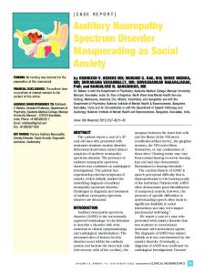
1. Test of cochlear hair cell function: evidence of normal outer hair cells in the cochlea: a. Otoacoustic emissions are, or have been, present (they sometimes disappear with time or with hearing aid use). Standard screening or diagnostic protocols using Transient-Evoked OAEs (TEOAEs) or Distortion Product OAEs (DPOAEs) may be used. b. Cochlear microphonics (CM): Auditory brainstem response (ABR) to high-level click stimuli (80-90 dB nHL) including positive and negative polarity clicks in separate trials through insert earphones.28,29 A trial run with the sounddelivery tube clamped will differentiate between the CM and the stimulus artifact.30 CMs generally remain present in individuals with ANSD even when there has been a loss of OAEs.3 The investigators reported that 80% of the patients had OAEs but that they disappeared over time in 11% of their patients and OAEs were not recorded in the other 9%.31 2. Evidence of neural impairment: ABR is absent or grossly abnormal. The ABR may also be mistakenly thought to be present when in fact a cochlear microphonic (CM) or hair cell response is masquerading as a neural response. Both rarefaction and condensation polarities should be evaluated to determine whether a cochlear microphonic is present (Figure 1). Insert earphones must be used and care taken to complete a “no-sound run” by clamping or disconnecting the sound tube from the transducer to eliminate the possibility of stimulus artifacts.32 3. Middle ear muscle reflexes (MEMRs) are absent. There are no normative data for acoustic reflex thresholds in very young infants using high
17/10/13 14:41
Auditory neuropathy
67
Figure 1 ABR of a child with an ANSD profile: presence of a cochlear microphonic at 100 dB nHL with rarefaction (R)/condensation (C) polarity in the absence of a recognisable neural response.
probe-tone frequencies (1000 Hz) and so this procedure is not required to diagnose ANSD in infants. Sininger and Oba (2001) reported that the vast majority of patients with ANSD have no auditory MEMRs, and about 20% have highly atypical, elevated, auditory MEMRs both ipsilaterally and contralaterally in the presence of recordable MEMRs to tactile stimulation.31 The presence of reflexes at levels near 90 dB HL in neonates and infants up to six months of age should in itself result in serious doubts about a diagnosis of ANSD.13,33,34 It has been argued that more standards are needed before we can rely on immittance audiometry as a tool in the diagnosis of ANSD in infants.34 Nevertheless, a complete test battery for ANSD should include middle ear muscle reflex testing whenever possible.
10-declau-.indd 67
4. Efferent suppression of otoacoustic emissions is absent. Although this test is not in widespread clinical use, it is a potential candidate for further diagnostic studies in individuals with reliably recorded OAEs.7 5. No masking level differences. Highly reduced binaural release from masking targeting a subject’s ability to detect interaural phase (timing) differences confirms the poor temporal processing of subjects with ANSD.5 6. Speech perception, which may be surprisingly good in some patients in a quiet environment, is seriously compromised in noisy surroundings.35 Most affected adults have perceptual deficits that are greater than would be expected from their audiometric (sound detection) levels. In addition, fluctuations in listening abilities have been reported, some associated with body
17/10/13 14:41
68
F. Declau et al. temperature and others with no clear precipitant.36,37 In order to understand running speech, or even to discriminate between sounds within individual words, a listener must be able to perceive the characteristic shape of individual phonemes, and be able to follow the rapid within-phoneme changes that give cues to coarticulation. It is this need to cope with the dynamic nature of speech that poses the greatest challenge for individuals with temporal processing problems.7
The initial diagnosis of an ANSD profile should be based on comprehensive electrophysiological assessment that includes, as a minimum, ABR testing, immittance measurements including tympanometry and acoustic reflex thresholds, and OAEs. Unlike “typical” sensorineural hearing loss, threshold estimation is virtually impossible using ABR or ASSR evaluation. The determination of thresholds for infants and young children with ANSD must therefore include developmentally appropriate behavioural measures such as visual reinforcement audiometry (VRA) or play audio metry.32 The degree of hearing loss in patients with ANSD varies from slight to profound; most losses are bilateral and symmetrical in configuration (66 to 82%), with some patients having normal hearing in either ear, or a unilateral disorder.13,15,38 In a review by Oba, audiogram configurations were usually flat. However, a smaller percentage (28%) had a rising audiometric configuration: thresholds were poorer in the low-frequency regions than the high. Hearing loss was considered stable in 36%, fluctuating by more than 10 dB pure tone average in 29%. In some cases, hearing thresholds shift from moment to moment, creating the illusion of a lack of cooperation or malingering. Hearing losses are found to be progressive in 14%, to have an undefined course in 14% and to apparently normalise in 3%.38 Numbers of subjects with ANSD who have undergone comprehensive psycho-acoustic studies are limited. Psychophysical measures have shown that disrupted neural activity has minimal effects on intensity-related perception such as loudness discrimination, pitch discrimination at high frequencies, and sound localisation using interaural level differences. By contrast, disrupted neural activity significantly impairs timing-related percep-
10-declau-.indd 68
tion such as pitch discrimination at low frequencies, temporal integration, gap detection, temporal modulation detection, backward and forward masking, signal detection in noise, binaural beats, and sound localisation using interaural time differences.39 In addition, the results of audiological tests can be very confusing in the context of diagnosing auditory neuropathy: OAEs may be absent in more than a third of patients on retesting, and any impairment of speech perception may be apparent only in the presence of noise.6 Although the detection of OAEs may indicate preserved OHC function, the physiological consequences of a lesion involving the afferent component of the auditory periphery cannot be evaluated effectively with far-field recording techniques such as ABR due to the low signal-to-noise ratio.28,40,41 ABR should be seen as an objective measure of auditory temporal processes. Both receptor (cochlear microphonic, CM; summating potential, SP) and auditory nerve activity (compound action potential, CAP) can be evaluated using a near-field recording technique such as transtympanic electrocochleography (ECochG42). Transtympanic electrocochleography can distinguish between presynaptic and postsynaptic dysfunction.40,43,44 Presynaptic dysfunction implicates the inner hair cell and its environs, while postsynaptic dysfunction implicates neural elements themselves. ECochG potentials have been recorded from children and young adults affected by ANSD.43,45 Cochlear microphonics, which are believed to arise almost exclusively from outer hair cells (OHCs) in the basal portion of the cochlea, were normal.46 Moreover, Santarelli et al.45 showed that the cochlear potentials obtained after CM cancellation from one group of subjects with ANSD consisted only of the SP not followed by a CAP. Since it is acknowledged that the SP is generated primarily by inner hair cells (IHCs) in the basal portion of the cochlea47,48, this was thought to be consistent with a pre-synaptic IHC disorder. In other patients, the SP was normal and was followed by a delayed CAP or sustained low-amplitude negative activity consistent with a dendritic origin.49 In addition, unlike acoustic ABRs, electrical auditory brainstem responses (EABR) can differentiate between auditory dyssynchrony and true auditory neuropathy.50 Results for cortical auditory event-related potentials (CAEPs) could provide a way of predicting
17/10/13 14:41
69
Auditory neuropathy perceptual skills in children since the presence of CAEPs (with age-appropriate latency and morphology) was correlated with significant openset-speech-perception abilities and amplification benefit. By contrast, the absence of CAEPs indicated profound hearing disability evidenced by profound hearing loss and/or extremely poor speech perception.51 ANSD symptomatology can resemble central auditory dysfunction or ‘central auditory processing disorder’52; an absence of, or a severe abnormality in, ABRs beginning at wave I in the presence of preserved otoacoustic emissions is required for a diagnosis of ANSD and can be used in differential diagnosis with central auditory processing disorders.3 Vestibular dysfunction has been reported in some patients with ANSD. Absent caloric responses were reported to be more likely if the patient had concomitant peripheral neuropathies. However, most patients with ANSD do not have symptomatic vestibular complaints.15 Regardless of current technological limitations, it is clear that the standard ABR, OAE and speech reception threshold and supra-threshold speech comprehension measures are inadequate to identify ANSD in the strict sense from hair cell, retro- cochlear/brainstem pathologies and thalamo- cortical disorders.53 Neonatal hearing screening Universal newborn hearing screening has now been implemented in many countries. In the JCIH 2007 position statement, screening with ABR was recommended for NICU infants who receive care for five days or more so that neural hearing loss will not be missed.22 Children diagnosed with ANSD require a different, multidisciplinary, approach and management, and so it is essential to distinguish between these children and those with other aetiologies of hearing loss.54 Screening well babies for ANSD is more problematic. Typically, screening programmes use a two-stage screening approach (either OAEs repeated twice, OAEs followed by ABR, or automated ABR repeated twice).55 Screening with ABR only or otoacoustic emissions only will introduce diagnostic errors: children who fail ABR-based screening and continue to have poor or no ABR may have ANSD and not respond well to hearing aids. Conversely,
10-declau-.indd 69
children with ANSD will have normal otoacoustic emissions and cannot therefore be identified with an OAE-only screening test. In an analysis of risk factors in NICU infants, Berg et al.19 failed to predict which infants would be at risk for the ANSD profile either unilaterally or bilaterally. The absence of ABRs and presence of OAEs suggests an ANSD profile. However, infants should be fully assessed before the diagnosis of ANSD can be firmly established. Many of the assessments recommended for infants with ANSD are similar to assessments recommended for infants with SNHL.22 The recommended assessments for infants with ANSD include7: 1. paediatric and developmental evaluation and history; 2. otological and audiological evaluation with imaging of the cochlea and auditory nerve (CT and MRI); 3. medical genetics evaluation; 4. ophthalmologic assessment; 5. neurological evaluation to assess peripheral and cranial nerve function; and 6. communication assessment. Improved auditory function and even “recovery” may be seen during ABR testing in some infants with an initial diagnosis of ANSD.56,57 Particularly in high-risk neonates, repeat ABR testing at the age of 6 months is recommended. Transient hearing losses may also be related to concomitant periods of secretory otitis media. In those infants who “recover” from ANSD, the regular surveillance of developmental milestones, auditory skills, parental concerns, and middle ear status is recommended in line with the Joint Committee on Infant Hearing (JCIH) 2007 Position Statement.22,56 Despite the total absence of an ABR, approximately 7% of infants and children will act as if they have no hearing problems. These children will usually have had neonatal hyperbilirubinaemia, and some actually develop with no apparent speech or hearing problems despite the absence of an ABR.27 Imaging CT/MRI ANSD may be unilateral or bilateral. When there is electrophysiological evidence of unilateral ANSD in association with a profound hearing loss, the
17/10/13 14:41
70
a
F. Declau et al.
b
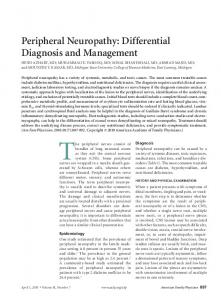
Figure 2 (a) T2-weighted CISS axial image of a patient with unilateral cochlear nerve dysplasia at the right side. (A) Right internal auditory canal (IAC) with evidence of superior vestibular and facial nerves. C: Cochlea. (b) Reconstructed sagittal T2 weighted CISS image through the right IAC demonstrates the absence of the right cochlear nerve. FN: facial nerve; VN: superior and inferior vestibular nerves.
c linician should be very alert to the possibility of cochlear nerve deficiency.23 Levi et al.58 suggested that a nerve that is 50% smaller than the adjacent facial nerve on MRI constitutes a diagnostic criterion for cochlear nerve hypoplasia. A normal bony internal auditory canal and inner ear morphology are not reliable surrogate markers of cochlear nerve integrity.59,60 MRI is therefore superior to CT as a diagnostic tool and should be performed in all paediatric patients with an ANSD profile (Figure 2). Between 40% and 85% of patients with a cochlear nerve deficiency have associated inner ear abnormalities.26,59-62 There is a higher rate of malformations, both labyrinthine and in the hindbrain, in bilateral cochlear nerve deficiency than in unilateral cases.26,58 In unilateral cochlear dysplasia/aplasia, the left side is involved slightly more frequently.58 Causes of ANSD A variety of risk factors and aetiologies are associated with this heterogeneous form of hearing loss, which may explain the variation in auditory skills in ANSD patients. ANSD may be congenital or acquired. Congenital ANSD affects the development of language, an ability that is strictly related to a period of sensitivity that declines with age.63 During
10-declau-.indd 70
this period, the development of language skills is strictly dependent on cortical plasticity and efficient auditory input is required. When the onset of ANSD is delayed until childhood or adult life (acquired ANSD), abnormalities of auditory input lead to the severe impairment of speech perception and progressive deterioration. Patients can range from newborns to adults. Sininger and Oba reviewed their patients in 2001,38 finding that onset of the condition was before 10 years in 75% of the cases, and before 2 years in about 40%. Of the children in whom onset was before the age of 2 years, 44% had potential genetic or familial risk factors. Of the other children, 12% had histories of hyperbilirubinaemia, 8% were born prematurely, 28% had multiple neonatal risk factors, and 52% had no other risk factors. A proportion of the unknown causes are also thought to have an underlying genetic aetiology. Approximately 40% of auditory neuropathy cases may therefore have a genetic basis. A study by Starr et al.64 which also looked into the causes of ANSD shows that the disorder in 42% of patients was associated with hereditary neurological disorders; in 10% of patients, it was associated with toxic, metabolic, immunological, and infectious factors, and the cause was unknown in 48% of patients. The majority of cases with ANSD are sporadic: there is only one affected
17/10/13 14:41
71
Auditory neuropathy
Immune/Metabolic
Table 1 Non-genetic causes of ANSD, modified after11,31,78,79
Hyperbilirubinaemia/ Kernicterus
Polyarteritis nodosa
Anoxia /hypoxia
Sarcoidosis
Alcoholism/nutritional Diabetes mellitus Uraemia
Infection/Inflammatory Leprosy
Borreliosis Toxins
Paraproteinaemias
Rheumatoid arthritis
Guillain-Barré Syndrome21 AIDS
Ramsey Hunt syndrome
Heavy metals (lead, cobalt, mercury)80-82 Drugs (Carboplatin)83,84 Trauma
Vascular diseases
individual. However, research has identified many familial cases with two or more members in a family a ffected.65 Non-genetic causes of ANSD Although Berg et al.19 were unable to determine a specific risk profile, the identification of risk factors for hearing loss by using the guidelines of the Joint Committee on Infant Hearing 2000 position statement is highly recommended.55,66 Retrospective clinical studies have suggested that perinatal problems like anoxia/hypoxia, hyperbilirubinaemia, dysmaturity and prematurity in particular are non-genetic risk factors for ANSD.67-69 A more comprehensive list of non-genetic causes is given in Table 1. The causes of acquired ANSD are poorly understood because there are only a few relevant temporal bone studies.70,71 Amatuzzi provided a histopathological demonstration in a temporal bone study that the mean gestation period for infants with selective inner hair cell loss was 32 weeks, and 36 weeks in infants with normal ears or with more common histopathological patterns of OHC loss or combined OHC and IHC loss.72 Hyperbilirubinaemia is a risk factor for ANSD. Wennberg suggests ≥ 1.2 μg/dl free (unbound) bilirubin as a possible criterion for further studies.73 One of the cardinal clinical signs of acute or chronic classical kernicterus is auditory impair-
10-declau-.indd 71
Neoplasma/ intracranial cystic lesions
ment, and all but one of the kernicterus subtypes also include auditory impairment.74 Shapiro and Popelka suggest that bilirubin exposure results in auditory system damage, initially at the level of the brainstem and progressing to the level of the VIII cranial nerve and then to greater neural centres.74 There is no evidence of neural damage at the level of the cochlea. Auditory neural damage from bilirubin toxicity ranges from neural timing deficits, including neural firing delays and dyssynchrony, to neural response reduction and even the elimination of auditory neural responses. Chisin et al.75 were the first to record both the CM and the ABR in patients with hyperbilirubinaemia; they found that the majority of their cases had a CM but an atypical or absent ABR, suggesting auditory nerve or brainstem dysfunction. More recently, Rhee et al.76 reported normal OAEs and absent or atypical ABR findings in 4 out of 11 infants with severe hyperbilirubinaemia. Amin et al.77 recorded changes in ABR maturity over the first 5 days in premature infants that were correlated with unconjugated bilirubin levels. In this subgroup of ANSD, there is a tendency towards spontaneous improvement: Rhee et al.,76 and others, have stressed that most of the initially atypical ABR findings reverse or improve with time, suggesting that neural/brainstem pathology tends to normalise after treatment with exchange transfusion or phototherapy.76
17/10/13 14:41
72
Non-syndromic
F. Declau et al.
Transmission
Locus
Disease name
Autosomal dominant
13q21-q24
AUNA1
Moderate to profound deafness DIAPH365
Autosomal recessive
2p23-p22
DFNB9
Congenital profound deafness
OTOF86
DFNB1A
Congenital profound deafness
GJB288
Mild deafness with peripheral neuropathy
GJB390
Mild to severe deafness; demyelinating neuropathy
MPZ94
Autosomal recessive Autosomal recessive Syndromic
Table 2 Genetic causes of ANSD, modified after79 and20
Mitochondrial
2q31.1-q31.3
DFNB59
13q12.11
12S rRNA
MTRNR1
Autosomal dominant
17p11.2-p12
CMT 1A
Autosomal dominant
1q22
CMT 1B
Autosomal dominant
8p21
CMT 2E
Autosomal dominant
3q28-q29
Autosomal recessive
8q24.3
Autosomal recessive
11q14.111q22.3
Autosomal recessive
9q13
Autosomal recessive
10p13
X-linked recessive
Xq22.1
X-linked recessive
Xq23-q27.3
Autosomal dominant
X-linked dominant
Mitochondrial
1p34.3
Xp13
MTND4
Congenital profound deafness Moderate to severe deafness
CMT 4D
Genetic markers linked to ANSD
PJVK87
T1095C mutation89
Mild to severe deafness; demyelinating neuropathy
PMP2291,92,93
Normal hearing; axonal neuropathy
NF-L95
Autosomal Optic neuropathy; moderate Dominant Optic deafness Atrophy (ADOA) or Kjer’s Disease Mild to severe deafness; axonal/demyelinating neuropathy
OPA196
NDRG197
Autosomal Optic neuropathy; mild hearing TMEM126A98 Recessive Optic loss Atrophy (AROA)
Friedreich’s Ataxia Ataxia; axonal neuropathy; optic neuropathy; cardiomyopathy; normal hearing threshold to mild deafness Refsum Disease CMT 1X
Progressive deafness
Demyelinating neuropathy
FXN99
PHYH100 GJB1101
Mohr-Tranebjaerg Progressive deafness; dystonia, DDP1 Syndrome (MTS) optic neuropathy; dementia TIMM8A102 or DeafnessDystonia Syndrome DFNX5
Sensory axonal neuropathy; mild-to-severe deafness
Leber Hereditary Optic neuropathy; mild-toOptic Neuropathy moderate deafness (LHON)
However, the decision to treat hyperbilirubinaemia is itself controversial in terms of the criteria and the threshold for treatment.15 Genes involved in ANSD Genetic studies of hearing loss make it clear that the majority (up to 80%) of hereditary deafness is
10-declau-.indd 72
Phenotype
AUNX1103 11778mtDNA mutation104
non-syndromic.85 Even though the exact percentage of non-syndromic occurrence of ANSD is not known, a large number of patients with ANSD are non-syndromic. The identification of several genes involved in the pathogenesis of both presynaptic and postsynaptic AN has greatly contributed to the diagnosis and a better understanding of the mechanisms underlying this disorder.20 The inheritance
17/10/13 14:41
73
Auditory neuropathy pattern can include all the main types of inheritance, such as autosomal dominant, autosomal recessive, X-linked and mitochondrial. About one third to 40% of subjects with ANSD are affected by peripheral neuropathies.13,64 In addition, optic neuropathies and other CNS disorders have been found in association with ANSD. In this group of subjects the hearing disorder is caused by several genetic defects, all resulting in neuronal loss and demyelination in peripheral and/or cranial nerves, and the site of the lesion is invariably postsynaptic. ANSD can also be associated with hereditary syndromes such as Charcot-Marie-Tooth (CMT) Disease,93-95,97 Leber’s Hereditary Optic Neuropathy (LHON),104 Autosomal Dominant Optic Atrophy (ADOA)96, Autosomal Recessive Optic Atrophy (AROA),98 Fredreich’s Ataxia,99 Mohr-Tranebjaerg Syndrome (MTS),102 Refsum Disease,100 and X-linked disease.103 For a comprehensive overview, the reader is referred to Manchaiah et al.79 (2011) and Santarelli (2010).20 Treatment options Auditory impairment ranges across a continuum from mild to severe. A recent evidence-based systematic review by Roush et al.105 (2011) found that the clinical evidence related to intervention for ANSD is in the very preliminary stages. Generally, behavioural audiograms are not valuable in ANSD patients as a management tool for treatment. In contrast to children with most forms of sensorineural hearing loss, perceptual ability in ANSD children cannot be predicted with reasonable accuracy on the basis of the behavioural audiogram. According to Rance and Barker, CAEPs could be used to predict in advance children’s linguistic abilities: their results showed that approximately 50% of children with ANSD had speech-recognition scores similar to those of children with sensory hearing loss; the other 50% essentially had no open-set speech-perception ability. Interestingly, the children with no open-set speech perception had no cortical evoked potentials, while the children who had measurable speech-recognition scores did have CAEPs.106 Hearing aids Success with hearing aids in quiet as reported by Deltenre et al.106, and Rance et al.107 did not lead to
10-declau-.indd 73
age-appropriate language acquisition in the majority of patients.13 However, there appears to be a subset of children whose aided speech-perception abilities benefit, and in whom there is improvement in puretone detection thresholds.105 Berlin et al.13 (2010) summarised outcome management in 260 patients with ANSD. Approximately 15% reported some benefits with hearing aids in terms of language learning, while improvement in speech comprehension and language acquisition was reported in 85% of patients with implants. Approximately 5% of the total population developed normal speech and language without intervention. Current evidence-based protocols for fitting hearing aids rely on estimates of hearing threshold levels to determine the recommended gain and output variables. In children with ANSD, the determination of hearing thresholds is typically delayed until the infant is developmentally able to perform reliable behavioural testing using VRA. Recommendations for management include the use of low-gain hearing aids or FM systems, the use of low-gain amplification in one ear only, or the avoidance of hearing aids altogether, in combination with visual and/or sign language exposure. 7,108 Relentless mouth-covered auditory-verbal therapy should be avoided. Usually, hearing aids make the child more aware of environmental sounds but do not result in auditory learning language. The drawback to using powerful hearing aids is that they may destroy outer hair cell function and, if the child is on the road to recovery, may cause a preventable high-frequency sensory loss.109 This population is also unique because of the high rate of comorbidities of up to 54%, which can be very demanding for the therapeutic team.7 A speech language team should monitor the progress of the child quarterly to ensure that appropriate progress is being achieved. Cochlear implants Behavioural pure-tone audiograms are of limited prognostic value in predicting aided benefit in children with ANSD, and so cochlear implantation should be considered even if audiometric thresholds are better than they would be when typically considering implants after progress with conventional amplification has proven inadequate. However, this step should be taken only after adequate attempts have been made to address the case with
17/10/13 14:41
74 amplification, appropriate early-intervention services, and a comprehensive evaluation by a team of professionals.7 No child should receive an implant before a behavioural measure of hearing has been obtained. Audiometric criteria used to select paediatric cochlear implant candidates with sensorineural hearing loss can also be applied in ANSD cases. Young ANSD candidates should therefore be considered for the procedure as soon as ‘stable’ hearing thresholds in the severe/profound range can be established. The age at which this is possible will be determined to some extent by the child’s ability to respond reliably to conditioned audio metric testing. Another consideration, however, should be the possibility of improvement in auditory neural function, which has been demonstrated both in the emergence of a repeatable ABR and a decrease in hearing thresholds in some ANSD infants (usually in the first year of life).56,57,110 This finding therefore raises issues relating to implantation before the age of 1 year.111 However, the outcome of cochlear implantation varies widely among patients and even between cochlear implant teams.7 According to Breneman et al.112 (2012), the expected outcomes of cochlear implantation for children with ANSD, excluding children with cochlear nerve deficiency, are no different than for children with non-ANSD SNHL. Raveh et al.111 examined the effect of CI intervention on speech and language outcomes in children with ANSD. The authors noted that children with ANSD using CI achieved results in speech production tests that were comparable with age-matched CI users with SNHL. Electrically evoked compound action potentials (ECAPs) in children with implants were not predictors of outcome.113 While it is generally acknowledged that patients with presynaptic AN due to mutations in the OTOF gene invariably benefit49, poor outcome has been reported for implant subjects with postsynaptic AN with an underlying axonal loss and degeneration of the entire nerve fibres, as in Friedreich ataxia114 and deafness dystonia syndrome.115 By contrast, as mentioned by Santarelli,20 two patients with the R445H mutation in the OPA1 gene116 and some subjects with mutations in the DIAPH3 gene8, recovered both speech perception and auditory brainstem responses following cochlear implantation.20 Recently, children with Friedreich ataxia have benefited
10-declau-.indd 74
F. Declau et al. significantly from an FM listening device in everyday life and in speech perception performance.117 There is lively debate about the optimal treatment for patients with cochlear nerve dysplasia, and the decision about the best therapy depends on whether there is a complete absence of nerve fibres or hypoplasia and on whether the condition is unilateral or bilateral, as well as on the possible presence of other abnormalities. Kutz et al.118 suggest that the presence of wave V on EABR may have predictive value for cochlear implantation. It is possible that, if some nerve fibres remain, wave V could be generated at high intensities and these patients could therefore benefit from CI to some extent. On the other hand, Carner et al.59 examined patients with cochlear nerve dysplasia who were given a CI and found no response. Colleti et al.119 stated that these patients may benefit from an auditory brainstem implant (ABI), although to a lesser extent than other patients. Merkus et al.120 and Sennaroglu et al.121 also stated that ABI could be indicated in the absence of the cochlear nerve when this has been proved by imaging and in testing since this is the only way to secure any chance of hearing. By contrast, Levi et al.58 recommended preferential seating (when CND is unilateral) or amplification as opposed to CI for patients with cochlear nerve dysplasia. However, two recent publications point out that the absence of a visible cochlear nerve on imaging does not preclude the auditory innervation of the cochlea.122,123 Electrically-evoked ABR testing should be performed since this factor is critical for the evaluation of this patient group.122,124 References 1. Hinchcliffe R, Osuntokun BO, Adeuja AO. Hearing levels in Nigerian ataxic neuropathy. Audiology. 1972; 11(3):218-230. 2. Kemp DT. Stimulated acoustic emissions from within the human auditory system. J Acoust Soc Am. 1978;64(5): 1386-1391. 3. Starr A, Picton TW, Sininger Y, Hood LJ, Berlin CI. Auditory neuropathy. Brain. 1996;119( Pt 3):741-753. 4. Berlin CI, Hood LJ, Cecola RP, Jackson DF, Szabo P. Does type I afferent neuron dysfunction reveal itself through lack of efferent suppression? Hearing research. 1993;65(1):40-50. 5. Rapin I, Gravel J. “Auditory neuropathy”: physiologic and pathologic evidence calls for more diagnostic
17/10/13 14:41
Auditory neuropathy s pecificity. Int J Pediatr Otorhinolaryngol. 2003;67(7): 707-728. 6. Starr A, Zeng F, Michalewski H, Moser T. Perspectives on Auditory Neuropathy: Disorders of Inner Hair Cell, Auditory Nerve, and Their Synapse. In: Basbaum AI, Kaneko A, Shepherd GM, Westheimer G, Eds. The Senses: A Comprehensive Reference. Academic Press, San Diego; Vol 3 Audition: 2008:397-412. 7. Hayes D, Sininger Y, Northern J. Guidelines for identification and management of infants and young children with auditory neuropathy spectrum disorder. Conference Proceedings. Guidelines Development Conference at NHS, Como, Italy. The Children’s Hospital, Colorado; 2008. 8. Starr A, Isaacson B, Michalewski HJ, Zeng FG, Kong YY, Beale P, Paulson GW, Keats BJB, Lesperance MM. A dominantly inherited progressive deafness affecting distal auditory nerve and hair cells. J Assoc Res Otolaryngol. 2004;5(4):411-426. 9. Rapin I, Gravel J. Auditory neuropathy: a biologically inappropriate label unless acoustic nerve involvement is documented. J Am Acad Audiol. 2006;17(2):147-150. 10. Shelton C, Luxford WM, Tonokawa LL, Lo WW, House WF. The narrow internal auditory canal in children: a contraindication to cochlear implants. Otolaryngol Head Neck Surg. 1989;100(3):227-231. 11. Sanyelbhaa Talaat H, Kabel AH, Samy H, Elbadry M. Prevalence of auditory neuropathy (AN) among infants and young children with severe to profound hearing loss. Int J Pediatr Otorhinolaryngol. 2009;73(7):937-939. 12. Maris M, Venstermans C, Boudewyns A. Auditory neuro pathy/dyssynchrony as a cause of failed neonatal hearing screening. Int J Pediatr Otorhinolaryngol. 2011;75(7): 973-975. 13. Berlin CI, Hood LJ, Morlet T, Wilensky D, Li L, Mattingly KR, Taylor-Jeanfreau J, Keats BJB, St John P, Montgomery E, Shallop JK, B Russell BA, Frisch SA. Multi-site diagnosis and management of 260 patients with auditory neuropathy/dys-synchrony (auditory neuropathy spectrum disorder). Int J Audiol. 2010;49(1):30-43. 14. Kirkim G, Serbetcioglu B, Erdag TK, Ceryan K. The frequency of auditory neuropathy detected by universal newborn hearing screening program. Int J Pediatr Oto rhinolaryngol. 2008;72(10):1461-1469. 15. Ngo RY, Tan HK, Balakrishnan A, Lim SB, Lazaroo DT. Auditory neuropathy/auditory dys-synchrony detected by universal newborn hearing screening. Int J Pediatr Otorhinolaryngol. 2006;70(7):1299-1306. 16. Dowley AC, Whitehouse WP, Mason SM, Cope Y, Grant J, Gibbin KP. Auditory neuropathy: unexpectedly common in a screened newborn population. Dev Med Child Neurol. 2009;51(8):642-646. 17. Foerst A, Beutner D, Lang-Roth R, Huttenbrink KB, von Wedel H, Walger M. Prevalence of auditory neuropathy/ synaptopathy in a population of children with profound hearing loss. Int J Pediatr Otorhinolaryngol. 2006;70(8): 1415-1422. 18. Sininger Y. A Sound Foundation Through Early Amplification. Proceedings of the Second International Conference. Section V, Chapter 15. In: Seewald R, Gravel J,
10-declau-.indd 75
75 Eds. Changing Considerations for Cochlear Implant Candidacy: Age, Hearing Level and Auditory Neuropathy. Immediate Proceedings, Ltd, Suffolk, 2001:187-194. 19. Berg AL, Spitzer JB, Towers HM, Bartosiewicz C, Diamond BE. Newborn hearing screening in the NICU: profile of failed auditory brainstem response/passed otoacoustic emission. Pediatrics. 2005;116(4):933-938. 20. Santarelli R. Information from cochlear potentials and genetic mutations helps localize the lesion site in auditory neuropathy. Genome Med. 2010;2(12):91. 21. Nelson KR, Gilmore RL, Massey A. Acoustic nerve conduction abnormalities in Guillain-Barré syndrome. Neurology. 1988;38(8):1263-1266. 22. American Academy of Pediatrics, Joint Committee on Infant Hearing. Year 2007 position statement: Principles and guidelines for early hearing detection and intervention programs. Pediatrics. 2007;120(4):898-921. 23. Buchman CA, Roush PA, Teagle HF, Brown CJ, Zdanski CJ, Grose JH. Auditory neuropathy characteristics in children with cochlear nerve deficiency. Ear Hear. 2006; 27(4):399-408. 24. Walton J, Gibson WP, Sanli H, Prelog K. Predicting cochlear implant outcomes in children with auditory neuropathy. Otol Neurotol. 2008;29(3):302-309. 25. Teagle HF, Roush PA, Woodard JS, Hatch DR, Zdanski CJ, Buss E, Buchman CA. Cochlear implantation in children with auditory neuropathy spectrum disorder. Ear Hear. 2010; 31(3):325-335. 26. Huang BY, Roche JP, Buchman CA, Castillo M. Brain stem and inner ear abnormalities in children with auditory neuropathy spectrum disorder and cochlear nerve deficien cy. AJNR Am J Neuroradiol. 2010;31(10):1972-1979. 27. Berlin CI, Morlet T, Hood LJ. Auditory neuropathy/ dyssynchrony: its diagnosis and management. Pediatr Clin North Am. 2003;50(2):331-40, vii-viii. 28. Starr A, Sininger Y, Nguyen T, Michalewski HJ, Oba S, Abdala C. Cochlear receptor (microphonic and summating potentials, otoacoustic emissions) and auditory pathway (auditory brain stem potentials) activity in auditory neuro pathy. Ear Hear. 2001;22(2):91-99. 29. Berlin CI, Bordelon J, St John P, Wilensky D, Hurley A, Kluka E, Hood LJ. Reversing click polarity may uncover auditory neuropathy in infants. Ear Hear. 1998;19(1):3747. 30. Rance G, Beer DE, Cone-Wesson B, Shepherd RK, Dowell RC, King AM, Rickards FW, Clark GM. Clinical findings for a group of infants and young children with auditory neuropathy. Ear Hear. 1999;20(3):238-252. 31. Sininger YS, Starr A. Auditory neuropathy: a new perspective on hearing disorders. Singular Publishing Group, San Diego; 2001. 32. Roush P. Auditory neuropathy spectrum disorder: evaluation and management. Hear J. 2008;61(11):36-41. 33. Hall JW, Smith SD, Popelka GR. Newborn hearing screening with combined otoacoustic emissions and auditory brainstem responses. J Am Acad Audiol. 2004;15(6): 414-425. 34. Sutton GJ. Usefulness of acoustic reflexes still unproven in newborns and young infants. J Am Acad Audiol. 2007; 18(2):187.
17/10/13 14:41
76 35. Rance G. Auditory neuropathy/dys-synchrony and its perceptual consequences. Trends Amplif. 2005;9(1):1-43. 36. Starr A, Sininger Y, Winter M, Derebery MJ, Oba S, Michalewski HJ. Transient deafness due to temperaturesensitive auditory neuropathy. Ear Hear. 1998;19(3):169179. 37. Berlin CI, Goforth-Barter L, Hood LJ. Auditory neuro pathy: three time courses after early identification. In: ARO Abstract Book. Association for research in Otolaryngology, St. Petersburg Beach, Florida, USA, 1999; Abstract # 668. 38. Sininger Y, Oba S. Patients with auditory neuropathy: who are they and what can they hear? In: Sininger YS, Starr A, Eds. Auditory neuropathy: a new perspective on hearing disorders. Singular Publishing Group, San Diego, 2001:15-36. 39. Zeng FG, Kong YY, Michalewski HJ, Starr A. Perceptual consequences of disrupted auditory nerve activity. J Neuro physiol. 2005;93(6):3050-3063. 40. Santarelli R, Arslan E. Electrocochleography in auditory neuropathy. Hear Res. 2002;170(1-2):32-47. 41. Santarelli R, Scimemi P, Dal Monte E, Arslan E. Cochlear microphonic potential recorded by transtympanic electrocochleography in normally-hearing and hearing-impaired ears. Acta Otorhinolaryngol Ital. 2006;26(2):78-95. 42. Eggermont JJ, and Odenthal DW. Methods in electro cochleography. Acta Otolaryngol Suppl. 1974;316:17-24. 43. McMahon CM, Patuzzi RB, Gibson WP, Sanli H. Frequency-specific electrocochleography indicates that presynaptic and postsynaptic mechanisms of auditory neuropathy exist. Ear Hear. 2008;29(3):314-325. 44. Gibson WP, Sanli H. Auditory neuropathy: an update. Ear Hear. 2007;28(2 Suppl):102S-106S. 45. Santarelli R, Starr A, Michalewski HJ, Arslan E. Neural and receptor cochlear potentials obtained by trans tympanic electrocochleography in auditory neuropathy. Clin Neurophysiol. 2008;119(5):1028-1041. 46. Withnell RH. Brief report: the cochlear microphonic as an indication of outer hair cell function. Ear Hear. 2001; 22(1):75-77. 47. Zheng XY, Henderson D, McFadden SL, Hu BH. The role of the cochlear efferent system in acquired resistance to noise-induced hearing loss. Hear Res. 1997;104(12):191-203. 48. Durrant JD, Wang J, Ding DL, Salvi RJ. Are inner or outer hair cells the source of summating potentials recorded from the round window? J Acoust Soc Am. 1998;104(1): 370-377. 49. Santarelli R, Del Castillo I, Rodríguez-Ballesteros M, Scimemi P, Cama E, Arslan E, Starr A. Abnormal cochlear potentials from deaf patients with mutations in the otoferlin gene. J Assoc Res Otolaryngol. 2009;10(4):545556. 50. Gardner-Berry K, Gibson WP, Sanli H. Pre-operative testing of patients with neuropathy or dys-synchrony. Hear J. 2005;58(11):24-25. 51. Rance G, Cone-Wesson B, Wunderlich J, Dowell R. Speech perception and cortical event related potentials in children with auditory neuropathy. Ear Hear. 2002;23(3): 239-253.
10-declau-.indd 76
F. Declau et al. 52. Jerger J, Musiek F. Report of the Consensus Conference on the Diagnosis of Auditory Processing. J Am Acad Audiol. 2000;11(9):467-474. 53. Bellis TJ. Assessment and management of central auditory processing disorders in the educational setting: from science to practice. Thomson, Delmar Learning, New York; 2003. 54. King AM. The national protocol for paediatric amplification in Australia. Int J Audiol. 2010;49 Suppl 1:S64-69. 55. Declau F, Boudewyns A, Van den Ende J, Peeters A, Van den Heyning P. Etiologic and audiologic evaluations after universal neonatal hearing screening: analysis of 170 referred neonates. Pediatrics. 2008;121(6):1119-1126. 56. Psarommatis I, Riga M, Douros K, Koltsidopoulos P, Douniadakis D, Kapetanakis I, Apostolopoulos N. Transient infantile auditory neuropathy and its clinical implications. Int J Pediatr Otorhinolaryngol. 2006;70(9): 1629-1637. 57. Attias J, Raveh E. Transient deafness in young candidates for cochlear implants. Audiol Neurootol. 2007;12(5):325333. 58. Levi J, Ames J, Bacik K, Drake C, Morlet T, O’Reilly RC. Clinical characteristics of children with cochlear nerve dysplasias. Laryngoscope. 2013;123(3):752-756. 59. Adunka OF, Roush PA, Teagle HF, Brown CJ, Zdanski CJ, Jewells V, Buchman CA. Internal auditory canal morphology in children with cochlear nerve deficiency. Otol Neurotol. 2006;27(6):793-801. 60. Carner M, Colletti L, Shannon R, Cerini R, Barillari M, Pozzi Mucelli R, Colletti V. Imaging in 28 children with cochlear nerve aplasia. Acta Otolaryngol. 2009;129(4): 458-461. 61. Laury AM, Casey S, McKay S, Germiller JA. Etiology of unilateral neural hearing loss in children. Int J Pediatr Otorhinolaryngol. 2009;73(3):417-427. 62. Roche JP, Huang BY, Castillo M, Bassim MK, Adunka OF, Buchman CA. Imaging characteristics of children with auditory neuropathy spectrum disorder. Otol Neurotol. 2010;31(5):780-788. 63. Uhler K, Heringer A, Thompson N, Yoshinaga-Itano C. A tutorial on auditory neuropathy/dyssynchrony for the speech-language pathologist and audiologist. Semin Speech Lang. 2012;33(4):354-366. 64. Starr A, Sininger YS, Pratt H. The varieties of auditory neuropathy. J Basic Clin Physiol Pharmacol. 2000;11(3): 215-230. 65. Kim TB, Isaacson B, Sivakumaran TA, Starr A, Keats BJ, Lesperance MM. A gene responsible for autosomal dominant auditory neuropathy (AUNA1) maps to 13q1421. J Med Genet. 2004;41(11):872-876. 66. Joint Committee on Infant Hearing; American Academy of Audiology; American Academy of Pediatrics; American Speech-Language-Hearing Association; Directors of Speech and Hearing Programs in State Health and Welfare Agencies. Year 2000 position statement: principles and guidelines for early hearing detection and intervention programs. Joint Committee on Infant Hearing, American Academy of Audiology, American Academy of Pediatrics, American Speech-Language-Hearing Association, and Directors of Speech and Hearing Programs in State
17/10/13 14:41
Auditory neuropathy Health and Welfare Agencies. Pediatrics. 2000;106(4): 798-817. 67. Madden C, Rutter M, Hilbert L, Greinwald JH, Choo DI. Clinical and audiological features in auditory neuropathy. Arch Otolaryngol Head Neck Surg. 2002;128(9):10261030. 68. Beutner D, Foerst A, Lang-Roth R, von Wedel H, Walger M. Risk factors for auditory neuropathy/auditory synaptopathy. ORL J Otorhinolaryngol Relat Spec. 2007; 69(4):239-244. 69. Xoinis K, Weirather Y, Mavoori H, Shaha SH, Iwamoto LM. Extremely low birth weight infants are at high risk for auditory neuropathy. J Perinatol. 2007;27(11):718723. 70. Slack RW, Wright A, Michaels L, Frohlich SA. Inner hair cell loss and intracochlear clot in the preterm infant. Clin Otolaryngol Allied Sci. 1986;11(6):443-446. 71. Amatuzzi MG, Northrop C, Liberman MC, Thornton A, Halpin C, Herrmann B, Pinto LE, Saenz A, Carranza A, Eavey RD. Selective inner hair cell loss in premature infants and cochlea pathological patterns from neonatal intensive care unit autopsies. Arch Otolaryngol Head Neck Surg. 2001;127(6):629-636. 72. Amatuzzi M, Liberman MC, Northrop C. Selective inner hair cell loss in prematurity: a temporal bone study of infants from a neonatal intensive care unit. J Assoc Res Otolaryngol. 2011;12(5):595-604. 73. Wennberg RP, Ahlfors CE, Bhutani VK, Johnson LH, Shapiro SM. Toward understanding kernicterus: a challenge to improve the management of jaundiced newborns. Pediatrics. 2006;117(2):474-485. 74. Shapiro SM, Popelka GR. Auditory impairment in infants at risk for bilirubin-induced neurologic dysfunction. Semin Perinatol. 2011;35(3):162-170. 75. Chisin R, Perlman M, Sohmer H. Cochlear and brain stem responses in hearing loss following neonatal hyperbilirubinemia. Ann Otol Rhinol Laryngol. 1978;88(3 Pt 1):352357. 76. Rhee CK, Park HM, Jang YJ. Audiologic evaluation of neonates with severe hyperbilirubinemia using transiently evoked otoacoustic emissions and auditory brainstem responses. Laryngoscope. 1999;109(12):2005-2008. 77. Amin SB, Ahlfors C, Orlando MS, Dalzell LE, Merle KS, Guillet R. Bilirubin and serial auditory brainstem responses in premature infants. Pediatrics. 2001;107(4):664-670. 78. Simmons J, McCreery R. Auditory neuropathy/dys- synchrony: trends in assessment and treatment. ASHA Lead. 2007;12(8):12-15. 79. Manchaiah VK, Zhao F, Danesh AA, Duprey R. The genetic basis of auditory neuropathy spectrum disorder (ANSD). Int J Pediatr Otorhinolaryngol. 2011;75(2):151158. 80. Wong VC, Ng TH, Yeung CY. Electrophysiologic study in acute lead poisoning. Pediatr Neurol. 1991;7(2):133136. 81. Apostoli P, Catalani S, Zaghini A, Mariotti A, Poliani PL, Vielmi V, Semeraro F, Duse S, Porzionato A, Macchi V, Padovani A, Rizzetti MC, De Caro R. High doses of cobalt induce optic and auditory neuropathy. Exp Toxicol Pathol. 2012;65 (6):719-727.
10-declau-.indd 77
77 82. Rice DC, Gilbert SG. Effects of developmental methylmercury exposure or lifetime lead exposure on vibration sensitivity function in monkeys. Toxicol Appl Pharmacol. 1995;134(1):161-169. 83. Harrison RV. An animal model of auditory neuropathy. Ear Hear. 1998;19(5):355-361. 84. El-Badry MM, McFadden SL. Evaluation of inner hair cell and nerve fiber loss as sufficient pathologies underlying auditory neuropathy. Hear Res. 2009;255(1-2):84-90. 85. Van Camp G, Smith RJ. Hereditary hearing loss homepage. Available at: http://hereditaryhearingloss.org. Accessed August 20, 2013. 86. Rodríguez-Ballesteros M, del Castillo FJ, Martín Y, Moreno-Pelayo MA, Morera C, Prieto F, Marco J, Morant A, Gallo-Terán J, Morales-Angulo C, Navas C, Trinidad G, Tapia MC, Moreno F, del Castillo I. Auditory neuro pathy in patients carrying mutations in the otoferlin gene (OTOF). Hum Mutat. 2003;22(6):451-456. 87. Delmaghani S, del Castillo FJ, Michel V, Leibovici M, Aghaie A, Ron U, Van Laer L, Ben-Tal N, Van Camp G, Weil D, Langa F, Lathrop M, Avan P, Petit C. Mutations in the gene encoding pejvakin, a newly identified protein of the afferent auditory pathway, cause DFNB59 auditory neuropathy. Nat Genet. 2006;38(7):770-778. 88. Cheng X, Li L, Brashears S, Morlet T, Ng SS, Berlin C, Hood L, Keats B. Connexin 26 variants and auditory neuropathy/dys-synchrony among children in schools for the deaf. Am J Med Genet A. 2005;139(1):13-18. 89. Wang Q, Li R, Zhao H, Peters JL, Liu Q, Yang L, Han D, Greinwald JH Jr, Young WY, Guan MX. Clinical and molecular characterization of a Chinese patient with auditory neuropathy associated with mitochondrial 12S rRNA T1095C mutation. Am J Med Genet A. 2005;133A(1):2730. 90. López-Bigas N, Olivé M, Rabionet R, Ben-David O, Martínez-Matos JA, Bravo O, Banchs I, Volpini V, Gasparini P, Avraham KB, Ferrer I, Arbonés ML, Estivill X. Connexin 31 (GJB3) is expressed in the peripheral and auditory nerves and causes neuropathy and hearing impairment. Hum Mol Genet. 2001;10(9):947-952. 91. Aarskog NK, Vedeler CA. Real-time quantitative polymerase chain reaction. A new method that detects both the peripheral myelin protein 22 duplication in CharcotMarie-Tooth type 1A disease and the peripheral myelin protein 22 deletion in hereditary neuropathy with liability to pressure palsies. Hum Genet. 2000;107(5):494-498. 92. Kovach MJ, Lin JP, Boyadjiev S, Campbell K, Mazzeo L, Herman K, Rimer LA, Frank W, Llewellyn B, Jabs EW, Gelber D, Kimonis VE. A unique point mutation in the PMP22 gene is associated with Charcot-Marie-Tooth disease and deafness. Am J Hum Genet. 1999;64(6):15801593. 93. Raeymaekers P, Timmerman V, Nelis E, Van Hul W, De Jonghe P, Martin JJ, Van Broeckhoven C. Estimation of the size of the chromosome 17p11.2 duplication in Charcot-Marie-Tooth neuropathy type 1a (CMT1a). HMSN Collaborative Research Group. J Med Genet. 1992;29(1):5-11. 94. Starr A, Michalewski HJ, Zeng FG, Fujikawa-Brooks S, Linthicum F, Kim CS, Winnier D, Keats B. Pathology
17/10/13 14:41
78 and physiology of auditory neuropathy with a novel mutation in the MPZ gene (Tyr145->Ser). Brain. 2003; 126(Pt 7):1604-1619. 95. Butinar D, Starr A, Zidar J, Koutsou P, Christodoulou K. Auditory nerve is affected in one of two different point mutations of the neurofilament light gene. Clin Neurophysiol. 2008;119(2):367-375. 96. Amati-Bonneau P, Guichet A, Olichon A, Chevrollier A, Viala F, Miot S, Ayuso C, Odent S, Arrouet C, Verny C, Calmels MN, Simard G, Belenguer P, Wang J, Puel JL, Hamel C, Malthièry Y, Bonneau D, Lenaers G, Reynier P. OPA1 R445H mutation in optic atrophy associated with sensorineural deafness. Ann Neurol. 2005;58(6):958-963. 97. Kalaydjieva L, Nikolova A, Turnev I, Petrova J, Hristova A, Ishpekova B, Petkova I, Shmarov A, Stancheva S, Middleton L, Merlini L, Trogu A, Muddle JR, King RH, Thomas PK. Hereditary motor and sensory neuropathy-Lom, a novel demyelinating neuropathy associated with deafness in gypsies. Clinical, electrophysiological and nerve biopsy findings. Brain. 1998;121( Pt 3):399-408. 98. Meyer E, Michaelides M, Tee LJ, Robson AG, Rahman F, Pasha S, Luxon LM, Moore AT, Maher ER. Nonsense mutation in TMEM126A causing autosomal recessive optic atrophy and auditory neuropathy. Mol Vis. 2010; 16:650-664. 99. Rance G, Fava R, Baldock H, Chong A, Barker E, Corben L, Delatycki MB. Speech perception ability in individuals with Friedreich ataxia. Brain. 2008;131(Pt 8):2002-2012. 100. Oysu C, Aslan I, Basaran B, Baserer N. The site of the hearing loss in Refsum’s disease. Int J Pediatr Otorhinolaryngol. 2001;61(2):129-134. 101. Bähr M, Andres F, Timmerman V, Nelis ME, Van Broeckhoven C, Dichgans J. Central visual, acoustic, and motor pathway involvement in a Charcot-Marie-Tooth family with an Asn205Ser mutation in the connexin 32 gene. J Neurol Neurosurg Psychiatry. 1999;66(2):202206. 102. Tranebjaerg L, Schwartz C, Eriksen H, Andreasson S, Ponjavic V, Dahl A, Stevenson RE, May M, Arena F, Barker D, Elverland HH, Lubs H. A new X linked recessive deafness syndrome with blindness, dystonia, fractures, and mental deficiency is linked to Xq22. J Med Genet. 1995;32(4):257-263. 103. Wang QJ, Li QZ, Rao SQ, Lee K, Huang XS, Yang WY, Zhai SQ, Guo WW, Guo YF, Yu N, Zhao YL, Yuan H, Guan J, Leal SM, Han DY, Shen Y. AUNX1, a novel locus responsible for X linked recessive auditory and peripheral neuropathy, maps to Xq23--27.3. J Med Genet. 2006;43(7):e33. 104. Ceranić B, Luxon LM. Progressive auditory neuropathy in patients with Leber’s hereditary optic neuropathy. J Neurol Neurosurg Psychiatry. 2004;75(4):626-630. 105. Roush P, Frymark T, Venediktov R, Wang B. Audiologic management of auditory neuropathy spectrum disorder in children: a systematic review of the literature. Am J Audiol. 2011;20(2):159-170. 106. Rance G, Barker EJ. Speech perception in children with auditory neuropathy/dyssynchrony managed with either hearing AIDS or cochlear implants. Otol Neurotol. 2008; 29(2):179-182.
10-declau-.indd 78
F. Declau et al. 107. Deltenre P, Mansbach AL, Bozet C, Christiaens F, Barthelemy P, Paulissen D, Renglet T. Auditory neuro pathy with preserved cochlear microphonics and secondary loss of otoacoustic emissions. Audiology. 1999;38(4): 187-195. 108. Rance G, Barker EJ. Speech and language outcomes in children with auditory neuropathy/dys-synchrony managed with either cochlear implants or hearing aids. Int J Audiol. 2009;48(6):313-320. 109. Berlin CI, Hood L, Morlet T, Rose K, Brashears S. Auditory neuropathy/dys-synchrony: diagnosis and management. Ment Retard Dev Disabil Res Rev. 2003;9(4):225231. 110. Madden C, Hilbert L, Rutter M, Greinwald J, Choo D. Pediatric cochlear implantation in auditory neuropathy. Otol Neurotol. 2002;23(2):163-168. 111. Raveh E, Buller N, Badrana O, Attias J. Auditory neuro pathy: clinical characteristics and therapeutic approach. Am J Otolaryngol. 2007;28(5):302-308. 112. Breneman AI, Gifford RH, Dejong MD. Cochlear implantation in children with auditory neuropathy spectrum disorder: long-term outcomes. J Am Acad Audiol. 2012; 23(1):5-17. 113. Jeong SW, Kim LS, Kim BY, Bae WY, Kim JR. Cochlear implantation in children with auditory neuropathy: outcomes and rationale. Acta Otolaryngol Suppl. 2007;(558): 36-43. 114. Miyamoto RT, Kirk KI, Renshaw J, Hussain D. Cochlear implantation in auditory neuropathy. Laryngoscope. 1999;109(2 Pt 1):181-185. 115. Brookes JT, Kanis AB, Tan LY, Tranebjaerg L, Vore A, Smith RJ. Cochlear implantation in deafness-dystoniaoptic neuronopathy (DDON) syndrome. Int J Pediatr Otorhinolaryngol. 2008;72(1):121-126. 116. Huang T, Santarelli R, Starr A. Mutation of OPA1 gene causes deafness by affecting function of auditory nerve terminals. Brain Res. 2009;1300:97-104. 117. Rance G, Corben LA, Du Bourg E, King A, Delatycki MB. Successful treatment of auditory perceptual disorder in individuals with Friedreich ataxia. Neuroscience. 2010;171(2):552-555. 118. Kutz Jr JW, Lee KH, Isaacson B, Booth TN, Sweeney MH, Roland PS. Cochlear implantation in children with cochlear nerve absence or deficiency. Otol Neurotol. 2011;32(6):956-961. 119. Colletti V, Shannon R, Carner M, Veronese S, Colletti L. Outcomes in nontumor adults fitted with the auditory brainstem implant: 10 years’ experience. Otol Neurotol. 2009;30(5):614-618. 120. Merkus P, Lella FD, Trapani GD, Pasanisi E, Beltrame MA, Zanetti D, Negri M, Sanna M. Indications and contraindications of auditory brainstem implants: systematic review and illustrative cases. Eur Arch Otorhinolaryngol. 2013; [Epub ahead of print]. Published online: DOI 10.1007/s00405-013-2378-3. 121. Sennaroglu L, Ziyal I, Atas A, Sennaroglu G, Yucel E, Sevinc S, Ekin MC, Sarac S, Atay G, Ozgen B, Ozcan OE, Belgin E, Colletti V, Turan E. Preliminary results of auditory brainstem implantation in prelingually deaf children with inner ear malformations including severe
17/10/13 14:41
Auditory neuropathy stenosis of the cochlear aperture and aplasia of the cochlear nerve. Otol Neurotol. 2009;30(6):708-715. 122. Warren III FM, Wiggins III RH, Pitt C, Harnsberger HR, Shelton C. Apparent cochlear nerve aplasia: to implant or not to implant? Otol Neurotol. 2010;31(7):1088-1094. 123. Song MH, Kim SC, Kim J, Chang JW, Lee WS, Choi JY. The cochleovestibular nerve identified during auditory brainstem implantation in patients with narrow internal auditory canals: can preoperative evaluation predict cochleovestibular nerve deficiency? Laryngoscope. 2011; 121(8):1773-1779. 124. Colletti V, Shannon RV, Carner M, Veronese S, Colletti L. Complications in auditory brainstem implant surgery
10-declau-.indd 79
79 in adults and children. Otol Neurotol. 2010; 31(4):558564.
Frank Declau M.D. Ph.D. University of Antwerp Department of Otorhinolaryngology, Head and Neck Surgery, and Communication Wilrijkstraat 10 2650 Edegem, Belgium E-mail:
[email protected]
17/10/13 14:41