855
Journal of Alzheimer’s Disease 41 (2014) 855–865 DOI 10.3233/JAD-140111 IOS Press
PY
Apolipoprotein C-III is an Amyloid--Binding Protein and an Early Marker for Alzheimer’s Disease
CO
Yao-Hsiang Shiha , Kuen-Jer Tsaia,b,c , Chu-Wan Leea , Shu-Chu Shiesha,b,d , Wei-Ting Chenf , Ming-Chyi Paib,e,∗ and Yu-Min Kuoa,b,f,∗ a Institute
of Basic Medical Sciences, National Cheng Kung University, Tainan, Taiwan Disease Research Center, National Cheng Kung University, Tainan, Taiwan c Institute of Clinical Medicine, National Cheng Kung University, Tainan, Taiwan d Department of Medical Laboratory Science and Biotechnology, National Cheng Kung University, Tainan, Taiwan e Division of Behavioral Neurology, Department of Neurology, National Cheng Kung University Hospital, College of Medicine, National Cheng Kung University, Tainan, Taiwan f Department of Cell Biology and Anatomy, National Cheng Kung University, Tainan, Taiwan
TH
Accepted 26 February 2014
OR
b Alzheimer
AU
Abstract. It has been demonstrated that peripheral injection of anti-amyloid- (A) antibodies to patients with Alzheimer’s disease (AD) and AD transgenic mice facilitate A clearance. We hypothesized that peripheral circulating A-binding proteins also possess the ability to enhance A clearance and the levels of circulating A-binding proteins could serve as early AD biomarkers. Circulating A-binding proteins were isolated from plasma and identified by LC-MS/MS. Their levels were compared among non-demented individuals without AD family history (ND), with AD family history (ND-FH), and patients with mild AD. The results showed that most of the identified A-binding proteins were apolipoproteins, i.e., apoA-I, apoB-100, apoC-III, and apoE. A bound preferentially to apoA-I-enriched HDL, followed by apoC-III- and apoE-enriched VLDL, and bound less favorably to apoB-100-enriched LDL. Levels of apoA-I were reduced in AD patients and could be used to discriminate AD from ND groups (AUC: 0.93); whereas levels of apoC-III were reduced in both ND-FH and AD groups and could be used to differentiate ND-FH from ND individuals (AUC: 0.81). Both the levels of apoA-1 and apoC-III positively correlated with CASI and MMSE scores. In conclusion, these results suggest that plasma apoA-I could be a sensitive AD biomarker and individuals with low plasma levels of apoC-III are at risk for AD. Keywords: Amyloid- binding protein, apolipoprotein, biomarker, family history, plasma
INTRODUCTION Alzheimer’s disease (AD) is the most common form of dementia among the elderly. In a small number of patients, there is a well-defined genetic etiology of this ∗ Correspondence
to: Dr. Ming-Chyi Pai, Department of Neurology, National Cheng Kung University. 1 Ta Hsueh Road, Tainan 70101, Taiwan. Tel.: +886 6 2353535/Ext. 5534; E-mail:
[email protected]; Dr. Yu-Min Kuo, Department of Cell Biology and Anatomy, National Cheng Kung University. 1 Ta Hsueh Road, Tainan 70101, Taiwan. Tel.: +886 6 2353535/Ext. 5294; Fax: +886 6 2093007; E-mail:
[email protected].
disease [1]. However, the etiology for the vast majority of late-onset sporadic AD patients remains unclear. Accumulated evidence suggests that the development of sporadic AD is also influenced by genetic factors. For example, the pairwise concordance rate for AD is much higher in aged monozygotic twins than in dizygotic twin pairs [2]. Furthermore, asymptomatic middle-aged individuals with a family history of AD (ADFH) exhibit worse episodic memory than similarly aged individuals without ADFH [3]. Amyloid- (A) is elevated in non-demented individuals who have at least one first-degree relative suffering from
ISSN 1387-2877/14/$27.50 © 2014 – IOS Press and the authors. All rights reserved
Y.-H. Shih et al. / ApoC-III as a Marker for Alzheimer’s Disease
to those who have no ADFH (ND) and those who have ADFH (ND-FH). The sensitivities and specificities of the isolated cirABPs as AD biomarkers were investigated among the ND, ND-FH, and AD groups. METHODS Subjects
All procedures were approved by the National Cheng Kung University Hospital Institutional Review Board for the Protection of Human Subjects. Participants were recruited from a dementia clinic at the National Cheng Kung University Hospital with informed consent between January 1, 2011 and November 30, 2012. Demographic data, including age, gender, years of education, medical history, current medication, and history of smoking and alcohol consumption were collected from patients or their caregivers. All participants received the Chinese version of the Mini-Mental State Examination (MMSE) [25], Cognitive Ability Screening Instrument (CASI) [26], Clinical Dementia Rating Scale [27], Consortium to Establish a Registry for Alzheimer’s Disease Assessment Packet [28], and other relevant neuropsychological tests. Patients with AD were diagnosed by a senior behavioral neurologist based on their history, neuropsychological tests, cerebral MRI, cerebral single photon emission computed tomography, and other relevant examinations. All AD participants fulfilled the Diagnostic and Statistical Manual of Mental Disorders (DSM-IV) criteria for dementia of Alzheimer type and the National Institute of Neurological and Communication Disorders and Stroke and the Alzheimer’s Disease and Related Disorders Association criteria for probable AD [29]. Only patients with very mild (Clinical Dementia Rating 0.5) and mild (Clinical Dementia Rating 1.0) AD were included. Subjects were excluded from the study if they met diagnostic criteria for any other functional psychiatric disorder, or if they showed any characteristic symptoms of diseases or abnormalities sufficient to cause memory impairment, such as those associated with normal pressure hydrocephalus, Parkinson’s disease, or progressive supranuclear palsy. Non-demented individuals were defined as having no previous history of neurological disease affecting cognition and daily activities, normal bodily condition in physical examinations, and scoring 28 or higher on the MMSE. A standardized interview was carried out for each individual to collect their ADFH or related dementia information. Non-demented
AU
TH
OR
CO
early-onset or late-onset AD [4]. Hippocampal activation [5] and functional magnetic resonance imaging (MRI) activation patterns [6] are also affected by ADFH. ADFH has been found to associate with changes in microstructural brain integrity in the cingulum, corpus callosum, tapetum, uncinate fasciculus, hippocampus, and adjacent white matter, all of which are regions known to be affected by AD [7]. A peptides, derived from amyloid- protein precursor, are normally present in the brain and physiological fluids [8–10]. Abnormal accumulation of the 40/42 amino acid long A peptides in the brain is one of the most important neuropathological hallmarks of AD. A recent study using metabolic labeling demonstrated that while the production rates of A40 and A42 were comparable between AD patients and agematched cognitively normal controls, the clearance rates of these two forms of A were reduced in the AD patients [11]. These findings suggest that the accumulation of A during the course of AD is likely caused by an impairment of A clearance. A milestone in treating AD is antibody-mediated removal of A from brain parenchyma. A immunization was found effectively reducing brain amyloid plaques [12, 13]. Several hypotheses have been proposed to account for the antibody-mediated clearance. The “Central Nervous System (CNS) Clearance Hypothesis” suggests that A antibodies enter the brain and facilitate A clearance via Fc receptormediated phagocytosis or transportation across the blood-brain barrier [14–16]. The “Peripheral Sink Hypothesis” proposes that the majority of antibodies remain in the periphery where they bind to A in circulation, thereby sequestering A in the blood. This hypothesis is based on findings of active A transport across the blood-brain barrier through low-density lipoprotein receptor-related protein (LRP) from the CNS into the periphery [17, 18]. Administration of A antibody into peripheral circulation has been shown to increase A efflux from brain to blood [19, 20]. The amphipathic property of A enables this peptide interacting with many serum proteins [21–24]. Based on the “Peripheral Sink Hypothesis”, we hypothesized that the peripheral circulating A-binding proteins (cirABPs) could shift the equilibrium favoring the export of A. Along this line, we further hypothesized that higher levels of cirABPs would have higher A-efflux capacity; hence, the levels of cirABPs could serve as AD biomarkers. In order to search for biomarkers that could distinguish between low- and high-risk individuals before the manifestation of AD symptoms, non-demented individuals were further divided
PY
856
Y.-H. Shih et al. / ApoC-III as a Marker for Alzheimer’s Disease
Protein identification by mass spectrometry analysis and database search
The procedure has been described previously with minor modifications [30]. In short, the tryptic digested peptides were separated using a C18 microcapillary column (75 m i.d. ×13.5 cm) at a flow rate of 0.5 L/min in a nano-High Performance Liquid Chromatography system (LC Packings, Netherlands) coupled to an ion trap mass spectrometer (LCQ DECA XP Plus, ThermoFinnigan, San Jose, CA) equipped with an electrospray ionization source. Chromatographic elution was performed using a 40 min solvent gradient from 0 to 60% buffer B (80% acetonitrile/0.1% formic acid). The elution buffer A solution contained 5% acetonitrile/0.1% formic acid. The eluted peptides were electrosprayed into the electrospray ionization-tandem mass spectrometry (MS/MS) with the application of a distal 1.3 kV spraying voltage. Each cycle of one full scan mass spectrum (mass-tocharge ratio: 400–2000) was followed by three data dependent tandem mass spectra and only the three most intense ion spectra were collected. The mass-to-charge ratios selected for MS/MS were dynamically excluded for 180 s. The electrospray voltage applied was 1.8 kV. The Mascot search engine’s database (version 2.2, Matrix Science, London, U.K.) was searched against Swiss-Prot (Version 57.7; release date, September 1, 2009; 497 293 sequences; human taxonomy) to analyze all the MS/MS spectra. The search parameter was ±0.1 Da for the MS and ±0.5 Da for the MS/MS mass tolerances. Modifications such as carbamidomethylation (Cys), deamidation (Asn, Gln), and oxidation (Met) and two missed cleavages from the trypsin digest were permitted. The false-discovery rate was set at 0.5 0.376
F/M, female/male number; Chol, total cholesterol; TG, triglyceride; HbA1c , glycosylated hemoglobin; CAD, coronary artery disease. *Significantly different from ND; # Significantly different from ND-FH.
OR
LDL; apoC-III is a VLDL apolipoprotein; apoE can be found in IDL and VLDL. Band #3, serum paraoxonase/arylesterase 1, is a HDL-associated protein. Characterization Aβ binding activity of the cirABPs
AU
TH
To validate the A binding activity of the identified cirABPs, three major forms of lipoproteins, HDL, LDL and VLDL, were used to perform the assay. In the presence of a high concentration of NaBr, lipoproteins float to the upper portion of the solution after ultracentrifugation. Our results showed that the majority of A40 co-migrated with all three lipoproteins to the upper fractions after centrifugation (Fig. 1B), whereas no such differential distribution was found if lipoprotein was absent in the preparation (data not shown). When equal amounts of A40 were added into equal protein concentrations of HDL, VLDL, and LDL, the relative amounts of A in the upper fractions were about 6:3:1, respectively (Fig. 1C). We have confirmed that apoA-I is mainly located in HDL, but not LDL or VLDL; whereas, apoC-III and apoE are present in VLDL, but not HDL or VLDL (data not shown). Quantifications of the cirABPs Forty mild AD patients and 80 non-demented participants, including 36 ND and 44 ND-FH individuals, were recruited for this study. Demographic data and subject characteristics are depicted in Table 2. The mild AD patients had higher age and lower CASI and MMSE scores than the ND and ND-FH individuals. The levels of total cholesterol, triglyceride, glucose, and glycosylated hemoglobin in the blood were similar in individuals among the three groups (Table 2).
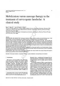
Furthermore, the ratios of individuals with diabetes, hypertension, hyperlipidemia, and coronary artery disease were also comparable among the three groups (Table 2). As shown in Fig. 2A, the mild AD patients had lower plasma levels of apoA-I and apoC-III, but higher levels of apoE, than the individuals of ND group. Lower levels of apoC-III and higher levels of apoE were also evident between the ND and ND-FH groups; whereas, the difference in apoA-I plasma concentrations between the ND and ND-FH groups was not significant (Fig. 2A). The levels of plasma apoB were comparable among the three study groups. These differences remained significant after adjustment for age and gender (Table 3). Validities of the cirABPs as AD biomarkers The utility of cirABPs as AD biomarkers was evaluated by ROC curve analyses using the age- and gender-adjusted values (Fig. 2B). Among them, apoAI showed the largest AUC: 0.93; 95% CI: 0.87 to 0.99) followed by apoC-III (AUC: 0.82; 95% CI: 0.69 to 0.94) and apoE (AUC: 0.72; 95% CI: 0.58 to 0.85). The AUC for apoB was relatively low (AUC: 0.52; 95% CI: 0.39 to 0.65). At the optimum cutoff level of the adjusted plasma apoA-I, the best sensitivity and specificity were 92% and 81%, respectively (Fig. 2B). We then examined the powers of the adjusted cirABPs levels in discriminating individuals of ND-FH from ND. The results showed that apoC-III and apoE gave acceptable AUCs, while the AUCs for apoA-I and apoB were relatively low (Fig. 2B). To validate the diagnostic power of the cirABPs, we performed correlations between the apolipoproteins and the cognitive scores, i.e., CASI and MMSE.
861
AU
TH
OR
CO
PY
Y.-H. Shih et al. / ApoC-III as a Marker for Alzheimer’s Disease
Fig. 2. The levels of plasma A-binding apolipoproteins of the non-demented subjects with no ADFH (ND), non-demented subjects with ADFH (ND-FH), and patients with mild AD (AD). A) The levels of apoA-I, apoB, apoC-III, and apoE of the ND, ND-FH, and mild AD groups. *p < 0.05), **p < 0.01), ***p < 0.001) versus ND; ### p < 0.001) versus ND-FH. B) Receiver operating characteristic analyses for gender- and gender-adjusted plasma apoA-I, apoB, apoC-III, and apoE concentrations and the prediction of AD. AUC, areas under the curve value.
862
Y.-H. Shih et al. / ApoC-III as a Marker for Alzheimer’s Disease Table 3 Comparisons of apolipoproteins between the ND, ND-FH and AD groups
ND versus ND-FH ND versus AD ND-FH versus AD
ApoA-I
ApoB
ApoC-III
ApoE
>0.5 (−5.24) 0.5 (−0.28) >0.5 (0.02)
0.001 (−10.91) 0.5 (−2.35)
The protective effects of apoA-I/HDL may result from several different mechanisms. ApoA-I/HDL could enhance A efflux and indirectly reduce the accumulation of A in brain. Alternatively, apoAI/HDL may reduce the risk of AD by preserving a healthier cardiovascular system [38, 39]. Accumulated evidence indicates that cardiovascular diseases and AD share many common risk factors [40, 41]. Overexpression of human apoA-I in APP/PS1 transgenic mice not only increases the levels of plasma HDL, but also restores the age-related deficits in learning and memory [42]. Although the quantities of A deposition are unaltered in the brains of these mice, overexpression of apoA-I reduces the degrees of cerebral amyloid angiopathy and glial activation. In addition to A sequestration, apoA-I/HDL is known to have many other beneficial effects against A-associated toxicities. For example, A-induced productions of proinflammatory chemokine and cytokine, i.e., MCP1 and IL-6, were reduced by apoA-I [42]. It has been demonstrated that ApoA-1 binds to A and inhibit its aggregation and neurotoxicity [43]. In addition, HDL has been shown to effectively inhibit amyloid formation [44]. These findings suggest that elevating plasma apoA-I/HDL levels may be an effective approach to preserve cognitive function in AD patients. Identification of plasma apoC-III levels as an early biomarker for AD, especially for the cognitive intact ND individuals, was a clinically significant and novel finding. Similar to apoA-I but to a lesser degree, the levels of apoC-III were also correlated with CASI and MMSE scores. The effect of apoC-III on brain function or AD was rarely investigated. In one of the few reports, plasma apoC-III levels were found reduced in carriers of the apoE 4 allele [19], the most potent genetic risk factor for sporadic AD. The potential of using plasma apoC-III as an early AD biomarker deserves further attention. Validation the potency of plasma apoC-III level as an early AD biomarker in a longitudinal followup and large cohort study is necessary. It is worthy to note that the diagnostic power of A-binding proteins as AD biomarkers may be reduced as age increased. The two major A efflux pathways are through the blood-brain barrier via the
CO
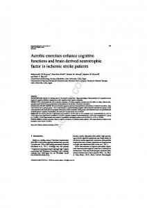
The results showed that the levels of plasma apoA-I and apoC-III positively correlated with both CASI and MMSE scores, while no correlation was found between the levels of apoB or apoE and the two cognitive test scores (Fig. 3).
PY
Multiple liner regression was performed adjusting for age and gender. Values represent as p value ( value).
DISCUSSION
AU
TH
OR
Biomarkers in the blood have already been used in a wide range of clinical diseases at high-throughput screening efficacy. However, clinically useful bloodbased biomarkers for AD are still lacking. In this study, we elaborated upon the “Peripheral Sink Hypothesis” and “AD family history” to search for early AD biomarkers. Based on the Peripheral Sink Hypothesis, we proposed that circulating A-binding proteins could facilitate the efflux of A from the brain. This hypothesis also implies that higher levels of the circulating A-binding proteins would have higher A-efflux capacity. Thus, we also proposed that the levels of plasma A-binding proteins could serve as AD biomarkers. To search for early AD biomarkers, we categorized the ND individuals into two separated cohorts based on their AD family history. In addition to other risk factors, first-degree AD family history is also known to increase the rate of brain atrophy, preclinical prevalence of AD-related pathologies and chance of developing AD [3–7]. Thus, the efficacies of the identified A-binding proteins as early AD biomarkers were assessed using the ND and ND-FH cohorts. Several plasma components, including lipoproteins and apolipoproteins, are known to bind A [22, 31–34]. In agreement with a previous finding [34], we also found that HDL exhibited the highest affinity for A. Among the many apolipoproteins of the HDL, e.g., apoA-I, apoA-II, apoE, and apoJ, most of the A was found to be associated with apoA-I [34]. Reduced levels of plasma apoA-I in subjects with mild cognitive impairment and late-onset AD patients have been previously reported in cross-section studies using community cohorts [35, 36] and longitudinal followup investigation (Honolulu-Asia Aging Study) [37]. These findings suggest that plasma apoA-I could serve as an AD biomarker.
863
AU
TH
OR
CO
PY
Y.-H. Shih et al. / ApoC-III as a Marker for Alzheimer’s Disease
Fig. 3. Scatter plots of the correlation between the four A-binding apolipoproteins (A-I, apoB, apoC-III, and apoE) and CASI (A) and MMSE (B).
Y.-H. Shih et al. / ApoC-III as a Marker for Alzheimer’s Disease
receptor-regulated mechanism [45] and periarterial interstitial fluid drainage [46]. Both pathways require a healthy cerebrovascular system to drain the A, e.g., expression level of LRP and an unimpeded conduit of the periarterial space. As cardiovascular diseases increases with age, the peripheral A-binding proteinfacilitated clearance may reduce it efficacy as age increases [47, 48].
[7]
[8]
[9]
CONCLUSION
[10]
ACKNOWLEDGMENTS
OR
CO
We have identified several apolipoproteins/ lipoproteins as cirABPs in the plasma. Among them, A preferentially bound to apoA-I-containing HDL. Although plasma apoA-I levels could be used to differentiate mild AD patients from ND subjects, plasma apoC-III levels gave better discrimination power in differentiating ND-FH individuals from ND subjects. The potential of using plasma apoC-III as an early AD biomarker deserves further investigation and confirmation.
AU
TH
This work was supported by National Cheng Kung University Hospital (NCKUH-10006002) and Department of Health, Taiwan (DOH101-TD-B-111-0002). The authors wish to thank National Cheng-Kung University Proteomics Research Core Laboratory for the assistance in mass spectrometry analyses and Professors Ih-Jen Su and Shan-Tair Wang for their support and suggestions. Authors’ disclosures available online (http://www. j-alz.com/disclosures/view.php?id=2180). REFERENCES [1] [2]
[3]
[4]
[5]
[6]
Bertram L (2009) Alzheimer’s disease genetics current status and future perspectives. Int Rev Neurobiol 84, 167-184. Bergem AL, Engedal K, Kringlen E (1997) The role of heredity in late-onset Alzheimer disease and vascular dementia. A twin study. Arch Gen Psychiatry 54, 264-270. La Rue A, Hermann B, Jones JE, Johnson S, Asthana S, Sager MA (2008) Effect of parental family history of Alzheimer’s disease on serial position profiles. Alzheimers Dement 4, 285290. Ertekin-Taner N, Younkin LH, Yager DM, Parfitt F, Baker MC, Asthana S, Hutton ML, Younkin SG, Graff-Radford NR (2008) Plasma amyloid beta protein is elevated in late-onset Alzheimer disease families. Neurology 70, 596-606. Johnson SC (2006) The influence of Alzheimer disease family history and apolipoprotein E 4 on mesial temporal lobe activation. J Neurosci 26, 6069-6076. Bassett SS, Yousem DM, Cristinzio C, Kusevic I, Yassa MA, Caffo BS, Zeger SL (2006) Familial risk for Alzheimer’s
disease alters fMRI activation patterns. Brain 129, 12291239. Bendlin BB, Ries ML, Canu E, Sodhi A, Lazar M, Alexander AL, Carlsson CM, Sager MA, Asthana S, Johnson SC (2010) White matter is altered with parental family history of Alzheimer’s disease. Alzheimers Dement 6, 394-403. Tamaoka A, Fukushima T, Sawamura N, Ishikawa K, Oguni E, Komatsuzaki Y, Shoji S (1996) Amyloid beta protein in plasma from patients with sporadic Alzheimer’s disease. J Neurol Sci 141, 65-68. Kuo YM, Emmerling MR, Vigo-Pelfrey C, Kasunic TC, Kirkpatrick JB, Murdoch GH, Ball MJ, Roher AE (1996) Water-soluble Abeta (N-40, N-42) oligomers in normal and Alzheimer disease brains. J Biol Chem 271, 4077-4081. Seubert P, Vigo-Pelfrey C, Esch F, Lee M, Dovey H, Davis D, Sinha S, Schlossmacher M, Whaley J, Swindlehurst C, et al. (1992) Isolation and quantification of soluble Alzheimer’s beta-peptide from biological fluids. Nature 359, 325-327. Mawuenyega KG, Sigurdson W, Ovod V, Munsell L, Kasten T, Morris JC, Yarasheski KE, Bateman RJ (2010) Decreased clearance of CNS -amyloid in Alzheimer’s disease. Science 330, 1774. Patton RL, Kalback WM, Esh CL, Kokjohn TA, Van Vickle GD, Luehrs DC, Kuo Y-M, Lopez J, Brune D, Ferrer I, Masliah E, Newel AJ, Beach TG, Casta˜no EM, Roher AE (2006) Amyloid- peptide remnants in AN-1792-immunized Alzheimer’s disease patients: A biochemical analysis. Am J Pathol 169, 1048-1063. Nicoll JAR, Wilkinson D, Holmes C, Steart P, Markham H, Weller RO (2003) Neuropathology of human Alzheimer disease after immunization with amyloid- peptide: A case report. Nat Med 9, 448-452. Schenk D, Barbour R, Dunn W, Gordon G, Grajeda H, Guido T, Hu K, Huang J, Johnson-Wood K, Khan K, Kholodenko D, Lee M, Liao Z, Lieberburg I, Motter R, Mutter L, Soriano F, Shopp G, Vasquez N, Vandevert C, Walker S, Wogulis M, Yednock T, Games D, Seubert P (1999) Immunization with amyloid-beta attenuates Alzheimer-disease-like pathology in the PDAPP mouse. Nature 400, 173-177. Bussiere T, Bard F, Barbour R, Grajeda H, Guido T, Khan K, Schenk D, Games D, Seubert P, Buttini M (2004) Morphological characterization of Thioflavin-S-positive amyloid plaques in transgenic Alzheimer mice and effect of passive Abeta immunotherapy on their clearance. Am J Pathol 165, 987-995. Deane R, Sagare A, Hamm K, Parisi M, LaRue B, Guo H, Wu Z, Holtzman DM, Zlokovic BV (2005) IgG-assisted agedependent clearance of Alzheimer’s amyloid beta peptide by the blood-brain barrier neonatal Fc receptor. J Neurosci 25, 11495-11503. Shibata M, Yamada S, Kumar SR, Calero M, Bading J, Frangione B, Holtzman DM, Miller CA, Strickland DK, Ghiso J, Zlokovic BV (2000) Clearance of Alzheimer’s amyloid-ss(1-40) peptide from brain by LDL receptor-related protein-1 at the blood-brain barrier. J Clin Invest 106, 14891499. Kang DE, Pietrzik CU, Baum L, Chevallier N, Merriam DE, Kounnas MZ, Wagner SL, Troncoso JC, Kawas CH, Katzman R, Koo EH (2000) Modulation of amyloid -protein clearance and Alzheimer’s disease susceptibility by the LDL receptorrelated protein pathway. J Clin Invest 106, 1159-1166. DeMattos RB, Bales KR, Cummins DJ, Paul SM, Holtzman DM (2002) Brain to plasma amyloid-beta efflux: A measure of brain amyloid burden in a mouse model of Alzheimer’s disease. Science 295, 2264-2267.
PY
864
[11]
[12]
[13]
[14]
[15]
[16]
[17]
[18]
[19]
Y.-H. Shih et al. / ApoC-III as a Marker for Alzheimer’s Disease
[24]
[25]
[26]
[27]
[28]
[29]
[30]
[31]
[32]
[33]
[34]
[37]
[38]
PY
[36]
high density lipoprotein: Association with apolipoprotein and lipids. Clin Chim Acta 270, 75-84. Song F, Poljak A, Crawford J, Kochan NA, Wen W, Cameron B, Lux O, Brodaty H, Mather K, Smythe GA, Sachdev PS (2012) Plasma apolipoprotein levels are associated with cognitive status and decline in a community cohort of older individuals. PLoS One 7, e34078. Kawano M, Kawakami M, Otsuka M, Yashima H, Yaginuma T, Ueki A (1995) Marked decrease of plasma apolipoprotein AI and AII in Japanese patients with late-onset non-familial Alzheimer’s disease. Clin Chim Acta 239, 209-211. Saczynski JS, White L, Peila RL, Rodriguez BL, Launer LJ (2007) The relation between apolipoprotein A-I and dementia: The Honolulu-Asia aging study. Am J Epidemiol 165, 985992. Anantharamaiah GM, Mishra VK, Garber DW, Datta G, Handattu SP, Palgunachari MN, Chaddha M, Navab M, Reddy ST, Segrest JP, Fogelman AM (2007) Structural requirements for antioxidative and anti-inflammatory properties of apolipoprotein A-I mimetic peptides. J Lipid Res 48, 1915-1923. Barter P, Gotto AM, LaRosa JC, Maroni J, Szarek M, Grundy SM, Kastelein JJ, Bittner V, Fruchart JC (2007) HDL cholesterol, very low levels of LDL cholesterol, and cardiovascular events. N Engl J Med 357, 1301-1310. Gorelick PB, Scuteri A, Black SE, Decarli C, Greenberg SM, Iadecola C, Launer LJ, Laurent S, Lopez OL, Nyenhuis D, Petersen RC, Schneider JA, Tzourio C, Arnett DK, Bennett DA, Chui HC, Higashida RT, Lindquist R, Nilsson PM, Roman GC, Sellke FW, Seshadri S (2011) Vascular contributions to cognitive impairment and dementia: A statement for healthcare professionals from the American Heart Association/American Stroke Association. Stroke 42, 2672-2713. Daviglus ML, Bell CC, Berrettini W, Bowen PE, Connolly ES Jr, Cox NJ, Dunbar-Jacob JM, Granieri EC, Hunt G, McGarry K, Patel D, Potosky AL, Sanders-Bush E, Silberberg D, Trevisan M (2010) NIH state-of-the-science conference statement: Preventing Alzheimer’s disease and cognitive decline. NIH Consens State Sci Statements 27, 1-30. Lewis TL, Cao D, Lu H, Mans RA, Su YR, Jungbauer L, Linton MF, Fazio S, LaDu MJ, Li L (2010) Overexpression of human apolipoprotein A-I preserves cognitive function and attenuates neuroinflammation and cerebral amyloid angiopathy in a mouse model of Alzheimer disease. J Biol Chem 285, 36958-36968. Paula-Lima AC, Tricerri MA, Brito-Moreira J, Bomfim TR, Oliveira FF, Magdesian MH, Grinberg LT, Panizzutti R, Ferreira ST (2009) Human apolipoprotein A-I binds amyloidbeta and prevents Abeta-induced neurotoxicity. Int J Biochem Cell Biol 41, 1361-1370. Olesen OF, Dago L (2000) High density lipoprotein inhibits assembly of amyloid beta-peptides into fibrils. Biochem Biophys Res Commun 270, 62-66. Zlokovic B (2005) Neurovascular mechanisms of Alzheimer’s neurodegeneration. Trends Neurosci 28, 202-208. Weller RO, Massey A, Kuo YM, Roher AE (2000) Cerebral amyloid angiopathy: Accumulation of A in interstitial fluid drainage pathways in Alzheimer’s disease. Ann N Y Acad Sci 903, 110-117. Banks W (2003) Efflux of human and mouse amyloid  proteins 1–40 and 1–42 from brain: Impairment in a mouse model of Alzheimer’s disease. Neuroscience 121, 487-492. Pascale CL, Miller MC, Chiu C, Boylan M, Caralopoulos IN, Gonzalez L, Johanson CE, Silverberg GD (2011) Amyloidbeta transporter expression at the blood-CSF barrier is agedependent. Fluids Barriers CN 8, 21.
CO
[23]
[35]
[39]
OR
[22]
TH
[21]
Lemere CA, Spooner ET, LaFrancois J, Malester B, Mori C, Leverone JF, Matsuoka Y, Taylor JW, DeMattos RB, Holtzman DM, Clements JD, Selkoe DJ, Duff KE (2003) Evidence for peripheral clearance of cerebral Abeta protein following chronic, active Abeta immunization in PSAPP mice. Neurobiol Dis 14, 10-18. Kuo YM, Kokjohn TA, Kalback W, Luehrs D, Galasko DR, Chevallier N, Koo EH, Emmerling MR, Roher AE (2000) Amyloid- peptides interact with plasma proteins and erythrocytes: Implications for their quantitation in plasma. Biochem Biophys Res Commun 268, 750-756. Biere AL, Ostaszewski B, Stimson ER, Hyman BT, Maggio JE, Selkoe DJ (1996) Amyloid beta-peptide is transported on lipoproteins and albumin in human plasma. J Biol Chem 271, 32916-32922. Chuang J-Y, Lee C-W, Shih Y-H, Yang T, Yu L, Kuo Y-M (2012) Interactions between Amyloid- and hemoglobin: Implications for amyloid plaque formation in Alzheimer’s disease. PLoS One 7, e33120. Kuo YM, Emmerling MR, Lampert HC, Hempelman SR, Kokjohn TA, Woods AS, Cotter RJ, Roher AE (1999) High levels of circulating A42 are sequestered by plasma proteins in Alzheimer’s disease. Biochem Biophys Res Commun 257, 787-791. Chuo LJ, Sheu WH, Pai MC, Kuo YM (2007) Genotype and plasma concentration of cystatin C in patients with late-onset Alzheimer disease. Dement Geriatr Cogn Disord 23, 251-257. Teng EL, Hasegawa K, Homma A, Imai Y, Larson E, Graves A, Sugimoto K, Yamaguchi T, Sasaki H, Chiu D, et al. (1994) The Cognitive Abilities Screening Instrument (CASI): A practical test for cross-cultural epidemiological studies of dementia. Int Psychogeriatr 6, 45-58. Hughes CP, Berg L, Danziger WL, Coben LA, Martin RL (1982) A new clinical scale for the staging of dementia. Br J Psychiatry 140, 566-572. Morris JC, Heyman A, Mohs RC, Hughes JP, van Belle G, Fillenbaum G, Mellits ED, Clark C (1989) The Consortium to Establish a Registry for Alzheimer’s Disease (CERAD). Part I. Clinical and neuropsychological assessment of Alzheimer’s disease. Neurology 39, 1159-1165. McKhann G, Drachman D, Folstein M, Katzman R, Price D, Stadlan EM (1984) Clinical diagnosis of Alzheimer’s disease: Report of the NINCDS-ADRDA Work Group under the auspices of Department of Health and Human Services Task Force on Alzheimer’s Disease. Neurology 34, 939-944. Liao PC, Yu L, Kuo CC, Lin C, Kuo YM (2007) Proteomics analysis of plasma for potential biomarkers in the diagnosis of Alzheimer’s disease. Proteomics-Clin Appl 1, 506-512. Sanan DA, Weisgraber KH, Russell SJ, Mahley RW, Huang D, Saunders A, Schmechel D, Wisniewski T, Frangione B, Roses AD, Rall SC Jr (1994) Apolipoprotein E associates with beta amyloid peptide of Alzheimer’s disease to form novel monofibrils. Isoform apoE4 associates more efficiently than apoE3. J Clin Invest 94, 860-869. Zlokovic BV, Martel CL, Mackic JB, Matsubara E, Wisniewski T, McComb JG, Frangione B, Ghiso J (1994) Brain uptake of circulating apolipoproteins J and E complexed to Alzheimer’s amyloid . Biochem Biophys Res Commun 205, 1431-1437. Koudinov A, Matsubara E, Frangione B, Ghiso J (1994) The soluble form of Alzheimer’s amyloid  protein is complexed to high density lipoprotein 3 and very high density lipoprotein in normal human plasma. Biochem Biophys Res Commun 205, 1164-1171. Koudinov AR, Berezov TT, Kumar A, Koudinova NV (1998) Alzheimer’s amyloid  interaction with normal human plasma
AU
[20]
[40]
[41]
[42]
[43]
[44]
[45] [46]
[47]
[48]
865