DOI: 10.5152/eurjrheum.2014.005
Original Investigation
Autonomic functions in acrocyanosis assessed by heart rate variability Sedat Yılmaz1, Mehmet Yokuşoğlu2, Muhammet Çınar1, İsmail Şimşek1, Oben Baysan2, Bilgehan Savaş Öz3, Hakan Erdem1, Salih Pay1, Ayhan Dinç1
Abstract Objective: To evaluate the autonomic activity of patients with acrocyanosis by using heart rate variability indices. Material and Methods: The study group consisted of 24 patients with acrocyanosis and the control group contained 22 sex- and age-matched healthy subjects. All subjects underwent 24-hour Holter monitoring. Among the heart rate variability (HRV) parameters, time-domain and frequency-domain indices were analysed. Results: The time-domain indices of HRV indicating global autonomic functions were found to be increased, and indices indicating parasympathetic activity showed a significant decrease in the patient group. Power-spectral analysis of HRV revealed that the low frequency and high frequency power were higher in the patient group than in controls. However, the ratio of Low Frequency/High Frequency was found to be lower in the patient group than in controls. Conclusion: In acrocyanosis, both sympathetic and parasympathetic systems seem to be disrupted. Therefore, we may conclude that acrocyanosis may be resulted of systemic autonomic imbalance rather than pure sympathetic over-activation. Also, these results suggest that acrocyanosis is not a localised disorder; on the contrary, it is associated with various abnormalities of the systemic autonomic nervous system. Key words: Acrocyanosis, heart rate variability, autonomic nervous system
Introduction
1 Department of Rheumatology, Gülhane Military Medical Academy, Ankara, Turkey 2 Department of Cardiology, Gülhane Military Medical Academy, Ankara, Turkey 3 Department of Cardiovascular Surgery, Gülhane Military Medical Academy, Ankara, Turkey Address for Correspondence: Sedat Yılmaz, Department of Rheumatology, Gülhane Military Medical Academy, Ankara, Turkey E-mail:
[email protected] Submitted: 26.12.2013 Accepted: 23.01.2014 Copyright 2014 © Medical Research and Education Association
18
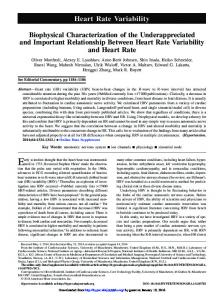
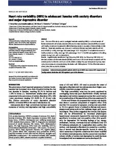
Acrocyanosis (AC) was first described by Crocq in 1896 and is a disorder of the peripheral circulation. It is characterised by the following symptoms and signs; permanent and painless cyanosis of the acral parts, local hypothermia, permanent local hyperhidrosis, elastic infiltration of the integument and a general lack of pain (Figure 1). Acrocyanosis is mostly included in the differential diagnosis of Raynaud’s phenomenon (RP) and can be differentiated from RP by the relatively permanent occurrence rather than frequent attacks and the absence of a sharp line separating normal skin from the abnormal areas (1). The age of onset is often during adolescence and early adult life, and it is relatively common among women. Although the disease is temperature-dependent and generally worsens with cold exposure, the aetiopathogenesis is poorly understood, and several hypotheses have been suggested. Firstly, it was believed that the main pathology is excessive sensitivity to cold on the part of the small vessels of the extremities (2). On the other hand, some investigators have proposed that it was caused by hypertonicity (hyperactivity) of the sympathetic nervous system (3, 4). Also, local disorder of the small vessels with the contribution of centrally arising impulses has also been suggested as an underlying mechanism (5). However, it was mostly suggested that the pathogenesis was the result of the sympathetic hyperactivity at the acral parts leading to the increased tone and spasm of the arterioles associated with dilation of capillaries and venules. Based on these suggestions, several investigators have applied sympathectomy for treatment, with very poor success rates. Despite the existence of a hypothesis citing the sympathetic system as the cause of AC, it is interesting to note that no study, to our knowledge, has evaluated the autonomic functions in patients with AC. The electrical and contractile activity of the heart is largely modulated by the autonomic nervous system (ANS). Heart rate variability (HRV), is a reliable and non-invasive electrocardiographic method to measure the autonomic nervous system, and consists of a series of measurements of successive RR interval variations of sinus origin (intervals between QRS complexes of normal sinus depolarization). Analysis of HRV consists of a series of measurements of successive RR interval variations of sinus origin, which provide information about tonic baseline autonomic function. In normal conditions, there will be continuous physiological variations of the sinus cycles, while there will be a sympatho-vagal imbalance reflected by a diminished HRV if there is an abnormality in ANS (6). The detection of such changes may be used as a marker of underlying
Eur J Rheum 2014; 1: 18-20 pathology, especially in autonomic nervous system dysfunctions. HRV parameters can be obtained with time domain, spectral domain, non-linear and mathematical modelling methods. It decreases with age and shows a circadian variation, being maximal during sleep (7, 8). In this prospective, cross-sectional study, by using time-domain and power spectral analysis of HRV, we aimed to determine whether there is an autonomic abnormality and whether it is localised or generalised in patients with AC. To the best of our knowledge, there has been no study evaluating the autonomic nervous system in AC by using HRV parameters.
Material and Methods Twenty-four consecutive patients with AC (Male/Female ratio: 18/6, mean age: 27±10 years) compromised the study group. The control group was composed of 22 age- and gender-matched healthy subjects (Male/Female ratio: 16/6, mean age: 25±9 years). Concomitant diseases were investigated by a detailed medical history, physical examination, whole blood count, and routine blood chemistry in all participants. Patients with obesity, current smokers, diabetes mellitus, anaemia, hypertension, any sign of infection, taking any medication, and with a suspicion of coronary heart disease and hypersensitivity were excluded from the study. All subjects underwent 24-hour Holter monitoring. The study was reviewed and approved by local ethics committee of Gülhane Military Medical School, and all participants signed written informed consent. Diagnosis of acrocyanosis The diagnosis of AC is based on mainly clinical symptoms, including persistently cyanotic and hyperhydrotic hands and/or feet with a lack of pain. Also, if one hand is kept in a dependent position and the other is kept raised, the colour of the latter becomes normal in patients (Figure 1). Heart rate variability Twenty-four-hour ambulatory electrocardiographic recordings were obtained from each subject with a Rozinn RZ 152 digital Holter recorder (Rozinn Electronics, Inc., Glendale, NY, USA) with the sampling frequency of 1024 Hz. All cases were strictly advised to maintain the normal course of their daily life. Their obedience to this advice was confirmed again while removing the device from the cases. The HRV was determined by the software of the same device. The time domain HRV measures employed in our study were the standard deviation of all normal sinus R-R intervals over 24 hours (SDNN), the standard deviation of all averaged
Yılmaz et al. Autonomic functions in acrocyanosis a
b
Figure 1. a, b. A globally bluish appearance of hands without any separation line from normal skin (a). Difference between the hands; one kept in a dependent position and the other kept raised (b) normal sinus R-R intervals for each 5-minute segment in the 24-hour recordings (SDANN), root mean square of successive differences between normal sinus R-R intervals (RMSSD), the ratio of number of all R-R intervals to the height of histogram created by the charting all the RR intervals (HRV triangular index), the number of R-R intervals exceeding 50 ms (SNN50 count), and the percentage of difference between adjacent normal R-R intervals that are greater than 50 ms computed over the entire 24-hour ECG recording (PNN50). Among them, RMSSD, SNN50, and PNN50 primarily reflect parasympathetically-mediated changes in heart rate (9). The other time-domain variables reflect a mixture of parasympathetic, sympathetic, and other physiologic influences. We used power spectral analysis of heart rate with the parameters of low frequency (low frequency (LF): 0.04-0.15 Hz), which is related to baroreceptor control and is dually mediated by vagal and sympathetic systems; a high frequency (high frequency (HF): 0.15-0.5 Hz) band reflects respiratory sinus arrhythmia and, thus, cardiac vagal activity (10, 11). Also, the most indicative parameter of LF/HF is used for assessing the autonomic balance. In addition to HRV variables, basic rhythm and associated problems such as atrial or ventricular arrhythmias were carefully evaluated. Statistical analysis A normality test was done using the Shapiro-Wilk test and graphically for continuous variables. Comparisons between the groups were carried out with 2-tailed Student’s t test for normally distributed continuous variables, Mann-Whitney U test for data without normal distribution and chi-square test for dichotomous variables. A p value below 0.05 was considered to be significant.
Results Demographic parameters including age, gender, haemoglobin levels and body mass in-
Table 1. Demographic and clinical characteristics of subjects and comparison of study and control groups
Study Control group group p (n=24) (n=22) value
Age (year) 27±10 25±9 NS Gender Male 18 (75%) 16 (73%) NS Female 6 (25%) 6 (27%) Hb value (g/dL) 12.9±0.6 13.1±0.4 NS Vit B12 level (pg/mL) 249±65 276±72 NS Folate (ng/mL) 6.1±1.2 7±1.9 NS BMI (kg/m2) 24.2±2.4 24.8±2.2 NS n: number of cases; Hb: haemoglobin; Hct: haematocrit; BMI: body mass index; NS: not significant
dex were similar in both groups (Table 1). All subjects were detected to be in sinus rhythm without any episode of sustained atrial or ventricular arrhythmias. The statistical comparisons of HRV parameters are presented in Table 2. When compared to the control group, the time-domain indices indicating global autonomic functions (SDNN, SDANN, and HRV-triangular index) were found to be increased, and other parameters indicating parasympathetic activity (RMSSD, SNN 50 count, and PNN50) showed a significant decrease in the patient group. Power-spectral analysis of HRV revealed LF and HF power were higher in the patient group than in controls (p=0.00 for both). However, LF/HF was found to be significantly lower in patient group than in controls.
Discussion This study indicates that the disrupted part of the autonomic nervous system is not only the sympathetic system, but also the parasympathetic system in AC. Therefore, we may conclude that AC may be the result of systemic autonomic imbalance rather than pure sympathetic over-activation. The imbalance we observed between sympathetic and parasympathetic nerve activities
19
Yılmaz et al. Autonomic functions in acrocyanosis
Eur J Rheum 2014; 1: 18-20
Table 2. Statistical comparison of HRV variables of study and control groups
Unit
Study group
Control group
SNN50 count 14404±7487 14357±7308 RMSSD ms 56±33 31±19 PNN50 % 17±9 23±9 HRV-Triangular Index 42±11 38±8 SDANN ms 146±62 123±36 SDNN ms 168±50 145±42 LF 1.8±0.3 1.2±0.4 HF 1.5±0.4 1.0±0.2 LF/HF 1.3±0.2 1.5±0.2
p value NS 0.003 0.044 0.048 0.021 0.012 0.032 0.004 0.041
SDNN: the standard deviation of all normal sinus R-R intervals over 24 hours; SDANN: the standard deviation of all averaged normal sinus R-R intervals for each 5-minute segment in the 24-hour recordings; RMSSD: root mean square of successive differences between normal sinus R-R intervals; HRV triangular index: the ratio of number of all R-R intervals to the height of histogram created by the charting all the RR intervals; SNN50 count: number of R-R intervals exceeding 50 ms; PNN50: the percentage of difference between adjacent normal R-R intervals that are greater than 50 ms computed over the entire 24-hour ECG recording; LF: low frequency; HF: high frequency; NS: not significant
can disturb the electrophysiological properties of the heart. Some investigators have suggested that a decrease in HRV was associated with morbidity and mortality in patients with coronary heart disease after an acute coronary event (12). Moreover, HRV parameters were also found to be altered in various disorders and there are no data concerning increased mortality in these disorders (13-20). There could be an association between HRV alterations seen in AC and increased mortality, although there are no data confirming or even suggesting this inference. To the best of our knowledge, this is the first study evaluating the autonomic functions in AC. In AC, sympathectomy has been applied since 1929 and was based on the hypothesis of sympathetic over-activation. However, the lower success rate of this approach leads us to evaluate the autonomic functions in AC. A recent report declared that sympathectomy might cause cold sensitivity as a side effect (21). In addition, diminished capillary density was shown in AC (22). Considering these findings, the results of our study contradict the suggested physiopathological mechanism by which pure sympathetic over-activation plays a role in AC. On the other hand, these results also suggest that AC is not a localised disorder that was previously considered; on the contrary, it is associated with various abnormalities of the systemic autonomic nervous system. Since patients with AC often report the nose, ears, lips, and nipples being affected, this is not an unexpected finding. However, one can claim that it can also be explained by the local disturbances in the aforementioned areas; thus, our results have confirmed the systemic autonomic feature of AC.
20
In conclusion, despite acrocyanosis having been known for over a century, its pathophysiologic mechanisms remain poorly understood. While autonomic dysfunction is not sufficient to explain all aspects of acrocyanosis, our findings nonetheless reflect the indirect evidence of the impaired function of autonomic nervous system. Since most of the studies evaluating the pathophysiological mechanisms operating in this entity date back to the beginning of the 20th century, further studies with modern approaches are needed to better define the specific abnormalities of the autonomic nervous system in patients with acrocyanosis. Ethics Committee Approval: Ethics committee approval was received for this study from the ethics committee of Gülhane Military Medical Academy. Informed Consent: Written informed consent was obtained from patients who participated in this study. Peer-review: Externally peer-reviewed. Author contributions: Concept - A.D., M.Y.; Design - I.S.; Supervision - A.D., S.P., O.B.; Resource - B.S.O., O.B.; Materials - B.S.O., M.Y., O.B.; Data Collection&/or Processing - S.Y., M.Ç.; Analysis&/or Interpretation - H.E., İ.Ş., S.P.; Literature Search - S.Y., M.Ç.; Writing - S.Y.; Critical Reviews - A.D., I.S. Conflict of Interest: No conflict of interest was declared by the authors. Financial Disclosure: The authors declared that this study has received no financial support.
References 1. Belch J. Raynaud’s phenomenon. Cardiovasc Res 1997; 33: 25-30. [CrossRef] 2. Lewis T, Landis EM. Vascular mechanisms in acrocyanosis. Heart 1930; 15: 229. 3. Villaret M, Justin-Besangon L, Cachera R, Boucomont R. Etude critique sur la pathogenie des troubles circulatoires peripheriques. Premiere Partie: Les Acrocyanoses. Arch d mal du Coeur 1934; 27: 725. 4. Kreindler A, Elias H. Zur Klinik und Pathogenese der juvenilen Akrocyanose. Ztschr f Kinderh 1930; 50: 608. [CrossRef]
5. Elliott AH, Evans RD, Stone CS. Acrocyanosis: A study of the circulatory fault. Am Heart J 1936; 11: 431. [CrossRef] 6. Van Ravenswaaij-Arts CMA, Kollée LAA, Hopman JCW, Stoelinga GBA, van Geijn HP. Heart rate variability. Ann Intern Med 1993; 118: 436-47. [CrossRef] 7. O’Brien IA, O’Hare P, Corrall RJ. Heart rate variability in healthy subjects: effect of age and the derivation of normal ranges for tests of autonomic function. Br Heart J 1986; 55: 348-54. [CrossRef] 8. Malpas SC, Purdie GL. Circadian variation of heart rate variability. Cardiovasc Res 1990; 24: 210-3. [CrossRef] 9. Kleiger RE, Stein PK, Bosner MS, Rottman JN. Time domain measurements of heart rate variability. Cardiol Clin 1992; 10: 487-98. 10. Akselrod S, Gordon D, Ubel FA, Shannon DC, Berger AC, Cohen RJ. Power spectrum analysis of heart rate fluctuation: a quantitative probe of beat-to-beat cardiovascular control. Science 1981; 213: 220-2. [CrossRef] 11. Pomeranz B, Macaulay RJ, Caudill MA, Kutz I, Adam D, Gordon D, et al. Assessment of autonomic function in humans by heart rate spectral analysis. Am J Physiol 1985; 248: H151-3. 12. Odemuyiwa O, Malik M, Farrell T, Bashir Y, Poloniecki J, Camm J. Comparison of the predictive characteristics of heart rate variability index and left ventricular ejection fraction for all-cause mortality, arrhythmic events and sudden death after acute myocardial infarction. Am J Cardiol 1991; 68: 434-9. [CrossRef] 13. Kitney RI, Byrne S, Edmonds WE, Watkins PJ, Roberts VC. Heart rate variability in the assessment of autonomic diabetic neuropathy. Automedica 1982; 4: 155-67. 14. Saul JP, Arai Y, Berger RD, Lilly LS, Colucci WS, Cohen RJ. Assessment of autonomic regulation in chronic congestive heart failure by heart rate spectral analysis. Am J Cardiol 1988; 61: 1292-9. [CrossRef] 15. Nevruz O, Yokusoglu M, Uzun M, Demirkol S, Avcu F, Baysan O, et al. Cardiac autonomic functions are altered in patients with acute leukemia, assessed by heart rate variability. Tohoku J Exp Med 2007; 211: 121-6. [CrossRef] 16. Yokusoglu M, Nevruz O, Baysan O, Uzun M, Demirkol S, Avcu F, et al. The altered autonomic nervous system activity in iron deficiency anemia. Tohoku J Exp Med 2007; 212: 397-402. [CrossRef] 17. Yokusoglu M, Ozturk S, Uzun M, Baysan O, Demirkol S, Çalışkaner AZ, et al. Heart rate variability in patients with allergic rhinitis. Mil Med 2007; 172: 98-101. 18. Dundaroz MR, Denli M, Uzun M, Aydin HI, Sarici SU, Yokuşoğlu M, et al. Analysis of heart rate variability in children with primary nocturnal enuresis. Int Urol Nephrol 2001; 32: 393-7. [CrossRef] 19. Yokusoglu M, Dede M, Uzun M, Baysan O, Koz C, Yenen MC, et al. Cardiac autonomic balance is impaired in preeclampsia. J Med Sci 2009; 29: 605-10. 20. Tascilar E, Yokusoglu M, Dundaroz R, Baysan O, Ozturk S, Yozgat Y, et al. Cardiac autonomic imbalance in children with allergic rhinitis. Tohoku J Exp Med 2009; 219: 187-91. [CrossRef] 21. Lowe EG, Allmendinger PD, Lowe R. Cold sensitivity as a new side effect after sympathicotomy for hyperhidrosis. Ann Thorac Surg 2005; 80: 2356-8. [CrossRef] 22. Monticone G, Colonna L, Palermi G, Bono R, Puddu P. Quantitative nail fold capillary microscopy findings in patients with AC compared with patients having systemic sclerosis and control subjects. J Am Acad Dermatol 2000; 42: 787-90. [CrossRef]