INFECTION AND IMMUNITY, May 2005, p. 2999–3006 0019-9567/05/$08.00⫹0 doi:10.1128/IAI.73.5.2999–3006.2005 Copyright © 2005, American Society for Microbiology. All Rights Reserved.
Vol. 73, No. 5
B-Cell and T-Cell Immune Responses to Experimental Helicobacter pylori Infection in Humans Zhannat Z. Nurgalieva,1 Margaret E. Conner,2 Antone R. Opekun,1 Carl Q. Zheng,2 Susan N. Elliott,3 Peter B. Ernst,3† Michael Osato,1 Mary K. Estes,2 and David Y. Graham1,2* Department of Medicine1 and Department of Molecular Virology and Microbiology,2 Veterans Affairs Medical Center and Baylor College of Medicine, Houston, Texas, and Department of Pediatrics, University of Texas Medical Branch, Galveston, Texas3 Received 3 September 2004/Returned for modification 13 October 2004/Accepted 30 December 2004
The acute antibody and T-cell immune response to Helicobacter pylori infection in humans has not been studied systematically. Serum from H. pylori-naive volunteers challenged with H. pylori and cured after 4 or 12 weeks was tested by enzyme-linked immunosorbent assays for anti-H. pylori-specific immunoglobulin M (IgM) and IgA established using bacterial lysates from homologous (the infecting strain) and heterologous H. pylori. Proteins recognized by IgM antibody were identified by mass spectrometry of immunoreactive bands separated by two-dimensional gel electrophoresis. Mucosal T-cell subsets (CD4, CD8, CD3, and CD30 cells) were assessed by immunohistochemistry. All 18 infected volunteers developed H. pylori-specific IgM responses to both homologous or heterologous H. pylori antigens. H. pylori antigens reacted with IgM antibody at 4 weeks postinfection. IgM Western blotting showed immunoreactivity of postinfection serum samples to multiple H. pylori proteins with molecular weights ranging between 9,000 (9K) to 150K with homologous strains but only a 70K band using heterologous antigens. Two-dimensional electrophoresis demonstrated that production of H. pylori-specific IgM antibodies was elicited by H. pylori flagellins A and B, urease B, ABC transporter binding protein, heat shock protein 70 (DnaK), and alkyl hydroperoxide reductase. Mucosal CD3, CD4, and CD8 T-cell numbers increased following infection. IgM antibody responses were detected to a range of homologous H. pylori antigens 2 to 4 weeks postchallenge. The majority of H. pylori proteins were those involved in motility and colonization and may represent targets for vaccine development. The important human pathogen Helicobacter pylori causes a persistent gastroduodenal infection that produces a brisk humoral and cellular immune response. The histological characteristics of the mucosal inflammation contain features of both acute and chronic inflammation. Although much is known about the clinical manifestations of chronic H. pylori infection, there is little information regarding the immune response in the early phases of infection (11, 39). One major obstacle to the study of the early events in H. pylori infection in humans has been the difficulty in determining when an individual actually becomes infected. As such, the majority of the literature related to the immune response to the early phases of H. pylori infections has been extrapolated from data acquired from the screening of populations for the presence of anti-H. pylori immunoglobulin M (IgM), IgA, and IgG antibodies and from a few cases in which the acquisition of the infection was known with reasonable certainty (1, 2, 9). The initial humoral immune response to most bacterial infections involves a humoral IgM response. However, the available data regarding an IgM response among cases of acute H. pylori infection are both infrequent and inconsistent. For example, follow-up of two cases of acute H. pylori infection in adults reported no serologic IgM response at any time. How-
ever, one of the cases showed a local mucosal IgM response within the gastric mucosa at day 14 (18, 43). That patient developed a detectable serum H. pylori-specific IgG response by day 74 postinfection (43). A study of a family with intrafamilial transmission of H. pylori infection reported an IgM response in both the children and a parent (37). Their index case was an infant with a history of vomiting that settled spontaneously. The patient’s sibling was diagnosed with acute H. pylori infection 9 days later, based on histology. An IgM response was noted in both children that peaked at day 9 in the index case and rose over the first 63 days in the sibling. The infection was subsequently transmitted to their father, in whom a specific IgM was noted by day 63. Both children had a detectable serum anti-H. pylori IgG by day 30, whereas the father developed a serum IgG response between days 209 and 259. Finally, a serologic IgG response in one of the two reported cases of self-inoculation with H. pylori was noted between 22 and 33 days postingestion and was preceded by an IgM response (38) Longitudinal studies of the humoral immune response in several groups of children have also been reported (8, 19, 45). Czinn et al. noted H. pylori-specific IgM in 19% of symptomatic children with active H. pylori infection (8). A follow-up study of 80 Taiwanese infants showed that three of six Taiwanese infants with naturally acquired H. pylori infection developed a short-lived IgM response preceding development of an IgG antibody response (19). Finally, Gambian children were tested at 3-month intervals, and a rise in anti-H. pylori IgM antibodies was noted around the time of the first positive urea breath test
* Corresponding author. Mailing address: VAMC Rm. 3A-320 (111D), 2002 Holcombe Blvd., Houston, TX 77030. Phone: (713) 7950332. Fax: (713) 790-1040. E-mail:
[email protected]. † Present address: Department of Medicine, University of Virginia, Charlottesville, Va. 2999
3000
NURGALIEVA ET AL.
(UBT) (12). In those children, the IgG response was delayed until approximately 9 months after the first positive UBT (45). In 1999, we initiated studies aimed at establishing an experimental H. pylori infection in humans that could be used for future vaccine studies. These studies were based on the long history of clinical trials in which subjects are vaccinated and subsequently challenged to determine the protective activity of vaccine. Such candidates have been used in the development of vaccines and drugs against enteric and respiratory infections such as malaria, Q fever, cholera, Norwalk virus, rhinoviruses, influenza virus, dengue viruses, sand-fly fever virus, and respiratory syncytial virus and infections with Salmonella enterica serovar Typhi, enterotoxigenic Escherichia coli, Shigella, and Campylobacter jejuni (4, 5, 23, 36, 42, 44, 46). Details of the study design and results have been published elsewhere (20). The current study examined the antibody and T-cell immune response to acute H. pylori infection including data regarding kinetics, type, and duration of the humoral and cellular immune responses and the specific H. pylori proteins eliciting the response after an infection of known onset and duration. We also report the effects of using both homologous versus heterologous H. pylori strains as the antigen sources for antibody testing. MATERIALS AND METHODS A cag pathogenicity island-negative, OipA-positive, multiple-antibiotic-susceptible strain of H. pylori obtained from an individual with mild gastritis (Baylor strain 100 or ATCC BAA-945) was used for the oral challenge of 20 volunteers (9 women and 11 men, ages 23 to 33 years). Volunteers received 40 mg of famotidine at bedtime and 104 to 1010 CFU of H. pylori in beef broth the next morning. Infection was confirmed by 13C-UBT, culture, and histology. Antibiotic eradication therapy was given 4 (n ⫽ 12) or 12 (n ⫽ 4) weeks postchallenge, and eradication was confirmed by at least two separate UBTs, as well as culture and histology. Eighteen volunteers (90%) became infected. Mild to moderate dyspeptic symptoms occurred, peaked between days 9 and 12, and resolved. Gastric histology obtained 2 weeks postchallenge showed typical chronic H. pylori gastritis with intense acute and chronic inflammation. The density of H. pylori (as assessed by CFU/biopsy) was similar independent of the challenge dose (20). Briefly, 20 healthy volunteers were orally inoculated. H. pylori infection was established in 18 individuals as determined by histology, culture, and urea breath test. H. pylori eradication antibiotic therapy was given at 4 (n ⫽ 12) or 12 (n ⫽ 4) weeks postchallenge. Serum samples were collected at intervals before challenge and at intervals up to 39 weeks postchallenge. The current study presents data from the sera from the 18 individuals infected with H. pylori infection of known onset; neither of the two individuals who failed to become infected following challenged developed a humoral immune response. Antigen source. H. pylori strains ATCC 43504 (heterologous) and BCS-100 (homologous) were used in enzyme-linked immunosorbent assays (ELISAs) and Western blots. The BCS-100 challenge strain was genotype cagA negative and positive for vacA s1c-m1, iceA2, babA2. It contained a functional OipA protein, as there was no frameshift in the 5⬘ region of the oipA gene. The organism was susceptible to amoxicillin, tetracycline, metronidazole, and clarithromycin. The individual from whom the strain was obtained had mild nonatrophic gastritis, and this infection was eradicated with a 2-week course of standard anti-H. pylori triple (proton pump inhibitor, clarithromycin, amoxicillin) therapy. Antigen preparation. H. pylori was grown on brain heart infusion agar (Difco, Detroit, MI) with 7% horse serum (Cocalico, Reamstown, PA). Bacteria from 10 plates were scraped, suspended in 4 ml of 0.01 M phosphate saline buffer (PBS) (pH 7.4), and washed four times by pelleting the cells at 2,000 ⫻ g for 10 min, discarding the supernatant, and suspending the cell pellet. The final pellet was suspended in 5 ml of 0.01 M PBS and frozen (ethanol/dry ice) and thawed (37°C) four times (27). The antigen preparation was clarified by centrifugation at 2,000 ⫻ g for 20 min at 4°C, and the supernatant was aliquoted and stored at ⫺70°C until used. The protein concentration was determined by the Bradford protein assay (Sigma, Saint Louis, MO). Preparation of H. pylori antigen-coated microtiter plates. The optimal concentration of each reagent used in the ELISA was determined by checkerboard
INFECT. IMMUN. titration. Alternating wells of flexible polyvinyl chloride microtiter plates (Dynex Technologies, Inc., Chantilly, VA) were coated with 100-l/well H. pylori antigen (7.5 g/ml) diluted in 0.01 M PBS (antigen-positive well) or 0.01 M PBS (antigen-negative well). The plates were incubated at 37°C for 2 h. The plates were washed between every step with 200 l of 0.05% Tween 20 in 0.01 M PBS using an Ultrawash Plus plate washer (Dynatech Laboratories, Inc., Chantilly, VA). Following washing, all wells were blocked by the addition of 200 l of blocking buffer (10% powdered skim milk in 0.01 M PBS) at room temperature for 2 h and used for one of the following ELISAs. Serum IgM ELISA. Serum samples were collected from H. pylori-negative subjects at baseline, allowing each volunteer to serve as his/her own control. The ELISA optical density (OD) value at baseline was subtracted from ELISA OD values of all subsequent postchallenge serum samples. An IgM H. pylori-specific antibody response was identified based on the OD value of the baseline control sample with each case serving as their own control. A positive result was an OD value above the baseline OD value. Serum samples from volunteers or from known H. pylori-negative and H. pylori-positive subjects were serially diluted twofold starting at 1:50 and were added to both antigen-positive and antigen-negative wells. The negative and positive control sera used in all subsequent assays were selected from serum samples tested for the presence of anti-H. pylori IgM. Serum samples were incubated for 2 h at 37°C followed by overnight incubation at 4°C. Mouse anti-human IgM labeled with peroxidase (Southern Biotechnology Associates, Birmingham, AL) (100 l) diluted 1:2,500 in blocking buffer was added to each well and incubated for 1 h at 37°C. The sensitivity and specificity of the secondary antibody was confirmed against human IgG (Chemicon, Temecula, CA), human IgA (Cappel, Livermore, CA), human secretory IgA (sIgA) (Chemicon, Temecula, CA), and IgM (Cappel, Livermore, CA) by coating microtiter plates with the human antibodies that were serially diluted twofold starting at 10 g/ml with 0.078 g/ml being the last dilution on the plate. No reactivity was detected when the conjugate used in this ELISA was tested against human IgG. When the secondary antibodies were tested against human IgA and human sIgA, the detection limits were 0.15 g/ml and 1.25 g/ml, respectively. Finally, when the secondary antibodies used in this ELISA were tested against human IgM, the sensitivity was lower than 0.078 g/ml. Following washing, 100 l of TMB (tetramethylbenzidine) substrate (TMB Microwell Peroxidase Substrate System, Kirkegaard & Perry Laboratories, Gaithersburg, MD) was added to each well and plates were incubated at room temperature for 10 min. The reaction was stopped by addition of 100 l/well of 1 M phosphoric acid. The OD was read at 450 nm (Titertek Multiscan Plus plate reader; Labsystems, Finland). At each dilution, the OD value of the antigen-negative well was subtracted from the OD value of the antigen-positive well. Negative and positive control sera titers were determined by serial twofold dilution on each plate. If the end point titer of the positive control serum was less or greater than fourfold below or above the established titer, respectively, the assay was repeated. Serum IgA ELISA. For detection of anti-H. pylori IgA, all steps were performed identically as described for the anti-H. pylori IgM ELISA, except a 1:7,500 dilution of goat anti-human IgA horseradish peroxidase (HRP) was used (Sigma, St. Louis, MO). The sensitivity and specificity of the secondary antibody was confirmed against human IgG (Chemicon, Temecula, CA), human IgA (Cappel), human sIgA (Chemicon, Temecula, CA), and IgM (Cappel). When the secondary antibodies were tested against human IgG, the detection limit was 2.5 g/ml. When the secondary antibodies used in this ELISA were tested against human IgA and human sIgA, the sensitivity was lower than 0.078 g/ml. No reactivity was detected when the conjugate was tested against human IgM. A cutoff value of 0.27 was established for this assay based on prior testing of 30 H. pylori-negative serum samples as described for the IgM ELISA. We tested the serum samples for anti-H. pylori IgA collected at all time points from three volunteers who had the highest IgM antibody responses to further confirm the specificity of the IgM ELISA. Western blot analyses. Volunteer serum samples were tested for reactivity to H. pylori proteins by Western blotting as described previously, with slight modification (40). Briefly, sodium dodecyl sulfate-polyacrylamide gel electrophoresis was performed by the method of Laemmli with a 0.75-mm-thick 10% separating gel and a 4% running gel using a Mini Protean II Cell apparatus (Bio-Rad Laboratories, Hercules, CA). Solution A consisted of 5.98 g of Tris and 0.46 ml of TEMED (tetramethylethylenediamine) in 100 ml of distilled water adjusted to a pH of 6.6 with hydrochloric acid. To prepare the loading buffer, 5 ml of solution A, 4 ml of glycerol, 1 ml of distilled water, 600 l of 0.5% phenol red, 2.5 ml of 2-mercaptoethanol, and 0.6 g of sodium dodecyl sulfate were mixed and heated at 95°C for 10 min. One microgram of either homologous or heterologous H. pylori (freeze-thawed) antigen or 10 l of protein ladder (Bench Mark prestained protein ladder; Gibco BRL) were loaded per lane, and the electrophoresis was
VOL. 73, 2005
IMMUNE RESPONSE TO ACUTE H. PYLORI INFECTION
carried out for 1 h at 100 V. After electrophoresis, the separated proteins were transferred to a nitrocellulose membrane (Hybond-C pure; Amersham LifeScience, Little Chalfont, Buckingamshire, England) by using a Mini Trans-Blot transfer cell (Bio-Rad Laboratories, Hercules, CA) for 1 h at 200 mA with Towbin Western transfer buffer (15.15 g of Tris, 75.05 g of glycine, 1,000 ml of methanol, 5,000 ml of distilled water). Membranes were stained with Ponceau Red (Sigma, St. Louis, MO), and strips were cut, washed with 0.05% Tween 20 in 0.01 M PBS, and incubated in Blocker Casein in Tris-buffered saline (Pierce, Rockford, IL) for 2 h at room temperature to allow blocking of nonspecific sites. The serum samples from volunteers or negative and positive control sera were diluted 1:100 for total Ig and IgG detection or 1:50 for IgM detection and were incubated overnight at 4°C. The murine anti-H. pylori monoclonal antibodies (EPI, Stony Brook, NY) against H. pylori-specific heat shock protein, urease B, or urease A were diluted 1:100 and used as positive controls. The strips were washed between every step with 200 l of 0.05% Tween 20 in 0.01 M PBS at room temperature with shaking. Goat anti-human Ig total (IgM, IgG, IgA) (American Qualex, San Clemente, CA), mouse anti-human IgM-HRP (Southern Biotechnology Associates, Birmingham, AL), or goat anti-human IgG (Sigma, Saint Louis, MO), diluted 1:7,500 in blocking buffer, was added to each strip and incubated for 2 h at 37°C. The strips were incubated with ECL Western blotting detection reagents (Amersham Pharmacia Biotech, Piscataway, NJ) under the light for 1 min at room temperature, covered with Saran Wrap, and exposed on X-OMAT AR film for 15 s to 1 min (Eastman Kodak Company, Rochester, NY), and the film was processed using the Auto Automixer (Allied, Diagnostic Imaging Resources, Inc., Norcross, GA) and the Kodak M-35 X-Omat processor. Two-dimensional gel electrophoresis. H. pylori proteins were obtained following freeze-thaw of the whole-cell H. pylori used as a homologous antigen for ELISA and Western blotting as described above. The proteins were dialyzed in regenerated cellulose dialysis tubes (Fisher Scientific, Pittsburg, PA) against 0.05 M ammonium bicarbonate that was changed twice (Sigma, St. Louis, MO) during 12 h at 4°C and further concentrated using a Savant SpeedVac concentrator (Savant Instruments, Inc., Farmingdale, NY). Two-dimensional gel electrophoresis was performed using a ReadyPrep 2-D starter kit (Bio-Rad, Hercules, CA) according to the manufacturer’s instructions with modifications. Briefly, the dialyzed, lyophilized H. pylori protein samples were reconstituted in the rehydration sample buffer that consisted of 0.2% (wt/vol) of Bio-Lyte 3/10 Ampholyte, 10 mM of DDT (dichlorodiphenyltrichloroethane), 4% CHAPS {3-[(3cholamidopropyl)-dimethylammonio]-1-propanesulfonate}, and 10 M urea. The final concentrations of proteins in two samples were 1,000 g (intended for Coomassie blue staining) and 200 g (for immunoblotting). The prepared protein samples were subjected to isoelectric focusing (IEF) using a Protean IEF cell (Bio-Rad, Hercules, CA) with immobilized pH gradient (IPG) gel strips of 3 to 10 pH gradients. To achieve a better separation for lower-molecular-weight proteins, the IPG gel strips with pH gradients of 5 to 8 were used for later experiments (Bio-Rad, Hercules, CA) with 200 g protein per gel strip. The proteins from the gel used to separate the lower-molecular-weight proteins were transferred to a nitrocellulose membrane. The source of primary antibody was serum collected 4 weeks postinoculation. This serum was chosen because it had the highest OD values on the IgM ELISA and also showed good immunoreactivity on an IgM Western blot. This serum was used at a dilution of 1:100 in 5% BLOTTO. Mouse anti-human IgM-HRP (Southern Biotechnology Associates, Birmingham, AL) was used as a secondary antibody source (1:7,500). The enhanced chemiluminescence was performed as described above. The gel containing the higher protein concentration (1,000 g) was washed three times with distilled water for 15 min and was stained with Coomassie blue. The freshly stained protein bands were excised from the acrylamide gel, cut into small cubes, and analyzed by mass spectrometry at the Protein Chemistry Core Laboratory at Baylor College of Medicine. Mass spectrometry. Coomassie-stained gel bands or spots were rinsed in H2O for 10 min, cut with a scalpel blade into ⬃1-mm pieces, dehydrated with 0.2 M NH4HCO3/50% acetonitrile for ⬃30 min, and dried completely in a Speed-Vac. The gel pieces were then rehydrated in 0.1 M NH4HCO3 containing 0.5 to 1 g modified trypsin (Promega) and digested for 20 h at 37°C. The supernatant was removed to a clean centrifuge tube. The gel fragments were extracted with aqueous 50% methanol/2% formic acid for ⬃30 min, and this was then combined with the initial extract and was evaporated to ⬃30 l, acidified with formic acid to ⬃pH 3, and desalted on a C18 ZipTip column (Millipore) as recommended by the manufacturer. Peptides were eluted from the ZipTip with 3 to 6 l of an aqueous solution of 50% methanol and 2% formic acid. One microliter was spotted on a matrix-assisted laser desorption ionization target plate, dried, matrix (alpha-cyano-4-hydroxycinnamic acid) spotted and dried again, followed by analysis in the reflector mode on an Applied Biosystems Voyager DE-STR MALDI-
3001
TOF mass spectrometer. Monoisotopic peptide masses detected were sent to ProFound (PROWL, Rockefeller University) or MS-FIT (Protein Prospector, University of California, San Francisco) for protein database searches and protein identification by peptide mass fingerprinting. Immunohistochemistry. Formalin-fixed sections of biopsy specimens of the gastric antrum were processed by the immunohistochemistry service in the Department of Pathology at the University of Texas Medical Branch, Galveston, as previously described (3). Briefly, the biopsy specimens were obtained from study participants undergoing gastroesophageal endoscopy during the current study, as approved by the institutional review boards at Baylor College of Medicine and the University of Texas Medical Branch. The T-cell distributions were measured prior to the challenge and at 4 weeks postchallenge. T cells in three zones (neck, pit, and gland) were counted in seven (7/18) infected volunteers. Three of these volunteers were infected for 4 weeks and four were infected for 12 weeks. Adjacent sections were stained using an automated staining process in which they were labeled with antibodies recognizing human CD3, CD4, CD8 (Ventana, Tucson, AZ), CD30 (Dako Corp., Carpinteria, CA), or an appropriate immunoglobulin isotype control (Ventana). Slides were counterstained with hematoxylin and eosin. Tissue sections were examined by two readers who did not have prior knowledge of the stain, although the state of infection was self-evident. Cell counts are expressed as the number of positive cells in five high-powered fields. Statistical analyses. For the ELISAs, the mean OD values and standard deviations were determined and compared using the Student t test, which was used to calculate a P value using the STATA statistical software package (College Station, Texas). T-cell counts were compared using the Wilcoxon rank sum test.
RESULTS Anti-H. pylori IgM response. Because the ability to detect serum IgM responses can be compromised if other Ig isotype antibodies are also present in a sample, we tested whether the presence of IgG to H. pylori interfered with the detection of anti-H. pylori IgM. Anti-H. pylori total Ig-positive and anti-H. pylori IgM-negative or anti-H. pylori Ig total-negative and anti-H. pylori IgM-positive serum samples were mixed in equal volumes to a final dilution of 1:50, and IgM and total antibody titers were compared with the results of the unmixed sample. The H. pylori-specific IgM titers were identical for the unmixed and mixed IgM-positive samples, indicating that anti-H. pylori Ig total antibodies did not interfere with anti-H. pylori-specific IgM detection (data not shown). The volunteers from both groups, infected for 4 weeks and infected for 12 weeks, developed systemic anti-H. pylori IgM antibodies (Fig. 1). All infected volunteers had detectable anti-H. pylori antibodies by 4 weeks postchallenge. In general, the ELISA OD values of serum samples collected from volunteers infected for 12 weeks were higher compared to the OD values of serum samples collected from volunteers infected for 4 weeks, but the difference was not statistically significant. H. pylori-specific IgM antibodies were detectable as early as 2 weeks in 22% of volunteers. Comparison of results between heterologous and homologous H. pylori antigen by ELISA. Anti-H. pylori IgM responses were identified by ELISA in all 18 infected volunteers, irrespective of the antigen used. While the OD values of H. pylorispecific IgM antibodies were higher with homologous than with heterologous antigen, the difference was not statistically significant. In addition, the kinetics of the cumulative seroconversion rates was similar irrespective of the antigen used (Fig. 2) and the duration of the IgM response after H. pylori eradication was also similar (data not shown). The H. pylori-specific IgM antibodies persisted longer (17.7 weeks) after the curing
3002
NURGALIEVA ET AL.
INFECT. IMMUN.
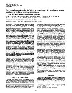
FIG. 3. Western blot analysis. IgM antibodies from H. pylori-infected volunteer serums recognized a single heterologous H. pylori protein with a MW of 70K and multiple homologous H. pylori proteins with MW of 9, 19 to 40, 60, 80, and 140K. The source of primary antibody was serum collected 4 weeks postinfection. This serum was chosen because it had the highest OD values by IgM ELISA.
FIG. 1. (a) Mean OD values of anti-H. pylori IgM antibodies in four volunteers infected for 12 weeks. Homologous (BCS-100) and heterologous (ATCC 43504) H. pylori whole-cell frozen-thawed antigens were used. The OD values were higher when homologous antigen was used. (b) Mean OD values of anti-H. pylori IgM antibodies in the 14 volunteers infected for 4 weeks. OD values were similar irrespective of the antigen used. The vertical dashed lines indicate the end of H. pylori eradication therapy.
of volunteers infected for 12 weeks compared to those infected for only 4 weeks (9.7 weeks). H. pylori-specific IgA and IgG responses. To examine whether IgA antibodies developed during acute H. pylori infection, we tested serum samples from the three infected vol-
FIG. 2. The cumulative proportion of volunteers developing seroconversion to anti-H. pylori IgM. Anti-H. pylori IgM was detected at similar rates irrespective of whether homologous or heterologous H. pylori antigens were used. The majority of volunteers seroconverted to H. pylori by 4 weeks postchallenge.
unteers with the highest IgM antibody OD values at all time points (pre- and postchallenge) for H. pylori-specific IgA antibodies. H. pylori-specific IgA antibodies were not detected in any of the three volunteers tested (data not shown). We found that a specific anti-H. pylori IgG response began to appear about 4 weeks postchallenge and peaked 12 to 19 weeks postchallenge. Of note, 33% (n ⫽ 12) of the volunteers infected for only 4 weeks failed to develop IgG antibodies against H. pylori infection (data not shown). Western blotting. We used Western blotting to examine which H. pylori proteins were responsible for eliciting H. pylorispecific IgM antibody. Sera from volunteers who developed H. pylori-specific serum IgM as detected by ELISA were tested against both the homologous and the heterologous H. pylori antigens by Western blotting (Fig. 3). Antibodies present in all sera collected before challenge recognized proteins with molecular weights (MW) of 50 and 55, and these proteins were considered to be cross-reactive antigens (32). In addition to these cross-reactive antigens, IgM antibodies recognized a single heterologous H. pylori protein with a MW of 70,000 (70K). Sera tested with the homologous strain of H. pylori recognized multiple proteins with MW of 9, 19 to 40, 60, 70, and 150K. While the frequency of reactivity to the higher-molecularweight proteins was variable, all of the infected volunteer sera recognized a wide range of lower-molecular-weight H. pylori proteins with MW of 19 to 40K (Table 1). Reactivity to lowmolecular-weight proteins (e.g., MW 19 to 40K) was detected by 2 weeks postchallenge in 11 of the 18 volunteers. By 3 to 4 weeks postchallenge, the immune response broadened to include recognition of additional proteins with MW of 9, 60, 70, and 150K. IgM immunoreactivity of volunteer sera to heterologous and homologous H. pylori proteins peaked at approximately 4 weeks postchallenge. Generally, the intensity of bands increased from 2 through 4 weeks postchallenge and then remained approximately the same through 10 weeks postchallenge. Infection for 12 weeks was associated with a higher intensity and longer duration of immunoreactivity than those infected for 4 weeks. In volunteers infected for 12 weeks, the bands were faint but detectable at 26 weeks postchallenge (14 weeks after eradication therapy). In volunteers infected for 4 weeks, loss of immunoreactivity was observed approximately 16 weeks after eradication therapy.
IMMUNE RESPONSE TO ACUTE H. PYLORI INFECTION
VOL. 73, 2005
3003
TABLE 1. All infected volunteers developed H. pylori-specific IgM that reacted with multiple H. pylori proteins, and patterns of reactivity were similar among volunteers MWa (103)
Frequencyb
19.5 25–26
18/18 17/18
29 35 50–60
18/18 18/18 3/18
61–63 77–80 150
8/18 12/18 8/18
Putative H. pylori antigen(s)c
Pfr, intracellular protein, protective function against iron toxicity (17) Laminin-binding protein (24), belongs to the class of adhesions (47); alkyl hydroperoxide reductase (51); UreA urease subunit (31) Hemagglutinin, an attachment protein (12) Outer membrane protein A (50) Catalase involved into antiphagocytic mechanism (22); AlpAB, belongs to the class of adhesions (41); flagellar proteins (29); GroEl family of heat shock proteins (13); hemebinding outer membrane proteins (49) UreB urease subunit (31); exoenzyme S-like protein, belongs to the class of adhesins (33) Heme-binding outer membrane proteins (49); vacuolating cytotoxin (6) Neutrophil-activating factor (14)
a
MW determined by Western blotting using Bench Mark prestained protein ladder (Gibco BRL). Frequency was determined by the sum of the number of volunteers in which a specific band of corresponding molecular weight (numerator) was identified by Western blotting among all of the infected volunteers (denominator ⫽ 18). c Based on the presence of proteins with MW in the range indicated. b
Identification of immunogenic proteins of H. pylori. We used two-dimensional gel electrophoresis and mass spectrometry to identify the most-prominent and well-separated bands detected by IgM Western blotting. Five proteins of higher molecular weight were identified as urease B, flagellin A and B, elongation factor TU (EF-TU), and the DNA K H. pylori protein (homolog of the heat shock protein). Four lower-molecular-weight H. pylori proteins were identified (Fig. 4 and Table 2), including alkyl hydroperoxide reductase (TsaA), hydrogenase expression protein (HypB), iron(III) ABC transporter periplasmic iron-binding protein, and superoxide dismutase (SodB). Cellular immune response. We also evaluated the cell-mediated response to acute H. pylori infection. Immunohistochemistry to detect CD3, CD4, CD8, and CD30 surface antigens was performed on sections of biopsy specimens obtained prior to and following H. pylori infections. CD30 cells were not detected among the T cells in the gastric mucosa (Table 3). Staining with the isotype control antibody did not identify
any immunoreactive cells. Specific antibodies for individual T-cell subsets confirmed previous reports that showed that infection with H. pylori was associated with a marked T-cell infiltrate in the epithelium as well as in the lamina propria (3). Cells that were positively stained for CD3, CD4, CD8, and CD30 were counted, and there was a statistically significant increase in all T-cell subsets examined, including the CD4 and CD8 subsets. These changes were most dramatic in the neck but were generally found throughout the gastric glandular structure (Table 3). DISCUSSION H. pylori challenge studies provided a unique opportunity to study some aspects of the H. pylori-host interactions from the time of colonization and establishment of the infection through the early phases of the infection, including the B- and T-cell immune response. We examined the kinetics and duration of the H. pylori-specific IgM response following infections lasting
FIG. 4. Western blot of a two-dimensional gel tested with a H. pylori-infected volunteer’s postinoculation serum sample shown with identified lower-molecular-weight proteins. Western blots were developed using the enhanced chemiluminescence detection system (Amersham Pharmacia). A 200-g portion of a whole-cell lysate of H. pylori was loaded onto the IEF gel. The identities of the proteins in order of spots: 1, superoxide dismutase (SodB); 2, hydrogenase expression/formation protein (HypB); 3, iron(III) ABC transporter periplasmic iron-binding protein (CeuE); 4, alkyl hydroperoxide reductase (TsaA).
3004
NURGALIEVA ET AL.
INFECT. IMMUN.
TABLE 2. Identity of proteins that elicited production of IgM antibodies during the early phase of H. pylori infection MW
22 26 32 34 47 56 58 65 67
Protein identity
Alkyl hydroperoxide reductase (TsaA) Superoxide dismutase (SodB) Hydrogenase expression/formation protein (HypB) Iron(III) ABC transporter, periplasmic iron-binding protein (CeuE) EF-TU Flagellin A Flagellin B Urease beta subunit (urea amidohydrolase) DnaK protein (heat shock protein 70)
No. of peptides matched
% Sequence coverage
5
40
13 12
53 42
5
21
20 16 6 12
62 39 10 19
6
12
up to 3 months with each volunteer serving as their own control. Additionally, we were able to compare the differences in humoral immune responses to antigens from the challenge strain as well as from a heterologous H. pylori strain. By 4 weeks postchallenge, up to 80% of infected volunteers had developed a specific anti-H. pylori IgM response. Although the response to specific proteins differed, the proportion of volunteers responding was similar irrespective of whether homologous or heterologous H. pylori antigens were used. IgM responses were greater in those with longer durations of infection; nonetheless, eradication of the infection prevented further rises in IgM levels. Existing literature regarding the IgM response during early H. pylori infection is scarce. The one reported voluntary ingestion with data reported that H. pylori-specific IgM antibodies are detected serologically early in the H. pylori infection (38). Two other studies reported serological evidence of anti-H. pylori IgM antibodies in the interval of 9 days to 3 months of infection in children (37, 45). Together these data support the notion that H. pylori infection elicits production of IgM antibodies, and these antibodies are developed within weeks postinfection. The use of the sera samples collected during the volunteer study allowed for the first time the determination of the kinetics of an IgM and IgG response to acute H. pylori infection in
more than several individuals with known initiation of infection. Based on previous reports, IgM responses in infected adults and children were inconsistent; few IgM seroconversions were observed in children (19%), and onset of appearance after presumed exposure differed greatly (from few weeks to 3 months) (8, 37, 38, 45). Our study, in which the onset of the infection was known with certainty, showed that up to 80% of adults infected with H. pylori developed an acute IgM antibody response within 2 to 4 weeks of infection and that the response could persist for more than 4 months after eradication of the infection. In children, IgM seroconversion to H. pylori has been reported for 19 to 50% of patients. However, the numbers of children tested were small and the onset of disease was rarely known. Although this alone may account for differences with our results, alternatively, the IgM responses might differ between children and adults. We also had the advantage of being able to use homologous antigens which resulted in an increase in reactivity (i.e., optical density). We collected serum samples regularly, which allowed for moreaccurate monitoring of IgM antibody development, and we found that the H. pylori-specific IgM antibodies peaked at 4 weeks postinfection and fell within 2 to 4 months after beginning of eradication therapy. In contrast, the anti-H. pylori IgG response began to appear about 4 weeks postchallenge and peaked 12 to 19 weeks postchallenge. These data are similar to the experimental inoculation of H. pylori in chimpanzees, where IgG seroconversion occurred at 3 weeks with a further rise of H. pylori-specific IgG antibodies by week 11 after the challenge (21). Rhesus monkeys have also been reported to develop seroconversion 2.5 to 3.5 months postchallenge (10). H. pylori proteins from the homologous H. pylori strain that elicited anti-H. pylori IgM antibodies in the volunteers included proteins with molecular weights of 19 to 40, 60, 70, and 150K. Previous reports of proteins recognized by H. pylorispecific IgM antibodies include proteins with MW of approximately 31K, 60K, 67K, and 90K (7, 16, 48). However, as the identity of the proteins was not determined, a direct comparison between studies of differences in proteins recognized by IgM is not possible. The majority of volunteers showed a response to the lower-molecular-weight proteins, which is similar to the pattern described following H. pylori infection of Mongolian gerbils (30). In contrast to the response with homologous antigens, only one H. pylori protein band with a MW of 70K reacted with volunteers’ sera when heterologous antigen
TABLE 3. Anti-H. pylori T-cell response in seven infected volunteers Median T-cell numbers (range)a Marker
CD3 CD4 CD8 CD30
Pit
Neck
Gland
Pre
Post
Pre
Post
Pre
Post
9.2 (4.8–18.2) 0.6 (0.2–1.6) 4.6 (2.6–14.4) 0.2 (0–2)
28.8ⴱ (13–39.2) 5.6ⴱ (1.4–11.8) 12ⴱ (10–33) 0.4 (0.2–1.4)
11 (6.4–19.8) 1.4 (0.4–3.6) 10.8 (4–12.6) 0.8 ⫾ 0.2
40.4ⴱ (21.6–48.2) 11ⴱ (2.4–19.8) 19.2ⴱ (11.8–32.6) 1 ⫾ 0.3
10.8 (5–21.8) 1.2 (0–2.6) 7.2 (3.6–17.6) 0.5 ⫾ 0.1
30.2ⴱ (2.6–47) 9.6ⴱ (4.2–22.4) 13.2 (7–14) 0.6 ⫾ 0.5
a n ⫽ 7; ⴱ, P value of ⬍0.05. Data are divided by the indicated sites and by relation to infection (pre- or postinfection). The T-cell distributions were measured prior to the challenge and at 4 weeks postchallenge. T cells in the three zones (neck, pit, and gland) were counted (n ⫽ 7) as described in Materials and Methods. The increase of CD3, CD4, and CD8 cells is statistically significant as assessed by the Wilcoxon rank sum test. CD3 marker reacts with T cells associated with the T-cell antigen receptor; CD4 marker reacts with a subset of T cells that carry the coreceptor protein CD4 that activates macrophages and B-cell responses to antigen; CD8 marker reacts with cytotoxic CD8 cells that recognize antigens; CD30 marker reacts with a tumor necrosis factor receptor family member that enhances B-cell proliferation.
VOL. 73, 2005
IMMUNE RESPONSE TO ACUTE H. PYLORI INFECTION
was used for Western blotting. The current study does not allow us to distinguish whether the 70K band represents one or several proteins of similar molecular weight. However, overall the immunoreactivity obtained with heterologous antigen was clearly restricted compared to that obtained using the homologous antigen. This difference is consistent with the organisms marked ability to undergo genetic rearrangements which may reduce the cross-reactivity between immunogenic epitopes on the homologous and heterologous H. pylori proteins (24, 25). Because Western blotting allows only presumptive identification of H. pylori proteins, we used two-dimensional gel electrophoresis and mass spectrometry to identify proteins that were prominent and well separated. Because of these limitations, we did not attempt to identify all of the protein bands identified by Western blot analysis using sera obtained 4 weeks after inoculation. Nine H. pylori proteins that elicited production of IgM antibodies were identified (Table 2). These proteins can be characterized as being generally involved in colonization and establishment of H. pylori infection. For example, flagella are thought to be essential for the ability of H. pylori to colonize the gastric mucosa (26). FlaA, elongation factor EFTU, and urease B have all been reported to be highly immunoreactive proteins (34). In addition, sera from volunteers also recognized heat shock protein 70 (35). We identified four lower-molecular-weight proteins, including hydrogenase expression/formation protein, known to be a housekeeping enzyme involved in energy metabolism; alkyl hydroperoxide reductase, involved in general cellular processes; superoxide dismutase, involved in combating host defenses; and iron(III) ABC transporter periplasmic iron-binding protein, which is likely important in iron homeostasis in what is generally an iron-poor environment. The hydrogenase expression protein and alkyl peroxidase reductase are among the immunodominant antigens of H. pylori based on reactivity to serum collected from patients infected with H. pylori infection (28). IgM antibody recognized additional H. pylori proteins that were not separated well enough to be clearly identified. Additionally, our use of antigens prepared in vitro may have limited the expression of important proteins and/or virulence factors expressed in vivo (e.g., host-bacteria cell contact is absent) and may have lead to an underestimation of the range of proteins eliciting the IgM immune response (28). Nonetheless, we were able to identify and characterize a number of antigens recognized in early infection. The pattern of T-cell subsets infiltrating the gastric mucosa is essentially identical to those observed in natural infection of unknown duration (3, 15). For example, cells expressing CD30 are rarely found in the gastric mucosa after natural infection, and they were also essentially absent from the mucosa of the volunteers after H. pylori challenge. Moreover, most of the T cells accumulated in the neck of the gastric gland. In previous reports, CD4-positive cells increased much more than CD8 cells (3). In this acute phase of the infection, this difference was less remarkable, and this may reflect the differences in a chronic versus a subacute infection as well as variation in patient populations, techniques, or the infecting strain. It is also possible that the relative contribution of intraepithelial lymphocytes is increased and, if so, this could inflate the percentage of CD8⫹ T cells. The current study did not separate the component of intraepithelial lymphocytes that would con-
3005
tribute to the CD8 counts, as only mucosal T-cell populations were recorded. We chose to assess total mucosal T-cell counts in an effort to avoid selective sampling and because of the difficulty in discerning the relationship of the T cells to the tortuous shape of the epithelial glandular unit. Nonetheless, the T-cell responses in these subjects as defined by their surface antigen expression are an excellent model of the changes observed in response to natural infection. The ontogeny of the cellular immune response to infection is often extrapolated from the observed response to immunization. CD4-positive helper T cells would be expected to expand in the first 42 to 72 h. Thereafter, the expression of various chemokines would recruit CD4- and CD8-bearing T cells. Our results suggest that the major changes in T-cell subset recruitment and expansion have occurred by 4 weeks postinfection. ACKNOWLEDGMENTS This material is based upon work supported in part by the Office of Research and Development Medical Research Service Department of Veterans Affairs and by Public Health Service grant DK56338 which funds the Texas Gulf Coast Digestive Diseases Center. We thank Kelly Warfield for her invaluable assistance in establishing the immunoassays. REFERENCES 1. Alem, M., N. Alem, H. Cohen, T. England, N. Hamedi, M. Moussazadeh, J. A. Roth, and G. Q. Shen. 2002. Diagnostic value of detection of IgM antibodies to Helicobacter pylori. Exp. Mol. Pathol. 72:77–83. 2. Andersen, L. P., S. J. Rosenstock, O. Bonnevie, and T. Jorgensen. 1996. Seroprevalence of immunoglobulin G, M, and A antibodies to Helicobacter pylori in an unselected Danish population. Am. J. Epidemiol. 143:1157–1164. 3. Bamford, K. B., X. Fan, S. E. Crowe, J. F. Leary, W. K. Gourley, G. K. Luthra, E. G. Brooks, D. Y. Graham, V. E. Reyes, and P. B. Ernst. 1998. Lymphocytes in the human gastric mucosa during Helicobacter pylori have a T helper cell 1 phenotype. Gastroenterology 114:482–492. 4. Bartelloni, P. J., and R. B. Tesh. 1976. Clinical and serologic responses of volunteers infected with phlebotomus fever virus (Sicilian type). Am. J. Trop. Med. Hyg. 25:456–462. 5. Black, R. E., M. M. Levine, M. L. Clements, T. P. Hughes, and M. J. Blaser. 1988. Experimental Campylobacter jejuni infection in humans. J. Infect. Dis. 157:472–479. 6. Cover, T. L., and M. J. Blaser. 1992. Purification and characterization of the vacuolating toxin from Helicobacter pylori. J. Biol. Chem. 267:10570–10575. 7. Crabtree, J. E., M. J. Mahony, J. D. Taylor, R. V. Heatley, J. M. Littlewood, and D. S. Tompkins. 1991. Immune responses to Helicobacter pylori in children with recurrent abdominal pain. J. Clin. Pathol. 44:768–771. 8. Czinn, S., H. Carr, L. Sheffler, and S. Aronoff. 1989. Serum IgG antibody to the outer membrane proteins of Campylobacter pylori in children with gastroduodenal disease. J. Infect. Dis. 159:586–589. 9. Czinn, S. J., H. S. Carr, and W. T. Speck. 1991. Diagnosis of gastritis caused by Helicobacter pylori in children by means of an ELISA. Rev. Infect. Dis. 13(Suppl. 8):S700–S703. 10. Dubois, A., D. E. Berg, E. T. Incecik, N. Fiala, L. M. Heman-Ackah, G. I. Perez-Perez, and M. J. Blaser. 1996. Transient and persistent experimental infection of nonhuman primates with Helicobacter pylori: implications for human disease. Infect. Immun. 64:2885–2891. 11. Ernst, P. B., P. Michetti, and P. D. Smith (ed.). 1997. The immunobiology of H. pylori: from pathogenesis to prevention. Lippincott-Raven Publishers, Philadelphia, Pa. 12. Evans, D. G., T. K. Karjalainen, D. J. Evans, Jr., D. Y. Graham, and C. H. Lee. 1993. Cloning, nucleotide sequence, and expression of a gene encoding an adhesin subunit protein of Helicobacter pylori. J. Bacteriol. 175:674–683. 13. Evans, D. J., Jr., D. G. Evans, L. Engstrand, and D. Y. Graham. 1992. Urease-associated heat shock protein of Helicobacter pylori. Infect. Immun. 60:2125–2127. 14. Evans, D. J., Jr., D. G. Evans, T. Takemura, H. Nakano, H. C. Lampert, D. Y. Graham, D. N. Granger, and P. R. Kvietys. 1995. Characterization of a Helicobacter pylori neutrophil-activating protein. Infect. Immun. 63:2213– 2220. 15. Fan, X. J., A. Chua, C. N. Shahi, J. McDevitt, P. W. Keeling, and D. Kelleher. 1994. Gastric T lymphocyte responses to Helicobacter pylori in patients with H. pylori colonisation. Gut 35:1379–1384. 16. Faulde, M., J. Cremer, and L. Zoller. 1993. Humoral immune response against Helicobacter pylori as determined by immunoblot. Electrophoresis 14:945–951.
3006
NURGALIEVA ET AL.
17. Frazier, B. A., J. D. Pfeifer, D. G. Russell, P. Falk, A. N. Olse¨n, M. Hammar, T. U. Westblom, and S. J. Normark. 1993. Paracrystalline inclusions of a novel ferritin containing nonheme iron, produced by the human gastric pathogen Helicobacter pylori: evidence for a third class of ferritins. J. Bacteriol. 175:966–972. 18. Frommer, D. J., J. Carrick, A. Lee, and S. L. Hazell. 1988. Acute presentation of Campylobacter pylori gastritis. Am. J. Gastroenterol. 83:1168–1171. 19. Gold, B. D., B. Khanna, L. M. Huang, C. Y. Lee, and N. Banatvala. 1997. Helicobacter pylori acquisition in infancy after decline of maternal passive immunity. Pediatr. Res. 41:641–646. 20. Graham, D. Y., A. R. Opekun, M. S. Osato, H. M. El-Zimaity, C. K. Lee, Y. Yamaoka, W. A. Qureshi, M. Cadoz, and T. P. Monath. 2004. Challenge model for Helicobacter pylori infection in human volunteers. Gut 53:1235– 1243. 21. Hazell, S. L., J. W. Eichberg, D. R. Lee, L. Alpert, D. G. Evans, D. J. Evans, Jr., and D. Y. Graham. 1992. Selection of the chimpanzee over the baboon as a model for Helicobacter pylori infection. Gastroenterology 103:848–854. 22. Hazell, S. L., D. J. Evans, Jr., and D. Y. Graham. 1991. Helicobacter pylori catalase. J. Gen. Microbiol. 137:57–61. 23. Hornick, R. B., S. E. Greisman, T. E. Woodward, H. L. DuPont, A. T. Dawkins, and M. J. Snyder. 1970. Typhoid fever: pathogenesis and immunologic control. 2. N. Engl. J. Med. 283:739–746. 24. Israel, D. A., N. Salama, C. N. Arnold, S. F. Moss, T. Ando, H. P. Wirth, K. T. Tham, M. Camorlinga, M. J. Blaser, S. Falkow, and R. M. Peek, Jr. 2001. Helicobacter pylori strain-specific differences in genetic content, identified by microarray, influence host inflammatory responses. J. Clin. Investig. 107: 611–620. 25. Israel, D. A., N. Salama, U. Krishna, U. M. Rieger, J. C. Atherton, S. Falkow, and R. M. Peek, Jr. 2001. Helicobacter pylori genetic diversity within the gastric niche of a single human host. Proc. Natl. Acad. Sci. USA 98:14625– 14630. 26. Kavermann, H., B. P. Burns, K. Angermuller, S. Odenbreit, W. Fischer, K. Melchers, and R. Haas. 2003. Identification and characterization of Helicobacter pylori genes essential for gastric colonization. J. Exp. Med. 197:813– 822. 27. Khanna, B., A. Cutler, N. R. Israel, M. Perry, A. Lastovica, P. I. Fields, and B. D. Gold. 1998. Use caution with serologic testing for Helicobacter pylori infection in children. J. Infect. Dis. 178:460–465. 28. Kimmel, B., A. Bosserhoff, R. Frank, R. Gross, W. Goebel, and D. Beier. 2000. Identification of immunodominant antigens from Helicobacter pylori and evaluation of their reactivities with sera from patients with different gastroduodenal pathologies. Infect. Immun. 68:915–920. 29. Kostrzynska, M., J. D. Betts, J. W. Austin, and T. J. Trust. 1991. Identification, characterization, and spatial localization of two flagellin species in Helicobacter pylori flagella. J. Bacteriol. 173:937–946. 30. Kumagai, T., J. Yan, D. Y. Graham, M. Tozuka, Y. Okimura, T. Ikeno, A. Sugiyama, T. Katsuyama, and H. Ota. 2001. Serum immunoglobulin G immune response to Helicobacter pylori antigens in Mongolian gerbils. J. Clin. Microbiol. 39:1283–1288. 31. Labigne, A., V. Cussac, and P. Courcoux. 1991. Shuttle cloning and nucleotide sequences of Helicobacter pylori genes responsible for urease activity. J. Bacteriol. 173:1920–1931. 32. Lee, A., S. M. Logan, and T. J. Trust. 1987. Demonstration of a flagellar antigen shared by a diverse group of spiral-shaped bacteria that colonize intestinal mucus. Infect. Immun. 55:828–831. 33. Lingwood, C. A., M. Huesca, and A. Kuksis. 1992. The glycerolipid receptor for Helicobacter pylori (and exoenzyme S) is phosphatidylethanolamine. Infect. Immun. 60:2470–2474.
Editor: F. C. Fang
INFECT. IMMUN. 34. Lock, R. A., G. W. Coombs, T. M. McWilliams, J. W. Pearman, W. B. Grubb, G. J. Melrose, and G. M. Forbes. 2002. Proteome analysis of highly immunoreactive proteins of Helicobacter pylori. Helicobacter 7:175–182. 35. McAtee, C. P., M. Y. Lim, K. Fung, M. Velligan, K. Fry, T. Chow, and D. E. Berg. 1998. Identification of potential diagnostic and vaccine candidates of Helicobacter pylori by two-dimensional gel electrophoresis, sequence analysis, and serum profiling. Clin. Diagn. Lab. Immunol. 5:537–542. 36. Mills, J., J. E. Van Kirk, P. F. Wright, and R. M. Chanock. 1971. Experimental respiratory syncytial virus infection of adults. Possible mechanisms of resistance to infection and illness. J. Immunol. 107:123–130. 37. Mitchell, J. D., H. M. Mitchell, and V. Tobias. 1992. Acute Helicobacter pylori infection in an infant, associated with gastric ulceration and serological evidence of intra-familial transmission. Am. J. Gastroenterol. 87:382–386. 38. Morris, A. J., M. R. Ali, G. I. Nicholson, G. I. Perez-Perez, and M. J. Blaser. 1991. Long-term follow-up of voluntary ingestion of Helicobacter pylori. Ann. Intern. Med. 114:662–663. 39. Nedrud, J. G., S. S. Blanchard, and S. J. Czinn. 2002. Helicobacter pylori inflammation and immunity. Helicobacter 7(Suppl. 1):24–29. 40. O’Neal, C. M., S. E. Crawford, M. K. Estes, and M. E. Conner. 1997. Rotavirus virus-like particles administered mucosally induce protective immunity. J. Virol. 71:8707–8717. 41. Odenbreit, S., M. Till, D. Hofreuter, G. Faller, and R. Haas. 1999. Genetic and functional characterization of the alpAB gene locus essential for the adhesion of Helicobacter pylori to human gastric tissue. Mol. Microbiol. 31:1537–1548. 42. Sabin, A. B. 1952. Research on dengue during World War II. Am. J. Trop. Med. Hyg. 1:30–50. 43. Sobala, G. M., J. E. Crabtree, M. F. Dixon, C. J. Schorah, J. D. Taylor, B. J. Rathbone, R. V. Heatley, and A. T. Axon. 1991. Acute Helicobacter pylori infection: clinical features, local and systemic immune response, gastric mucosal histology, and gastric juice ascorbic acid concentrations. Gut 32:1415– 1418. 44. Tacket, C. O., S. B. Binion, E. Bostwick, G. Losonsky, M. J. Roy, and R. Edelman. 1992. Efficacy of bovine milk immunoglobulin concentrate in preventing illness after Shigella flexneri challenge. Am. J. Trop. Med. Hyg. 47:276–283. 45. Thomas, J. E., A. Dale, M. Harding, W. A. Coward, T. J. Cole, and L. T. Weaver. 1999. Helicobacter pylori colonization in early life. Pediatr. Res. 45:218–223. 46. Tigertt, W. D., and A. S. Benenson. 1956. Studies on Q fever in man. Trans. Assoc. Am. Physicians 60:98–104. 47. Valkonen, K. H., T. Wadstrom, and A. P. Moran. 1997. Identification of the N-acetylneuraminyllactose-specific laminin-binding protein of Helicobacter pylori. Infect. Immun. 65:916–923. 48. von Wulffen, H., J. Heesemann, G. H. Butzow, T. Lo¨ning, and R. Laufs. 1986. Detection of Campylobacter pyloridis in patients with antrum gastritis and peptic ulcers by culture, complement fixation test, and immunoblot. J. Clin. Microbiol. 24:716–720. 49. Worst, D. J., B. R. Otto, and J. de Graaff. 1995. Iron-repressible outer membrane proteins of Helicobacter pylori involved in heme uptake. Infect. Immun. 63:4161–4165. 50. Yamaoka, Y., D. H. Kwon, and D. Y. Graham. 2000. A M(r) 34,000 proinflammatory outer membrane protein (oipA) of Helicobacter pylori. Proc. Natl. Acad. Sci. USA 97:7533–7538. 51. Yan, J., T. Kumagai, M. Ohnishi, I. Ueno, and H. Ota. 2001. Immune response to a 26-kDa protein, alkyl hydroperoxide reductase, in Helicobacter pylori-infected Mongolian gerbil model. Helicobacter 6:274– 282.