Oncogene (2006) 25, 2245–2253
& 2006 Nature Publishing Group All rights reserved 0950-9232/06 $30.00 www.nature.com/onc
ORIGINAL ARTICLE
BACH1 is a DNA repair protein supporting BRCA1 damage response M Peng1, R Litman1, Z Jin, G Fong and SB Cantor UMASS Medical School, Cancer Biology, Worcester, MA, USA
The link between defects in BRCA1 and breast cancer development may be best understood by deciphering the role of associated proteins. BRCA1 associated C-terminal helicase (BACH1) interacts directly with the BRCA1 Cterminal BRCT repeats, which are important for BRCA1 DNA repair and are mutated in the majority of BRCA1 familial cancers. Thus, BACH1 is a likely candidate for mediating BRCA1 DNA repair and tumor suppression functions. Although previous evidence using overexpression of a dominant negative BACH1 has suggested that BACH1 is involved in BRCA1-DNA repair function, our results using BACH1 deficient cells provide direct evidence for involvement of BACH1 in DNA repair as well as for localizing BRCA1. Following DNA damage BACH1 is modified by phosphorylation, displays a BRCA1-like nuclear foci pattern and colocalizes with c-H2AX. Given that the BACH1/BRCA1 complex is unaltered by DNA damage and the intensity of BRCA1 foci is diminished in BACH1 deficient cells, BACH1 may serve to not only facilitate DNA repair, but also maintain BRCA1 in DNA damage foci. Oncogene (2006) 25, 2245–2253. doi:10.1038/sj.onc.1209257; published online 6 February 2006 Keywords: BRCA1; BACH1; FA-J; BRIP1; DNA repair
Introduction The breast cancer susceptibility gene, BRCA1, is a nuclear phosphoprotein with an N-terminus that contains a zinc-finger RING domain, and a C-terminus that contains two BRCT (BRCA1 C-Terminal) repeats. BRCA1 has many protein partners and numerous ascribed functions including ubiquitin ligase activity, (Wu et al., 1996; Baer and Ludwig, 2002) transcriptional activity, (MacLachlan et al., 2002; Monteiro, 2002; Venkitaraman, 2002; Welcsh et al., 2002) DNA repair, cell cycle control, and chromatin remodeling functions (Scully and Livingston, 2000). However, it is not certain Correspondence: Professor S Cantor, UMASS Medical School, Cancer Biology, 364 Plantation St LRB 415, Worcester, MA 01605, USA. E-mail:
[email protected] 1 These two authors contributed equally to this work. Received 6 July 2005; revised 13 October 2005; accepted 13 October 2005; published online 6 February 2006
which protein partners contribute to BRCA1 function(s) and it is not clear which BRCA1 function(s) contribute to BRCA1 tumor suppression. BRCA1 is phosphorylated after geneotoxic stress in a manner dependent upon the DNA-damage-signaling proteins ATM, ATR, Chk1, and Chk2 (Cortez et al., 1999; Lee et al., 2000; Tibbetts et al., 2000; Zhou and Elledge, 2000). During S phase or after treatment with DNA-damaging agents, BRCA1 is in nuclear foci with several other DNA repair proteins including BRCA2, Rad51, and NBS1/Mre11/Rad50 complex (Maser et al., 1997; Scully et al., 1997b; Zhong et al., 1999; Paull et al., 2000). Recruitment and localization of BRCA1 in these foci is dependent on phosphorylation of the histone variant, H2AX-phosphate (g-H2AX) (Paull et al., 2000) and the MDC1 protein (Lou et al., 2003). In the absence of functional BRCA1 cells exhibit spontaneous and induced chromosomal aberrations, are sensitive to DNA-damaging agents, and demonstrate loss of cell cycle checkpoints (Venkitaraman, 2002). Interestingly, BRCA1 mutations that affect the ability of BRCA1 to localize in nuclear foci also affect the ability of BRCA1 to mediate an effective DNA damage response (Botuyan et al., 2004) suggesting that BRCA1 foci formation is critical for BRCA1 function. Evidence suggests that the C-terminus of BRCA1, including the BRCT domains, is important for BRCA1 focus formation (Botuyan et al., 2004; Au and Henderson, 2005). The integrity of the BRCA1 BRCT domains have also been shown to be critical for both BRCA1 DSBR function and homologous recombination (HR) (Moynahan et al., 1999; Scully et al., 1999; Snouwaert et al., 1999; Jasin, 2002). Moreover, nearly all BRCA1 germline mutations involve either truncation or loss of the C-terminal BRCT domains (Liu and West, 2002). These data suggest that BRCA1 DNA repair and tumor suppression functions are linked and highlight the importance of the BRCT domain. Proteins that bind the BRCT domains may participate in some or all of these BRCA1 BRCT domain specific activities including BRCA1 focus formation, DSBR, HR, and tumor suppression. In particular, a candidate mediator of BRCA1 function is the BRCA1 associated C-terminal helicase (BACH1) also called BRIP1, which was identified by its direct binding to the BRCT domains (Cantor et al., 2001). This binding was shown to be dependent on BACH1 phosphorylation at serine 990 and critical for BRCA1 to mediate the G2/M checkpoint (Rodriguez et al., 2003; Yu et al., 2003). BACH1 makes physical contacts with residues
BACH1 is a DNA repair protein M Peng et al
2246
located at the interface of the both BRCTs (Botuyan et al., 2004; Clapperton et al., 2004; Shiozaki et al., 2004), substantiating the finding that multiple clinical BRCT mutations disrupt BRCA1 binding to BACH1. We hypothesized that BACH1 is critical to mediate BRCA1 DNA damage response given its direct binding to BRCA1 and its helicase function. In fact, overexpression of a BACH1 helicase mutant disrupted the kinetics of DSBR in a BRCA1 binding dependent manner (Cantor et al., 2001). In addition, BACH1 helicase activity may also contribute to BRCA1 tumor suppression function given the identification of hereditary breast cancer patients with helicase disrupting mutations in BACH1 (Cantor et al., 2004). In this report, we provide evidence that BACH1 not only participates in the DNA damage response, but also supports BRCA1 localization in DNA damage foci.
Results Resolution of DSB is delayed in BACH1 deficient cells To directly test whether BACH1 function is required for efficient repair, we evaluated the ability of BACH1 deficient cells to repair IR-induced DNA breaks. We suppressed BACH1 protein expression with an shRNA vector targeting BACH1(Litman et al., 2005). Infection of MCF7 or SKOV3 cells with either vector led to substantial suppression of BACH1 expression (Figure 1a and b). However, shRNA directed against enhanced green fluorescent protein (eGFP) target sequence had no effect on the level of BACH1 protein expression (Figure 1a and b). Moreover, the suppression of BACH1
expression with the shRNA vectors is stable for as many as 4 months in culture with no detectable alterations in BRCA1 levels or in levels of b-actin (Figure 1a). Ionizing radiation (IR)-induced DSBs can be visualized by scoring phosphorylation with an antibody (Ab) specific to the variant histone H2AX at serine 139 (g-H2AX) (Rogakou et al., 1999). Phosphorylation of g-H2AX occurs rapidly after DSBs occur (Rogakou et al., 1998) and resolution of DSBs results in loss of g-H2AX phosphorylation similar to the timing of resolution of DSBs imaged by pulse field gel electrophoresis (Rothkamm et al., 2003). Here, we use g-H2AX foci to detect the presence of IR-induced DSBs. MCF7 cells were plated, either left untreated or treated with IR, collected at different time points post-IR, and prepared for immunofluorescence (IF) using a g-H2AX Ab (Upstate). We quantified the kinetics of repair by scoring the percent of cells containing damage-induced g-H2AX foci over total DAPI positive cells at several time points after a sublethal dose of IR, 0.5 Gray (Gy). g-H2AX irradiation-induced foci (IRIF) were detected initially at 30 min (min) (data not shown) and 1 h (hour) post-IR at similar levels in both cells stably expressing shRNA for BACH1 or eGFP control, suggesting that the accumulation of g-H2AX at the sites of DNA breaks was not affected by BACH1 deficiency (Figure 2a). However, BACH1 deficient cells maintained enhanced IR-induced foci at 4, 6 and 8 h (Figure 2b) compared to control cells. Also, in the BACH1 deficient cells, g-H2AX foci were not only found in more cells but these foci were brighter and larger (Figure 2a). This enhanced g-H2AX staining was reverted back to untreated cells 10–24 h post-IR. These results suggest that there is a delay in the resolution of DSBs generated
Figure 1 BACH1 protein is suppressed. (a) Effects of shRNA on protein expression in MCF7 and SKOV3 cells are shown by Western blot, whole-cell lysates (wce) (top) and BACH1 IPs (bottom). (b) MCF7 cells containing shRNA for eGFP or BACH1 were stained with BACH1 Abs. Oncogene
BACH1 is a DNA repair protein M Peng et al
2247
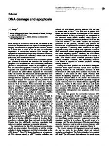
Figure 2 BACH1 deficient cells are delayed in DSBR and have sensitivity to IR. (a) MCF7 cells containing shRNA for eGFP or BACH1 were either left untreated or treated with IR (0.5 Gy) and were fixed at the indicated time points. Cells were then stained with g-H2AX Ab. The arrow denotes a single cell positive for g-H2AX (>10 foci per cell), which displays the variation in intensity of the g-H2AX signal. (b) The percent of MCF7 cells positive for g-H2AX foci are indicated for each time point with standard deviation based on three independent experiments. Over 500 cells were analyzed from each time point. (c) MCF7 cells containing shRNA for either eGFP or BACH1 were treated with increasing dose of IR. Cell growth was assessed 4 days later using the CellTiter glo viability assay (Promega). Each point represents an average of three data points from three independent experiments and a composite graph is shown.
by IR in cells deficient in BACH1 protein expression, but that breaks are eventually repaired. Consistent with a delay in DSBR, the BACH1 deficient cells demonstrated mild radiation sensitivity upon treatment with IR in a 4-day survival assay (Figure 2c). Suppression of BACH1 with RNAi reagents in SKOV3 and 293T led to similar IR sensitivity compared to their respective control cells containing luciferase RNAi (data not shown). BACH1 is phosphorylated after DNA damage and remains bound to BRCA1 The delay in DSBR detected in the BACH1 deficient cells is consistent with BACH1 functioning in DNA damage repair. In order to further evaluate BACH1’s role in DNA repair, we began by addressing whether BACH1 protein responded to DNA damage. First, we examined whether BACH1 demonstrated a post-DNA damage response similar to BRCA1 (Cortez et al., 1999; Lee et al., 2000; Kim et al., 2004). We hypothesized that if BACH1 was active in DNA repair similar to BRCA1, it would respond to DNA damage. We found that multiple forms of genotoxic stress previously shown to lead to BRCA1-induced phosphorylation (Scully and Livingston, 2000) resulted in the slower migration of BACH1 (Figure 3a). The change in BACH1 migration returned to its normal predamage migration as detected by sodium dodecyl sulfate–polyacrylamide gel electro-
phoresis (SDS–PAGE) after treatment with serine/ threonine phosphatases (lPpase) suggesting that the observed modification was a result of phosphorylation. The reversal in gel migration as a result of lPpase treatment occurred irrespective of the type of DNA damage (Figure 3b). Moreover, inclusion of phosphatase inhibitors (lPpase inhib.) with the serine/threonine phoshatases prevented the return of BACH1 to its normal predamage migration further supporting the finding that DNA-damage-induced change in BACH1 migration was due to phosphorylation (Figure 3b). The DNA-damage-induced phosphorylation appears to be both dose and time dependent. Maximum gel retardation is obtained by treating cells with 50 J/m2 of ultraviolet light (UV), 1 mM hydroxyurea (HU), or 20 Gy of IR, whereas 12 Gy of IR generates intermediate BACH1 migration that reaches maximal at 8 h post-IR (Figure 3c). The post-DNA damage BACH1 gel retardation was present in multiple cell lines including HeLa, SKOV3, and 293T as well as in cells that lack functional BRCA1 (HCC1937 cells) (data not shown) indicating that this is not a cell specific event and that BRCA1 is not required for BACH1 postdamage processing. We next addressed whether DNA damage changed the equilibrium of the BRCA1–BACH1 complex. Immunoprecipitation (IP) experiments followed by Western blot analysis demonstrated that co-precipitating BACH1/BRCA1 proteins were not noticeably Oncogene
BACH1 is a DNA repair protein M Peng et al
2248
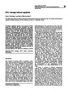
Figure 3 BACH1 is modified after DNA damage. (a) MCF7 cells were either left untreated or treated with the indicated type of DNA damage. Protein mobility was analyzed by Western blot with BRCA1 or BACH1 specific Abs. (b) MCF7 cells were either left untreated or treated with 50 J/m2 UV and subsequently BACH1 IPs were treated with l phosphatase and/or l phosphatase inhibitors as indicated. (c) For quantitative analysis of the BRCA1/BACH1 complex, MCF7 cells were either left untreated or treated with 12 Gy IR and collected 15 min, 4 and 8 h post-IR. The percentage of BRCA1 bound to BACH1 was quantified using a LAS-3000 image reader and Image Guage software. (d) SKOV3 cells were either left untreated or treated with IR (12 Gy), fixed 4 h later, and stained with antiBACH1 and anti-BRCA1 Abs as indicated.
changed by DNA damage (Figure 3c). Quantification of the percent BRCA1 in a BACH1 IP showed that the percentage of BRCA1-bound BACH1 was unchanged at 15 min to 8 h post-IR (Figure 3c) suggesting that neither DNA damage nor the DNA-damage-induced phosphorylation of BRCA1 or BACH1 affected the complex. Consistent with this finding, phosphorylation of BACH1 at S990, the site critical for BRCA1 binding, is not changed after DNA damage (data not shown). Moreover, colocalizing BRCA1 and BACH1 nuclear foci are easily detected before and after IR (Figure 3d). BACH1 and g-H2AX foci colocalize after DNA damage To better assess the role of BACH1 in the DNA repair response, we addressed if and when BACH1 localized to sites of DNA damage by testing whether BACH1 IRIF colocalize with sites of DSBs as has been shown for BRCA1 (Paull et al., 2000). MCF7 cells irradiated with 12 Gy IR demonstrated BACH1 IRIF pattern that overlapped with the g -H2AX IRIF pattern. Irradiationinduced colocalization of BACH1 and g-H2AX was detectable from 5 min to 24 h post-IR (Figure 4). These Oncogene
results suggest that BACH1 response to IR is similar to the published BRCA1 response (Paull et al., 2000) in that BACH1 and BRCA1 IRIF diffuse and reform with similar kinetics and both proteins form early and late IRIF costaining with g-H2AX. In addition, the pattern of BACH1 and BRCA1 foci was similar after DNA damage (Table 1). As reported previously, we found that the BRCA1 IRIF were disrupted within 1 h and then gradually reassembled into bright foci (Scully et al., 1997b). By 6–8 h after IR 70–90% of nuclei were positive for BRCA1 IRIF and remained until 12 h (Zhong et al., 1999). BACH1 foci pattern resembles the BRCA1 foci pattern overall. In untreated cells, BACH1 and BRCA1 foci are 35.9% and 36.5%, respectively, and BACH1 and BRCA1 IRIF reach maximum 6 h post-IR at 78.9% and 75.3%, respectively (Table 1). BACH1 deficient cells demonstrate reduced BRCA1 foci Since loss of BACH1, leads to enhanced cellular sensitivity to DNA damage and defects in DSBR kinetics, two consequences of BRCA1 deficiency, we reasoned that this result could be due to loss of BRCA1
BACH1 is a DNA repair protein M Peng et al
2249
Figure 4 BACH1 colocalizes with g-H2AX after IR. MCF7 cells were either left untreated or treated with IR (12 Gy). At the indicated time points, cells were stained with BACH1 and g-H2AX Abs.
Table 1 Focus formation of BRCA1 and BACH1 after IR Time after IR (h)
0 1.0 2 4 6 12
Foci contained cells (%) BRCA1
BACH1
36.575.6 17.773.7 44.775.1 60.275.4 75.377.0 69.076.6
35.974.9 24.872.9 43.276.5 60.678.6 78.976.1 52.076.8
MCF7 cells were either left untreated or treated with IR (12Gy), allowed to recover for the indicated time, and stained with antiBRCA1 or anti-BACH1 Abs. A cell nucleus displaying >10 BRCA1 or BACH1 foci was counted as a foci positive cell. At least 500 cells were analyzed for each time point, 7represents the standard deviation from the mean.
at sites of DNA damage. In fact, BACH1 has been proposed to localize BRCA1 into nuclear foci given the finding that BRCT mutations in BRCA1 disrupt both BACH1 binding and BRCA1 nuclear foci formation (Botuyan et al., 2004; Clapperton et al., 2004). These results are complicated by the fact that these BRCT mutations may also disrupt the binding of other BRCT interacting proteins like CTIP or RNA helicase A (Thompson and Schild, 2002), making it unclear whether BACH1 is truly directing BRCA1 to foci or whether other BRCT interacting proteins are directing BRCA1 to foci. To directly examine the contribution of BACH1 to BRCA1 localization, we analyzed BRCA1 foci formation in BACH1 deficient cells. BRCA1 foci were present, but appeared less intense overall in BACH1 deficient cells compared to control cells while g-H2AX signal was unaltered (Figure 5a). Nevertheless, these BRCA1 foci present in the BACH1 deficient cells colocalized with g-H2AX IRIF (Figure 5a). The reduced BRCA1 foci
present in BACH1 shRNA containing cells was not due to a decrease in BRCA1 protein levels (Figure 1a) and BRCA1 postdamage phosphorylation was unchanged (Figure 5b). Thus, we reasoned that the reduced BRCA1 staining was not due to changes in BRCA1 expression, we hypothesized that BACH1 deficiency may effect the kinetics of BRCA1 localization in IRIF. Cells expressing shRNA for BACH1 or eGFP control (Figure 5c) were left untreated or treated with low-dose IR (2.5 Gy), a sublethal dose that generates DNA-damage-induced BRCA1 and g-H2AX IRIF immediately post-IR. Cells were collected at the indicated time points post-IR and stained with BRCA1 and g-H2AX Abs (Figure 5d). As before, a visible reduction in BRCA1 staining was detectable in all BACH1 deficient cells (Figure 5d). In addition, a measurable reduction in cells positive for BRCA1 foci (>10 foci/cell) was clearly detected from 8–30 min post-IR (Figure 5c and d). Although complete recovery in BRCA1 foci intensity was not achieved in BACH1 deficient cells, the number of cells positive for greater than 10 BRCA1 foci per cell was similar to control cells by 1 h post-IR (Figure 5c and d). Together these results suggest that BRCA1 foci are dependent on BACH1 at early times post-IR for nucleation at sites of DNA damage and for maintenance of BRCA1 foci. To confirm our finding that BACH1 deficiency disrupted normal BRCA1 foci formation, BRCA1 foci were characterized in the BACH1 deficient cell line, EUFA30-F. This cell was derived from a Fanconi anemia (FA) patient previously assigned to the FA-J complementation group (Levitus et al., 2004). Recently, several groups have identified the gene defect in the FA-J complementation group as BACH1 (Bridge et al., 2005; Levitus et al., 2005; Levran et al., 2005; Litman et al., 2005). The FA-J cells, EUFA30-F, have a homozygous point mutation in the BACH1 gene encoding a premature stop codon resulting in undetectable BACH1 protein expression. Reintroduction of WT Oncogene
BACH1 is a DNA repair protein M Peng et al
2250
Figure 5 BRCA1 foci are diminished in BACH1 deficient cells. (a) SKOV3 cells containing shRNA for either eGFP or BACH1 were either left untreated or treated with IR (2.5 Gy), were fixed 4 h later, and stained with Abs for g-H2AX and BRCA1. BRCA1 foci colocalize with g-H2AX foci after DNA damage (merge). (b) MCF7 cells containing shRNA for either eGFP or BACH1 were either left untreated or treated with IR (10 or 20 Gy) and collected 1 or 3 h later, respectively. (c) The percent of SKVO3 cells positive for BRCA1 foci are indicated for each time point with standard deviation based on three independent experiments. A cell nucleus displaying >10 BRCA1 foci was counted as a foci-positive cell. Over 500 cells were analyzed from each time point. (d) Representative cell images from each time point are shown.
BACH1 corrects the post-DNA damage G2/M accumulation in these cells (Litman et al., 2005). Here, we have also reintroduced FA-J cells with either WT BACH1 or vector alone using lentiviral vectors (Litman et al., 2005). Cells stably expressing the vectors were drug selected and analyzed by Western blot to confirm BACH1 expression (Figure 6a). To investigate whether BACH1 is important for BRCA1 foci formation in these cells, we analyzed FA-J cells reconstituted with WT BACH1 or vector alone for the ability of these cells to form BRCA1 foci. FA-J cells with BACH1 or empty vector were seeded on coverslips, either left untreated or treated with 2.5 Gy IR, and cells positive (>10 foci/cell) for BRCA1 foci were counted. The cells reconstituted with WT BACH1 had a greater percentage of cells positive for BRCA1 foci than the cells reconstituted with empty vector alone, both before and after DNA damage. Approximately, 50% of FA-J cells reconstituted with WT BACH1 were positive for BRCA1 foci, however, only 25% of the FA-J cells reconstituted with the empty vector scored positive for BRCA1 foci (Figure 6b untreated). Following exposure to IR, both Oncogene
WT BACH1 and empty vector FA-J cells contained BRCA1 foci that colocalized with g-H2AX suggesting that BACH1 is not required for BRCA1 colocalization with g-H2AX (Figure 6c). However, WT BACH1 reconstituted cells maintained more detectable BRCA1 foci at all time points tested (Figure 6b and c) up to 24 h following IR treatment (data not shown). Perhaps the FA-J cells, unlike the shRNA BACH1-containing cells, have a greater disruption in BRCA1 foci formation due to the lack of BACH1 protein in the FA-J cells. Thus, similar to BACH1 deficient cells established by RNAi reagents, BACH1 mutant cells also demonstrate defects in the number and intensity of BRCA1 foci. Discussion In this report, we have shown that BACH1 deficient cells are sensitive to DNA damage and are delayed in the repair of DSBs. BRCA1 foci formation is not only diminished in BACH1 deficient cells, but also BRCA1 IRIF formation is delayed. BRCA1 eventually localizes
BACH1 is a DNA repair protein M Peng et al
2251
Figure 6 BRCA1 foci are diminished in BACH1 null cells. (a) FA-J cells containing either empty vector or WT BACH1 were stained with BRCA1 Abs for IF or analyzed by Western blot using BACH1 and b-actin Abs. (b) The percent of FA-J cells positive for BRCA1 foci are indicated for each time point with standard deviation based on three independent experiments. A cell nucleus displaying >10 BRCA1 foci was counted as a foci-positive cell. Over 600 cells were analyzed from each time point. (b) Representative cell images from the 2 h post IR time point are shown.
with g-H2AX at sites of breaks suggesting that BACH1 only contributes to recruitment of BRCA1 in IRIF or serves to maintain BRCA1 in IRIF. Our findings demonstrate that BACH1 is a DNA repair protein and suggest that BRCA1 and BACH1 functions are linked. The same DNA-damage-signaling pathways that modify BRCA1 also modify BACH1. First, similar to BRCA1, BACH1 post-DNA damage modification is most likely phosphorylation since lPpase treatment of post-DNA damage BACH1 IPs led to a reversion of BACH1 post-DNA damage slower gel migration to a pre-DNA damage gel migration. Second, similar to BRCA1, BACH1 IRIF form immediately after DNA damage and maximum IRIF is obtained at 6 h post-IR. Third, BACH1 mimics BRCA1 postdamage colocalization with g-H2AX. Given that BACH1 DNA damage response mimics BRCA1 DNA damage response, it is not surprising that BACH1 binding to BRCA1 is unaltered by DNA damage. While BRCA1 binding to BACH1 may be important for localization of BRCA1 in foci, (Botuyan et al., 2004; Clapperton et al., 2004), BACH1 may only contribute to BRCA1 localization since both N- and C-terminal portions of BRCA1 have been shown to be required for BRCA1 localization (Au and Henderson, 2005). Our
results in BACH1 deficient cells suggest that BACH1 contributes to BRCA1 localization, but is not ultimately required. We predict that BACH1 serves to maintain BRCA1 in IRIF and not directly recruit BRCA1 to IRIF. Unlike MDC1 deficient cells in which BRCA1 foci do not form in the absence or presence of DNA damage (Lou et al., 2003), in BACH1 deficient cells BRCA1 foci have reduced intensity, but are present. Moreover, BRCA1 IRIF reform in BACH1 deficient cells by 1 h post-IR. In contrast to BACH1 deficient cells in which BRCA1 foci returned by 1 h post-IR, in FA-J cells BRCA1 foci did not fully return even at later times post-IR. This difference may result from residual BACH1 expressed in the BACH1 RNAi treated cells compared to the FA-J cells used in this study, which have no detectable BACH1 protein (Litman et al., 2005). Alternatively, differences in the cell type or transformation state of the transformed MCF7 cells compared to immortalized FA-J fibroblasts may influence the BRCA1 foci formation. Finally, BRCA1 appears to be appropriately modified in BACH1 deficient cells following IR unlike in MDC1 deficient cells in which BRCA1 post-IR modification is disrupted (Lou et al., 2003). Perhaps the mild radiosensitivity found in BACH1 deficient cells reflects the finding that Oncogene
BACH1 is a DNA repair protein M Peng et al
2252
BACH1 deficient cells are capable of eventually localizing BRCA1 and repairing breaks. BACH1 may unwind the DNA in the vicinity of the DNA damage and facilitate access of BRCA1 to these sites. Previous results demonstrated that BACH1 is absent from nuclear foci in cells devoid of intact BRCA1 suggesting BRCA1 is upstream of BACH1 in the DNA damage response pathway. In these same cells, BACH1 foci will form when WT full-length BRCA1 is reconstituted (Cantor et al., 2001). One explanation for these findings may be that BRCA1 serves as a scaffold for which multiple repair proteins are localized and stabilized at the sites of breaks. Thus, in the absence of BRCA1, BACH1 and other repair proteins fail to form distinct repair foci. BACH1 may in turn stabilize BRCA1 at these sites. BRCA1 is a dynamic protein that relocalizes after DNA damage and associates with multiple repair proteins in multiple complexes. In doing so, BRCA1 may cycle between bound and unbound sites of DNA damage, replication, or transcription to mediate multiple functions. If BACH1 participates in maintaining BRCA1 at any of these sites, BRCA1 focus formation would be reduced overall as we detected. In addition, after low dose IR, BRCA1 IRIF would be reduced, but not impaired if BACH1 participates with other repair proteins to maintain BRCA1 binding to sites of DNA damage in BACH1 deficient cells. The direct and highly specific interaction between BACH1 and BRCA1 suggests that they are functionally linked. Consistent with this idea, our findings show that BACH1 resembles BRCA1 post-DNA damage response in its localization pattern, immediate presence at sites of DNA damage, and in the DNA-damage-induced phosphorylation pattern. Moreover, our findings suggest that BACH1 function is required for the timely arrival of BRCA1 into DNA damage foci, radioresistance, and for normal kinetics of DSBR. Recently, we demonstrated that BACH1, similar to BRCA1, is required for repair of DSBs by HR, MMC resistance, and for maintenance of chromosomal integrity (Litman et al., 2005). In addition, BACH1 is the gene defect in the FA-J complementation group of the FA disease in which patients are plagued by leukemia and squamous cell carcinomas (Kutler et al., 2003). This finding clearly demonstrates that BACH1 is a tumor suppressor gene. The next critical question is whether the BACH1– BRCA1 interaction is required for these mutual repair functions, whether BACH1 connects BRCA1 to the FA pathway, and whether ultimately BRCA1 and BACH1 tumor suppression functions are linked. Future experiments in which the BACH1–BRCA1 interaction is disrupted directly will clarify the importance of this complex formation for DNA repair and tumor suppression.
Materials and methods Cell cultures MCF7, SKOV3, and 293 cell lines were cultured in Dulbecco’s modified Eagle’s medium (DMEM) supplemented with 10% Oncogene
fetal bovine serum (Gibco) and antibiotics. FA-J fibroblasts (EUFA30-F) (Levitus et al., 2005) containing either empty pLenti vector or WT BACH1 pLenti vector (Yu et al., 2003) were grown in DMEM (Gibco) supplemented with 15% FBS, 7 mg/ml Blasticidin (Sigma) and antibiotics. Antibodies The monoclonal BACH1 Abs 2C10 and IG5 used for IF or as a monoclonal pool for Western blot (Cantor et al., 2001) and poloyclonal Ab E67 was used as previously described (Cantor et al., 2004). The phospospecific BACH1 serine 990 Ab (pser990) was generously provided by the Chen laboratory and was used as previously described (Yu et al., 2003). The monoclonal BRCA1 Abs SD118 and MS110 were used as previously described (Scully et al., 1997a) and the polyclonal BRCA1 Ab (Stressgen Bioreagents). The mouse monoclonal and polyclonal antiphospho-Histone H2A.X(Ser139) Abs were purchased from Upstate. Rhodamine Red-X conjugated AffiniPure Goat anti-rabbit IgG and Fluorescein (FITC)-conjugated AffiniPure Goat anti-mouse IgG (Jackson ImmunoReseach Laboratories Inc). Immunoprecipitations and Western blot assays Cells were harvested and lysed in lysis buffer (20 mMTris [pH8.0], 150 mM NaCl, 1 mMEDTA, 0.5%NP-40,1 mM phenylmethylsulfonyl fluoride,10 mg of leupeptin per ml, 10 mg of aprotinin per ml) for 30 min on ice. The extracts were clarified by centrifugation for 10 min at 13 200 rpm. The cell lysates were incubated with 3 ml E67, 30 ml protein A beads (Amersham) for 2 h at 41C and washed three times in IP washing buffer (20 mMTris[pH8.0], 150 mMNaCl, 1 mMEDTA, 0.5%NP-40). IPs were boiled in SDS-loading buffer and proteins were separated using (SDS–PAGE). Proteins were electrotransferred to nitrocellulose membranes and Western blot assays were carried out with the indicated primary Abs over night at 41C followed by horseradish peroxidaseconjugated secondary Abs for 1 h at room temperature. All blots were detected with ECL-plus Western Blotting Detection System (Amersham Biosciences) using Biomax MR or XAR film (Kodak). Bradford assays were used to determine protein content (Bio-Rad). For the DNA damage treatment experiments cells were treated with IR of 10 Gy, 20 Gy, 1 mM HU, and 50 J/M2 UV and then collected 1, 3, 24, and 3 h later, respectively. Proteins were separated on 4% tris-glycine gels. l-Phosphatase inhibitors included sodium orthovanadate and sodium fluoride (NEB). Cell survival assays MCF7 cells were either untreated or damaged with 5 or 10 Gy IR and plated at low density. A baseline reading was taken at the initial cell plating and total ATP was quantified as absorbance units after addition of Cell titer Glo reagent (Promega). Cells were allowed to grow for 4 days and an end line reading was taken to quantify total cellular ATP, which was detected as absorbance units. Total absorbance units were used to assess relative viability. Immunofluoresence Cells were grown on coverslips in a six-well plates. Before immunostaining, cells were irradiated or left untreated. At different time points, cells were fixed using 3%paraformalde/ 2%sucrose in PBS for 10 min, and permeabilized with 0.5% Triton X-100. Cells were then incubated with both primary and secondary Abs in six-well plates for 1 h at RT. Cells were washed five times for 5 min each in PBS after incubation with both primary and secondary Abs. Coverslips were mounted
BACH1 is a DNA repair protein M Peng et al
2253 using Vectashield with DAPI and sealed with nail polish, then stored in the dark at 41C. IF was performed by an inverted Olympus 1X71 fluorescence microscope. Retroviral or Lentiviral vectors and infection The lentiviral shRNA expression vector FSIPPW, (eGFP) targets the sequence 50 -AAGAACGGCATCAAGGTGAA CTT-30 and was described previously (Kanellopoulou et al., 2005). The FSIPPW targets BACH1 sequence 50 AAAGCUUACCCGUCACAGCUU-30 . The FSIPPW vectors were a generous gift of Dr Andrew Kung. The WT BACH1 pLenti vector was a generous gift of Dr Junjie Chen and Xiaochun Yu. 293TD retroviral packaging lines were used to produce all shRNA encoding lentivirus. 293TD cells were transfected with 2–4 mg of plasmid DNA using Fugenet (Roche) transfection reagent 48 h prior to harvesting retroviral
or lentiviral supernatants. Supernatants containing 8 mg/ml polybrene were added to recipient cell lines. Cells were transduced with lentivirus and were selected with either 2 mg/ ml of puromycin (Sigma) for cells containing FSIPPW shRNA vectors or 7 mg/ml Blasticidin for cells containing pLenti vectors (Sigma). Acknowledgements We would like to thank Tim Kowalik (UMASS) and Arthur Mercurio (UMASS) for critical review of the manuscript. Andrew Kung (DFCI) for the FSIPPW vectors. We thank Ronny Drapkin (DFCI) for sharing the initial observation that BACH1 is phosphorylated after DNA damage and Hans Joenje (UMC, Netherlands) for the FA-J fibroblasts (EUFA30-F).
References Au WW, Henderson BR. (2005). J Biol Chem 280: 6993–7001. Baer R, Ludwig T. (2002). Curr Opin Genet Dev 12: 86–91. Botuyan MV, Nomine Y, Yu X, Juranic N, Macura S, Chen J et al. (2004). Structure (Cambridge) 12: 1137–1146. Bridge WL, Vandenberg CJ, Franklin RJ, Hiom K. (2005). Nat Genet 37: 953–957. Cantor S, Drapkin R, Zhang F, Lin Y, Han J, Pamidi S et al. (2004). Proc Natl Acad Sci USA 101: 2357–2362. Cantor SB, Bell DW, Ganesan S, Kass EM, Drapkin R, Grossman S et al. (2001). Cell 105: 149–160. Clapperton JA, Manke IA, Lowery DM, Ho T, Haire LF, Yaffe MB et al. (2004). Nat Struct Mol Biol 11: 512–518. Cortez D, Wang Y, Qin J, Elledge SJ. (1999). Science 286: 1162–1166. Jasin M. (2002). Oncogene 21: 8981–8993. Kanellopoulou C, Muljo SA, Kung AL, Ganesan S, Drapkin R, Jenuwein T et al. (2005). Genes Dev 19: 489–501. Kim SS, Cao L, Li C, Xu X, Huber LJ, Chodosh LA et al. (2004). Mol Cell Biol 24: 9498–9507. Kutler DI, Singh B, Satagopan J, Batish SD, Berwick M, Glampietro PF, Averbach AD. (2003). Blood 101: 1249–1256. Lee JS, Collins KM, Brown AL, Lee CH, Chung JH. (2000). Nature 404: 201–204. Levitus M, Rooimans MA, Steltenpool J, Cool NF, Oostra AB, Mathew CG et al. (2004). Blood 103: 2498–2503. Levitus M, Waisfisz Q, Godthelp BC, de Vries Y, Hussain S, Wiegant WW et al. (2005). Nat Genet 37: 934–935. Levran O, Attwooll C, Henry RT, Milton KL, Neveling K, Rio P et al. (2005). Nat Genet 37: 931–933. Litman R, Peng M, Jin Z, Zhang F, Zhang J, Powell S et al. (2005). Cancer Cell 8: 255–265. Liu Y, West SC. (2002). Breast Cancer Res 4: 9–13. Lou Z, Chini CC, Minter-Dykhouse K, Chen J. (2003). J Biol Chem 278: 13599–13602. MacLachlan TK, Takimoto R, El-Deiry WS. (2002). Mol Cell Biol 22: 4280–4292. Maser RS, Monsen KJ, Nelms BE, Petrini JH. (1997). Mol Cell Biol 17: 6087–6096.
Monteiro AN. (2002). Cancer Biol Ther 1: 187–188. Moynahan ME, Chiu JW, Koller BH, Jasin M. (1999). Molecular Cell 4: 511–518. Paull TT, Rogakou EP, Yamazaki V, Kirchgessner CU, Gellert M, Bonner WM. (2000). Curr Biol 10: 886–895. Rodriguez M, Yu X, Chen J, Songyang Z. (2003). J Biol Chem 278: 52914–52918. Rogakou EP, Boon C, Redon C, Bonner WM. (1999). J Cell Biol 146: 905–916. Rogakou EP, Pilch DR, Orr AH, Ivanova VS, Bonner WM. (1998). J Biol Chem 273: 5858–5868. Rothkamm K, Kruger I, Thompson LH, Lobrich M. (2003). Mol Cell Biol 23: 5706–5715. Scully R, Chen JJ, Ochs RL, Keegan K, Hoekstra M, Feunteun J et al. (1997a). Cell 90: 425–435. Scully R, Chen JJ, Plug A, Xiao YH, Weaver D, Feunteun J et al. (1997b). Cell 88: 265–275. Scully R, Ganesan S, Vlasakova K, Chen JJ, Socolovsky M, Livingston DM. (1999). Molecular Cell 4: 1093–1099. Scully R, Livingston DM. (2000). Nature 408: 429–432. Shiozaki EN, Gu L, Yan N, Shi Y. (2004). Mol Cell 14: 405–412. Snouwaert JN, Pace AJ, Gowen LC, Xiao A, Nichols MA, Xiong Y et al. (1999). FASEB J 13: A1538–A1538. Thompson LH, Schild D. (2002). 509: 49–78. Tibbetts RS, Cortez D, Brumbaugh KM, Scully R, Livingston D, Elledge SJ et al. (2000). Genes Dev 14: 2989–3002. Venkitaraman AR. (2002). Cell 108: 171–182. Welcsh PL, Lee MK, Gonzalez-Hernandez RM, Black DJ, Mahadevappa M, Swisher EM et al. (2002). Proc Natl Acad Sci USA 99: 7560–7565. Wu LC, Wang ZW, Tsan JT, Spillman MA, Phung A, Xu XL et al. (1996). Nat Genet 14: 430–440. Yu XC, Chini CCS, He M, Mer G, Chen JJ. (2003). Science 302: 639–642. Zhong Q, Chen CF, Li S, Chen Y, Wang CC, Xiao J et al. (1999). Science 285: 747–750. Zhou BB, Elledge SJ. (2000). Nature 408: 433–439.
Oncogene