Bactericidal Effect of ADP and Acetic Acid on Bacillus subtilis. Victor Asensi,1 Francisco Parra,2 Joshua Fierer,3 Eulalia Valle,2 Carmen Bordallo,2 Paz ...
CURRENT MICROBIOLOGY Vol. 34 (1997), pp. 61–66
An International Journal
R Springer-Verlag New York Inc. 1997
Bactericidal Effect of ADP and Acetic Acid on Bacillus subtilis Victor Asensi,1 Francisco Parra,2 Joshua Fierer,3 Eulalia Valle,2 Carmen Bordallo,2 Paz Rendueles,2 Santiago Gasco´n,2 Jose´ A. Carton,1 Jose´ A. Maradona,1 Jose´ M. Arribas1 1Department of Medicine, Division of Infectious Diseases, Covadonga Hospital, Oviedo University Medical School, Calle Celestino Villamil s/n, 33006 Oviedo, Spain 2Department of Biochemistry, Oviedo University Medical School, Oviedo, Spain 3Departments of Medicine and Pathology, Veterans Administration Medical Center and School of Medicine, University of California, San Diego, CA, USA
Received: 14 June 1996 / Accepted: 19 July 1996
Abstract. Bacillus subtilis is a ubiquitous soil bacterium used for measuring the b-lysin activity and in other bioassays. We observed a complete bactericidal effect of ADP on B. subtilis at concentrations of 50–100 µM at pH values ,5.5, which disappeared at pH values above 6. The effect was also found for acetic acid at concentrations .17.4 µM and similar pH values. ATP, adenosine, and HCl were not bactericidal. We used BCECF-AM, a pH-sensitive probe, and found that the killing of B. subtilis was due to a change in the intracellular pH caused by the passage across the cell membrane of these weak organic acids when incubated with B. subtilis at pH values near the pK. More experiments are needed to determine the biological meaning of these in vitro findings.
Bacillus subtilis is a ubiquitous soil bacterium, used as a model to study the metabolism of many other microorganisms. B. subtilis is also used in the determination of the biological activity of b-lysin, a platelet-derived antibacterial protein with a molecular weight of 9000 [6, 7, 16]. ADP induces the platelet aggregation and release of b-lysin and therefore can be used to improve the production of this protein [5]. However, we have noted an unexpected bactericidal effect of ADP when incubated alone with B. subtilis in in vitro assays. The present work was undertaken to measure the in vitro inhibitory effect of ADP on B. subtilis and to assess the effect of other weak and strong acids on the growth of this bacterium and other Gram-positive and Gramnegative microorganisms. Materials and Methods Bacteria. Bacillus subtilis, Staphylococcus aureus, Listeria monocytogenes serotype 4A, and Escherichia coli (ATCC 6633, 29213, 19114, Correspondence to: V. Asensi
and 25922) were purchased from the American Type Culture Collection (Rockville, Maryland). Isolates were grown on trypticase soy broth (TSB, Difco Laboratories, Detroit, Michigan) at 37°C overnight. Aliquots were made and stored at 270°C until used. Reagents preparation. ADP (Diagnostica Stago, Asnieres, France), ATP (Boehringer-Mannheim, Germany), and adenosine (Sigma, St. Louis, Missouri) were prepared as stock concentrations of 100 mM (ADP), 1000 mM (ATP), and 10 mM (adenosine) in physiological saline. Stocks were freshly diluted in saline for each experiment. HCl and acetic acid (CH3-COOH) were purchased from Merck (Darmstadt, Germany), and their respective dilutions were made in distilled water. Mes/HEPES/Tris buffer was made with 50 mM Mes and 50 mM HEPES (Sigma) in distilled water. The pH was adjusted to 4.6, 5, 5.5, and 6 with 1 M Tris buffer (Sigma). Bactericidal assay. Isolates of B. subtilis, S. aureus, L. monocytogenes, and E. coli were grown overnight at 37°C in TSB and adjusted to a final concentration of 103–104 bacteria ml21 for B. subtilis and 108 ml21 for the other bacteria. Each test dilution containing 125 µl of Mes/HEPES/ Tris buffer plus acid was inoculated with 12.5 µl of the respective bacterial suspension in 1.7-ml microfuge tubes. After 1, 2, or 3 h of incubation at 37°C in a water bath, serial tenfold dilutions of each sample were made in physiological saline, and 10 µl of each sample was
62
CURRENT MICROBIOLOGY Vol. 34 (1997)
Table 1. The bactericidal effect of ADP on B. subtilis is concentration dependent ADP (µM)
B. subtilis (CFU ml21)
0 6.3 12.5 25.0 50.0 100.0
2.7 3 104 1.3 3 104 1.8 3 104 2.9 3 103 0 0
ADP was dissolved in physiological saline (pH 5.5) and incubated for 2 h at 37°C with B. subtilis. The initial bacterial inoculum was 9.0 3 103 CFU ml21. Results are the mean value of three independent experiments.
plated on Trypticase soy agar (TSA, Difco) and incubated at 37°C overnight as previously described [1]. Colonies were counted the next morning. Measurement of the internal pH of B. subtilis after acid treatment. The procedure for determining the internal pH of B. subtilis after incubation with different acids was similar to that described by Molenaar et al. [11] and modified after Magill et al. [10]. To measure the internal pH of B. subtilis, we used the pH-sensitive fluorescent probe 28,78-bis (2-carboxyethyl)-5 (and-6)-carboxyfluorescein-acetoxymethyl ester (BCECF-AM, Sigma). BCECF-AM was dissolved in water-free dimethyl sulfoxide (Sigma) to a final concentration of 10 mM. The solution was stored at 220°C until use and always kept in the dark. B. subtilis, grown overnight and harvested in the logarithmic phase of growth, was adjusted to 107–108 ml21 in TSB, and 100 µl of this suspension was added to 900 µl of Mes/HEPES/Tris buffer plus ADP, acetic acid, or HCl in a 1.7-ml microfuge tube and incubated for 2 h in a water bath. Aliquots (100-µl) at different incubation times (5, 15, and 30 min) were taken if needed; 1 µl of BCECF-AM was simultaneously added to each sample. At the end of the incubation time, the samples were microfuged, washed twice in 50 mM potassium phosphate, pH 7.0, and resuspended in 20 µl of phosphate buffer. The spectra of the intracellular BCECF-AM were measured in a Perkin-Elmer MPF-44A fluorescence spectrophotometer. Excitation (emission at 525 nm) and emission (excitation at 502 nm) spectra of BCECF-AM were measured with slit widths of 5 and 10 nm, respectively. Fluorescence measurements with probe-loaded cells were performed in a 3-ml cuvette containing 3 ml of phosphate buffer, pH 7.0, and 10 µl of the cell suspension. The fluorescence signal was recorded in a millimetric paper and measured. Results of fluorescence are expressed as percentage of fluorescence uptake of the acid-treated test sample compared with the untreated control in Mes/HEPES/Tris buffer at different pH values. All the experiments performed in this paper were repeated three times.
Results Effect of ADP on the growth of B. subtilis. To determine the effect of ADP on the viability of B. subtilis, we incubated this microorganism with varying concentra-
tions of ADP. At 25 µM, ADP was bacteriostatic. At lower concentrations there was little effect on growth, but at 50 and 100 µM all the bacteria were killed (Table 1). ADP had no effect on the growth of S. aureus, L. monocytogenes, or E. coli (data not shown) at concentrations as high as 100 µM. Effect of the extracellular pH on the bactericidal activity of ADP on B. subtilis. We observed that the bactericidal effect of ADP on B. subtilis was present when ADP was diluted in physiological saline, which has a pH of 5.5, but not when the ADP was dissolved in phosphatebuffered saline (PBS), with a pH of 7. To determine whether changes in the extracellular pH were responsible for the killing effect of ADP, we prepared 100 µM ADP in Mes/HEPES/Tris buffer at different pH values (4.6, 5, 5.5, 6, and 7). The bactericidal effect of ADP was complete at pH values below 5, was small at pH 6, and disappeared at pH 7 (Fig. 1). However, B. subtilis alone grew well at pH values of 4.6, and therefore a bactericidal effect due to the acidification of the extracellular milieu of the bacteria was ruled out (Fig. 1). Effect of ATP and adenosine on the growth of B. subtilis. To determine whether the killing effect of ADP was specific for this nucleotide, we incubated B. subtilis with ADP, ATP, and adenosine at the same concentrations. No killing effect for adenosine or ATP was found, and we concluded that this effect was specific for the diphosphate (Fig. 1). Effect of different acids on the growth of B. subtilis. To determine whether other strong and weak acids could have a bactericidal effect on B. subtilis, we incubated this bacterium with 0.1% HCl and 1% acetic acid in Mes/ HEPES/Tris buffer at different pH values. Acetic acid was bactericidal for B. subtilis at concentrations above 17.4 µM at pH 4.6, and 34.9 µM at pH 5.0. Slight inhibition of growth was found at pH 6 for 34.9 µM of acetic acid (Fig. 2). The killing effect appeared after less than 5 min incubation. No bactericidal effect was found for HCl, even at pH 4.6 after 3 h of incubation (Fig. 3). Changes in the internal pH of B. subtilis after ADP and acetic acid treatment. To determine whether the bactericidal effect of ADP and acetic acid was due to a change in the internal pH of B. subtilis, we used BCECF-AM, a pH-sensitive fluorescence probe. After 5 min of incubation with 1% acetic acid, a change in the
V. Asensi et al.: ADP and Acetic Acid Kill Bacillus subtilis
63
Fig. 1. Effect of ADP, ATP, and adenosine at different pH values on the growth of B. subtilis. 100 µM of ADP, ATP, and adenosine made in Mes/HEPES/Tris buffer adjusted to pH values of 5, 6, and 7 were incubated for 2 h at 37°C with B. subtilis. The initial bacterial inoculum was 4.0 3 104 CFU ml21. Results are the mean values of three independent experiments.
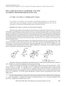
Fig. 2. Effect of different concentrations of acetic acid at different pH values on the growth of B. subtilis. Increasing concentrations of 1% acetic acid, 8.7, 17.4, and 34.9 µM, were added to Mes/HEPES/Tris buffer adjusted to pH values of 4.6, 5, 5.5, and 6 in a final volume of 125 µl and incubated for 2 h at 37°C with B. subtilis. The initial bacterial inoculum was 5.0 3 103 CFU ml21. Results are the mean values of three independent experiments.
intracellular pH of B. subtilis was observed at fixed extracellular pH values of 4.6 and 5. The penetration of acetic acid decreased, and therefore the fluorescence uptake was higher at pH above 5. No change in the fluorescence uptake was found at any pH value for HCl (Fig. 4).
Discussion The present work was aimed at study of the surprising and previously unreported bactericidal effect of ADP on B. subtilis. Our results showed that this effect was
concentration dependent, increased at external pH values below 5.5, and disappeared at pH values above 6. No killing effect was found for ATP or adenosine. On the other hand, acetic acid was also bactericidal for B. subtilis, whereas no killing activity was found for HCl. Neither ADP nor acetic acid inhibited the growth of S. aureus, L. monocytogenes, or E. coli. We propose that the bactericidal activity of ADP and acetic acid was due to the decrease in the bacterial intracellular pH promoted by the intracellular dissociation of ADP or acetic acid, both organic acids. We demonstrated the drop in pH, using BCECF-AM. We
64
CURRENT MICROBIOLOGY Vol. 34 (1997)
Fig. 3. Effect of different acids at different pH values on the growth of B. subtilis. 17.4 µM of acetic acid and 1.01 µM of HCl were added to Mes/HEPES/Tris buffer adjusted to pH values of 4.6, 5, and 6 in a final volume of 125 µl and incubated for 1 h at 37°C with B. subtilis. The initial bacterial inoculum was 6.0 3 103 CFU ml21. Results are the mean values of three independent experiments.
found that the intracellular pH of B. subtilis fell to pH values below 5.5 after incubation with acetic acid and ADP, but not with HCl. This effect was almost instantaneous (appeared in less than 5 min of incubation). The common link between ADP and acetic acid is the fact that both are weak organic acids (pK values of 3.9 and 4.7 for ADP and acetic acid, respectively). That means that, following the Henderson-Hasselbach equation, at pH 4.6, 16.6% of ADP and 44.5% of acetic acid are not dissociated and therefore can pass through the B. subtilis bacterial membrane, while at pH 5 only 7.4% of ADP and 33.5% of acetic acid are not dissociated. This fact can explain that a high concentration of ADP (50 µM) is needed to observe a complete bactericidal effect on B. subtilis, while for acetic acid the killing concentration is lower (17.4 µM). On the other hand, HCl, a strong acid, is completely dissociated at pH 4.6 and therefore cannot be found in the protonated state in significant amount. We were surprised that the bactericidal effect was specific for ADP and not for ATP or adenosine. Adenosine (9-a-ribofuranosyladenine) and, therefore, the purine base adenine do not seem to play a role in the killing of B. subtilis, and probably the first phosphate group of ATP dissociates at lower pH values than that of ADP and therefore is negatively charged at pH 4.6 and cannot enter in the bacteria. We are uncertain why ADP and acetic acid are toxic for B. subtilis but not for the other organisms we tested. It is a well-known fact that bacteria that maintain a high intracellular pH, near neutral, such as E. coli, are
sensitive to fermentation acids when the extracellular pH drops [2, 3]. The flux of undissociated acid, lipid soluble when protonated, into the cell and the release of protons in the more alkaline interior dissipate the chemical gradient of protons (ZDpH). On the other hand, fermentative bacteria that maintain a small ZDpH with declining pH, such as Streptococcus bovis, are not sensitive to acetate at low pH. Streptococcus bovis has been shown to grow at pH 4.5 in the presence of 100 mM acetate [4, 8, 12, 13]. It is surprising that in our system we did not observe any growth inhibition of E. coli due to ADP or acetic acid. We did, however, observe a clear bactericidal effect of small amounts of these weak acids (50 µM ADP, 17.4 µM acetic acid) on B. subtilis. This effect is even more surprising knowing that B. subtilis has one of the highest buffering capacities of any bacterium, 400–1000 nmol of H1/pH unit per mg of protein [3, 9]. One possibility is that B. subtilis may have a selective transport mechanism to facilitate the passage of acetic acid through the bacterial membrane at pH values near 7. At pH values near the pK (4.7), a massive entry of not dissociated acetic acid could occur, decrease the intracellular pH, and kill the bacteria. It is also possible that ADP may be transported via an ADP/ATP translocator in B. subtilis. E. coli, a Gram-negative rod, may lack this translocator. One of the main criticisms of our paper is the use of a fluorescent probe to measure intracellular pH in B. subtilis. Previous unsuccessful assays with radioactively labeled benzoate [15] and BCECF led us to use
65
V. Asensi et al.: ADP and Acetic Acid Kill Bacillus subtilis
acids, including ADP and acetic acid, have an in vitro bactericidal effect on B. subtilis . The mechanism of this killing effect is a change in the intracellular pH. More experiments are needed to determine if the killing mechanism is finally due to a break in the cell wall or to a blockade of the respiratory cycle of the bacteria mediated by the action of these weak acids as uncouplers, as suggested by Russell [14]. ACKNOWLEDGMENTS We are most grateful to Dr. P. Setlow from Connecticut University, USA, and to Dr. R. van Veen and Dr. W. Konings from Groningen University, Holland, for useful information regarding the measurement of the intracellular pH of B. subtilis. Fig. 4. Changes in the internal pH of B. subtilis after acetic and HCl treatment. 10.4 µM of acetic acid and 0.6 µM of HCl acid were added to Mes/HEPES/Tris buffer in a final volume of 100 µl with a B. subtilis inoculum of 2.0 3 107 CFU sample21. Each sample was incubated for 5 min with 1 µl (0.01 µM) of BCECF-AM, washed, centrifuged, and the cell pellet resuspended in 20 µl of 50 mM potassium phosphate buffer, pH 7.0, and measured in a fluorescence spectrophotometer as described in Materials and Methods. Results of fluorescence are expressed as percentage of fluorescence uptake of the acid-treated test sample compared with the untreated control in Mes/HEPES/Tris buffer at different pH values. Results are the mean values of three independent experiments.
BCECF-AM instead [10]. Fluorescent probes can have some problems measuring intracellular pH. The cytoplasmic membrane of bacteria is very permeable to the fluorescent probe carboxyfluorescein, and the resultant signal depends on a number of factors: intracellular pH, pH gradient, the intracellular pH optimum of the esterases in the cytoplasm, and the concentration of released probe at the instant of measurement, and therefore it is difficult to interpret the significance of the signal [17]. On the other hand, with our technique we measure only changes in the intracellular pH rather than quantitative pH values. ADP (200 µM) and thrombin have been classically used to induce platelet aggregation and release of b-lysin and other platelet microbicidal proteins [5, 16, 18]. If ADP is used and the b-lysin assay is made in physiologic saline (pH 5.5) as described originally by Donaldson et al. [6], the killing activity of B. subtilis could be falsely attributed to the platelet protein when it is an ADPmediated effect. Therefore, ADP is not recommended for b-lysin release and purification unless a solvent with a pH near 7, such as phosphate-buffered saline (PBS), is used in the dilutions of the assay. In summary, weak organic
Literature Cited 1. Asensi V, Fierer J (1991) Synergistic effect of human lysozyme plus ampicillin or b-lysin on the killing of Listeria monocytogenes. J Infect Dis 163:574–578 2. Bakker EP, Mangerich WE (1982) The effects of weak acids on potassium uptake by Escherichia coli K-12, inhibition by low cytoplasmic pH. Biochim Biophys Acta 730:379–386 3. Booth IR (1985) Regulation of cytoplasmic pH. Microbiol Rev 49:359–378 4. Cook GM, Russell JB (1994) The effect of extracellular pH and lactic acid on pH homeostasis in Lactococcus lactis and Streptococcus bovis. Curr Microbiol 28:165–168 5. Czuprinski CJ, Balish E (1981) Interaction of rat platelets with Listeria monocytogenes. Infect Immun 33:103–108 6. Donaldson DM, Ellsworth B, Matheson A (1964) Separation and purification of b-lysin from normal serum. J Immunol 92:896–901 7. Johnson FB, Donaldson DM (1968) Purification of staphylocidal b-lysin from rabbit serum. J Bacteriol 96:589–595 8. Kashkett ER (1987) Bioenergetics of lactic acid bacteria: cytoplasmic pH and osmotolerance. FEMS Microbiol Rev 46:233–244 9. Krulwich TA, Agus R, Scheier M, Guffanti AA (1985) Buffering capacity that grows in different pH ranges. J Bacteriol 162:768–772 10. Magill NG, Cowan AE, Koppel DE, Setlow P (1994) The internal pH of the forespore compartment of Bacillus megaterium decreases by about 1 pH unit during sporulation. J Bacteriol 176:2252–2258 11. Molenaar D, Abee T, Konings WN (1991) Continuous measurement of the cytoplasmic pH in Lactococcus lactis with a fluorescent pH indicator. Biochim Biophys Acta 1115:75–83 12. Russell JB (1991a) Resistance of Streptococcus bovis to acetic acid at low pH: relationship between intracellular pH and anion accumulation. Appl Environ Microbiol 57:255–259 13. Russell JB (1991b) Intracellular pH of acid-tolerant ruminant bacteria. Appl Environ Microbiol 57:3383–3384 14. Russell JB (1992) Another explanation for the toxicity of fermentation acids at low pH: anion accumulation versus uncoupling. J Appl Bacteriol 73:363–370 15. Russell JB, Hino T (1985) Regulation of lactate production in Streptococcus bovis: a spiraling effect that leads to rumen acidosis. J Dairy Sci 68:1712–1721
66 16. Tew TG, Roberts RR, Donaldson DM (1974) Release of b-lysin from platelets by thrombin and by a factor produced in heparinized blood. Infect Immun 9:179–186 17. Thomas JA, Kolbeck PC, Langworthy TA (1982) Spectrophotometric determination of cytoplasmic and mitocondrial pH transitions using trapped pH indicators. In: Nutticelli R, Deamer DW (eds)
CURRENT MICROBIOLOGY Vol. 34 (1997) Intracellular pH: its measurement, regulation and utilization of cellular functions. New York: Alan R Liss, Inc, pp 105–123 18. Yeaman MR, Puentes SM, Norman DC, Bayer AS (1992) Partial characterization and staphylocidal activity of thrombin-induced platelet microbicidal protein. Infect Immun 60:1202–1209