bs_bs_banner
Environmental Microbiology (2012) 14(1), 162–176
doi:10.1111/j.1462-2920.2011.02576.x
Phosphate transporters in marine phytoplankton and their viruses: cross-domain commonalities in viral-host gene exchanges emi_2576
162..176
Adam Monier,1 Rory M. Welsh,1 Chelle Gentemann,2 George Weinstock,3 Erica Sodergren,3 E. Virginia Armbrust,4 Jonathan A. Eisen5 and Alexandra Z. Worden1* 1 Monterey Bay Aquarium Research Institute, 7700 Sandholdt Road, Moss Landing, CA 95039, USA. 2 Remote Sensing Systems, 444 Tenth Street, Suite 200, Santa Rosa, CA, 95401, USA. 3 The Genome Center, Washington University School of Medicine, 4444 Forest Park Avenue, St. Louis, MO 63108, USA. 4 School of Oceanography, University of Washington, Seattle, WA 98195, USA. 5 University of California Davis, Davis, CA 95616; DOE Joint Genome Institute Walnut Creek, CA, USA. Summary Phosphate (PO4) is an important limiting nutrient in marine environments. Marine cyanobacteria scavenge PO4 using the high-affinity periplasmic phosphate binding protein PstS. The pstS gene has recently been identified in genomes of cyanobacterial viruses as well. Here, we analyse genes encoding transporters in genomes from viruses that infect eukaryotic phytoplankton. We identified inorganic PO4 transporter-encoding genes from the PHO4 superfamily in several virus genomes, along with other transporter-encoding genes. Homologues of the viral pho4 genes were also identified in genome sequences from the genera that these viruses infect. Genome sequences were available from host genera of all the phytoplankton viruses analysed except the host genus Bathycoccus. Pho4 was recovered from Bathycoccus by sequencing a targeted metagenome from an uncultured Atlantic Ocean population. Phylogenetic reconstruction showed that pho4 genes from pelagophytes, haptophytes and infecting viruses were more closely related to homologues in prasinoReceived 15 April, 2011; accepted 27 July, 2011. *For correspondence. E-mail
[email protected]; Tel. (+1) 831 775 2122; Fax (+1) 831 775 1620. Re-use of this article is permitted in accordance with the Terms and Conditions set out at http://wileyonlinelibrary.com/ onlineopen#OnlineOpen_Terms
© 2011 Society for Applied Microbiology and Blackwell Publishing Ltd
phytes than to those in what, at the species level, are considered to be closer relatives (e.g. diatoms). We also identified PHO4 superfamily members in ocean metagenomes, including new metagenomes from the Pacific Ocean. The environmental sequences grouped with pelagophytes, haptophytes, prasinophytes and viruses as well as bacteria. The analyses suggest that multiple independent pho4 gene transfer events have occurred between marine viruses and both eukaryotic and bacterial hosts. Additionally, pho4 genes were identified in available genomes from viruses that infect marine eukaryotes but not those that infect terrestrial hosts. Commonalities in marine host-virus gene exchanges indicate that manipulation of host-PO4 uptake is an important adaptation for viral proliferation in marine systems. Our findings suggest that PO4-availability may not serve as a simple bottom-up control of marine phytoplankton. Introduction Marine phytoplankton play a major role in global photosynthesis and CO2 capture from Earth’s atmosphere (Field et al., 1998). Phytoplankton populations are controlled by grazing and viral mortality as well as nutrient availability and other biological and physico-chemical factors. How these different forces control individual taxa remains poorly understood, see, e.g. (Azam et al., 1983; Rohwer and Thurber, 2009). A major challenge for understanding eukaryotic phytoplankton controls is their tremendous diversity and the fact that many remain uncultured. The smallest of these are the picoeukaryotes (ⱕ 2–3 mm diameter), a broad assemblage composed of picoprasinophytes, small haptophytes and stramenopiles (Worden and Not, 2008; Shi et al., 2009; Cuvelier et al., 2010). Picoprasinophytes in the order Mamiellales are widespread and genome sequences are available for two common genera, Ostreococcus (Derelle et al., 2006; Palenik et al., 2007) and Micromonas (Worden et al., 2009), but not yet for Bathycoccus. Ostreococcus is seen in tropical and temperate waters while Bathycoccus and Micromonas extend to polar systems (Not et al., 2005; Worden et al., 2009). While complete genome sequences
Phosphate and commonalities in marine viral-host exchanges are not yet available for haptophyte and stramenopile picoplankton, the picoprasinophyte genomes provide information on the molecular underpinnings of nutrient uptake and insights on the relative importance of bottom-up controls. Concentrations of the major nutrients nitrate, ammonium and phosphate (PO4) vary seasonally in the euphotic zone where they influence the dynamics and successional patterns of resident phytoplankton communities. PO4 depletion in particular is thought to cause intense competition between different taxa in the North Atlantic Ocean and to have influenced gene content in different strains of the picocyanobacteria Prochlorococcus and Synechococcus through selective adaptations (Scanlan et al., 2009). Indeed, Prochlorococcus populations were recently reported to have different frequencies of PO4-uptake related genes depending on whether they were from the North Pacific or the North Atlantic Gyre characterized by much lower PO4 concentrations (Coleman and Chisholm, 2010). Similar results were reported for the SAR11 clade, a lineage of widely distributed marine heterotrophic bacteria. These findings were hypothesized to reflect the influence of water mass characteristics on the gene repertoires of resident taxa (Coleman and Chisholm, 2010). The two main types of inorganic PO4 transporters in cultured microbial taxa are encoded by genes in the PHO4 superfamily (Pfam PF01384) and Pst genes. Many eukaryotes and bacteria encode PHO4 superfamily members. This superfamily includes high- and low-affinity inorganic phosphate transporters. In eukaryotic phytoplankton a pho4 gene has been identified in expressed sequence tags from the haptophyte Emiliania huxleyi (Dyhrman et al., 2006a) as well as in the genome sequence of a virus that infects it, EhV-86 (Wilson et al., 2005). Comparative analysis of putative transporterencoding genes in Micromonas and Ostreococcus indicate that PHO4 superfamily members are present in these taxa as well (Worden et al., 2009), but their relatedness and PO4 affinities are not yet known. The marine picocyanobacteria use the Pst system, a multi-unit highaffinity inorganic PO4 transport system that depends on the high-affinity periplasmic PO4-binding protein encoded by the gene pstS/phoS (Diaz et al., 2005). PstS homologues are present in all sequenced Prochlorococcus and Synechococcus genomes, sometimes in multiple copies (Martiny et al., 2006; Scanlan et al., 2009). Interestingly, some marine cyanophages also encode pstS genes and the presence of pstS in cyanophage genomes appears to be linked to whether the phage source waters were PO4deplete or not (Sullivan et al., 2005; Sullivan et al., 2010). Phage-mediated gene transfer has been hypothesized to be responsible for the fact that many PO4-uptake related genes in Prochlorococcus ecotypes do not appear to follow the ‘species’ tree (Martiny et al., 2006).
163
Here, we analyse phosphate and other transporterencoding genes in published genomes from viruses that infect eukaryotic phytoplankton. Ecological and evolutionary aspects of PHO4 superfamily members were investigated in the viruses and their hosts, including a ‘wild’ Bathycoccus population. The distribution and diversity of this transporter gene was evaluated in metagenomes from a North Pacific Ocean transect and other marine environments.
Results and discussion Virally encoded transporter genes We analysed viruses infecting unicellular and/or marine eukaryotes for transporter-encoding genes, including the pho4 gene previously reported in EhV-86 (Wilson et al., 2005). Analysis of open reading frames (ORFs) in published viral genomes showed that several eukaryotic viruses encoded pho4 genes, as well as other putative transporter genes (Table 1). Apart from the amoebainfecting mimivirus, all of these viruses belong to the Phycodnaviridae (Wilson et al., 2005; Derelle et al., 2008; Weynberg et al., 2009; 2011; Moreau et al., 2010), a family of nucleocytoplasmic large double-stranded DNA viruses. Genes from the different viruses that encoded PHO4 superfamily members had higher overall similarities to each other (ranging from 59% to 94% at the aminoacid level) than did the genes that encoded each of the other transporter types identified in the viral genomes (Table 1). Apart from EhV-86, the other identified pho4 genes were in Mamiellales-infecting viruses. Specifically, they were found in one of the two viruses infecting Bathycoccus, two of the four infecting Ostreococcus but not in MpV-1, which infects Micromonas. Some of these Mamiellales-infecting viruses contained the pho4 gene but no other known transporter genes (Table 1). Although genomic data are limited, it is possible that the presence or absence of pho4 genes reflects the influence of the environment, or environmental stresses acting on their hosts, in shaping viral genomes. For example, the pho4 gene-encoding virus OtV-2 infects Ostreococcus RCC393 (Weynberg et al., 2011), a strain that belongs to an Ostreococcus clade (OII) known to inhabit warm, oligotrophic waters where PO4 is often depleted (Demir-Hilton et al., 2011). In contrast, the Ostreococcus viruses OtV-1 and OtV-5 do not encode pho4 and their host, O. tauri, appears to be restricted to higher nutrient systems, such as bays, lagoons and brackish waters. Two different Bathycoccus viruses, BpV-1 and BpV-2, were isolated against the same Bathycoccus strain (RCC1105), but the source waters the viruses came from were collected at different times of the year (fall and winter). Hence, different nutrient conditions associated with seasonal changes
© 2011 Society for Applied Microbiology and Blackwell Publishing Ltd, Environmental Microbiology, 14, 162–176
164 A. Monier et al. Table 1. Transporter and phoH sequences detected in all published Mamiellales viruses, other representative eukaryotic viruses and mimivirus (as an outgroup). Transporter Virus Phycodnaviridae BpV-1 BpV-2 MpV-1 OtV-1 OtV-2 OtV-5 OlV-1 PbCV-1 EhV-86 EsV-1 FsV Mimivirus AcpV-1
Other
Host genus
PHO4
ABC
MC
MFS
VIC
phoH
Bathycoccus Bathycoccus Micromonas Ostreococcus Ostreococcus Ostreococcus Ostreococcus Chlorella* Emiliania Ectocarpus Feldmannia
YP_004061633 – – – YP_004063655 – YP_004061866 – YP_002296186 – –
– – – – – – – NP_049022 – – –
– – – – – – – – – – –
– – – – – – – – YP_293932 – –
YP_004061440 ADQ91178 YP_004062056 – – – – NP_048599 – NP_077708 –
YP_004061453 ADQ91193 YP_004062114 YP_003494870 YP_004063457 YP_001648107 YP_004061669 – – – –
Acanthamoeba*
–
YP_003987262
YP_003986777
–
–
–
The asterisk indicates a non-marine host; – indicates not found. Accessions provided were retrieved by blasting predicted ORFs against GenBank NR. PHO4, inorganic phosphate transporter family (PiT); ABC, ATP-binding cassette superfamily; MC, mitochondrial carrier family; MFS, the major facilitator superfamily; VIC, the voltage-gated ion channel family; phoH, phosphate starvation induced ATPase. BpV-1/2, Bathycoccus RCC1105 viruses 1 and 2; MpV-1, Micromonas pusilla virus 1; OtV-1/2/5, Ostreococcus tauri viruses 1, 2 and 5; OlV-1, Ostreococcus lucimarinus virus 1; PbCV-1, Paramecium bursaria Chlorella virus 1; EhV-86, Emiliania huxleyi virus 86; EsV-1, Ectocarpus siliculosus virus 1; FsV, Feldmannia sp. virus; mimivirus, Acanthamoeba polyphaga mimivirus.
may explain the fact that only BpV-1 encodes the pho4 gene. Different Micromonas clades also vary along environmental gradients (Foulon et al., 2008) and the distribution of the strain infected by MpV-1 is unknown. Lack of pho4 in MpV-1 is however consistent with Micromonas being most abundant in coastal settings, at least in temperate and subtropical systems (Foulon et al., 2008), that are usually PO4 replete. Putative functions of other virally encoded transporters were broader and more difficult to interpret ecologically. A single member of the ATP-Binding Cassette superfamily was found in the Paramecium bursaria Chlorella virus (PbCV-1, which infects a Chlorella alga that lives in a symbiotic association with P. bursaria) and one Major Facilitator Superfamily (small solute transporters) member was identified (Table 1). The latter was found in EhV-86 and an apparently unrelated Major Facilitator Superfamily member has been reported in a dsDNA virus infecting the moth Helicoverpa zea. A putative ion channel transporter gene (belonging to the VIC Superfamily) was detected in several of the Mamiellales viruses (Table 1). A homologue of this gene was first reported in PbCV-1 where it was shown to act as a potassium selective channel and appeared to be essential to the viral life cycle (Plugge et al., 1999). It was later described in EsV-1, which infects the multicellular brown alga Ectocarpus siliculosus (Chen et al., 2005). Members of the Cation Channel-forming Heat Shock Protein-70 family were also identified in the Bathycoccus viruses (YP_004061438, ADQ91175) and mimivirus (YP_003986897, YP_
003986752). A putative ATPase-encoding gene, phoH, was also identified in most of the viral genomes (Table 1, see below). The only viral sequence included in the full PHO4 Pfam alignment at the time of our analysis was from EhV-86. Pho4 was not present in the 50 nucleocytoplasmic large double-stranded DNA viruses (Asfarviridae, Iridoviridae, Mimiviridae, Phycodnaviridae and Poxviridae) and 46 herpesvirus genomes sequences available when that Pfam alignment was generated. The majority of these viruses infect terrestrial hosts including mammals, nonmammalian vertebrates and invertebrates. Furthermore, pho4 genes were not detected in newly available genomes from viruses that infect other hosts, including non-marine unicellular eukaryotes. In environments like the mammalian cellular milieu or many freshwater systems, PO4 is not considered limiting and therefore selection pressure favouring viruses that encode a PO4 transporter (or retention of this gene) is presumably weak. In contrast, manipulation of PO4-uptake in marine hosts that often encounter PO4-deplete conditions could ensure enough PO4 is available for viral replication. Origins of virally encoded pho4 genes Many previous studies have suggested that viruses acquire genes from their hosts (studies by, for example, Monier et al., 2007; Sharon et al., 2009; Colson and Raoult, 2010). We screened eukaryotic algal genomes for the pho4 gene, including hosts for the analysed viruses. In
© 2011 Society for Applied Microbiology and Blackwell Publishing Ltd, Environmental Microbiology, 14, 162–176
Phosphate and commonalities in marine viral-host exchanges
165
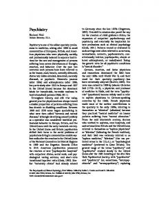
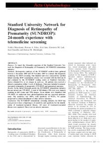
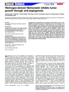
Fig. 1. Atlantic Ocean site (pink star) from which a natural population of Bathycoccus was sorted and sequenced. Ocean colour represents Aqua MODIS Atlantic Ocean chlorophyll concentration data (mg m-3) from 1 May to 5 September 2006, spanning the period of sample collection; black indicates land and white indicates missing data. Lower chlorophyll concentrations reflect lower phytoplankton biomass and occurs in regions with low nutrient availability. (Inset) phosphate (blue) and nitrate plus nitrite (yellow) concentrations as well as in vivo fluorescence from chlorophyll (green) at the sort site. The Y-axis represents depth (m). Arrow indicates the depth from which Bathycoccus was sorted.
symbiont or other material may have been present (Table S1). Therefore, only scaffolds with ⱖ half of predicted ORFs with best BLASTp hits to predicted protein sequences in other Mamiellales genomes (i.e. Micromonas and Ostreococcus), that fit other criteria as well (see Experimental procedures), were considered derived from the Bathycoccus nuclear genome and used in subsequent analyses. Two distinct G + C signatures were observed in the Bathycoccus nuclear metagenome similar to what has been seen in whole genome sequences from cultured members of the Mamiellales. The ‘anomalously lowG + C’ region of 38% G + C was about 10% lower than the overall metagenome average of 48%. These regions of considerably lower G + C content were first observed in O. tauri (58% for the whole genome and 52% for the low G + C region) and Micromonas sp. RCC299 (64% and 50% respectively) and have been hypothesized to serve as a sex chromosome that encodes convergent, overlapping genes (Derelle et al., 2006; Worden et al., 2009). The
addition to the previously described E. huxleyi pho4 gene (Dyhrman et al., 2006a), genes containing the PHO4 Pfam domain were found in Aureococcus anophagefferens (a pelagophyte) and all genome sequenced Ostreococcus strains (O. tauri, O. lucimarinus and Ostreococcus RCC809). They were also found in Micromonas RCC299 and CCMP1545 but with low similarity to the Ostreococcus pho4 genes (see discussion below). To better understand the distribution of pho4 genes in the Mamiellales and potential gene exchanges between viruses and hosts, we generated and screened a targeted metagenome for Bathycoccus. A natural population was sorted by flow cytometry (Fig. S1) from PO4-deplete waters (Fig. 1, Table 2) and the 18S rDNA clone library built from the sorted and multiple displacement amplification (MDA)-amplified material was composed solely of Bathycoccus sequences (Fig. S2; 99% nucleotide identity to Bathycoccus prasinos BLA77). Results for 16S rDNA clone sequences were less clear and an unknown endo-
Table 2. Summary of water samples from which metagenomes were sequenced and corresponding metadata. Site Atlantic Sta. 13 Sort Pacific H3 67–70 67–155 67–155
Date (d/m/y)
Location (lat.; long.)
Depth (m)
T (°C)
S (ppt)
PO4 (mM)
NO3 (mM)
NH4 (nM)
Chl a (mg m-3)
12/07/06
12.378; -27.241
64
21.76
35.89
0.04
0.20
na
na
10/10/07 09/10/07 07/10/07 06/10/07
36.740; 36.129; 33.286; 33.286;
5 10 5 86
12.28 15.57 19.02 13.39
33.47 33.12 33.19 33.13
1.121 0.609 0.655 0.579
8.860 0.511 0.013 0.397
na na 15 19
4.1966 2.7156 0.0998 0.9398
-122.020 -123.490 -129.428 -129.428
For Atlantic samples reported NO3 measurements reflect NO3 + NO2. T, temperature; S, salinity.
© 2011 Society for Applied Microbiology and Blackwell Publishing Ltd, Environmental Microbiology, 14, 162–176
166 A. Monier et al.
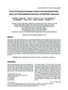
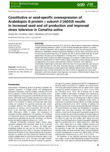
Fig. 2. Phylogeny of eukaryote and eukaryotic virus pho4 protein sequences, including the pho4 gene from the Bathycoccus targeted metagenome. While most prasinophyte gene sequences branched together in the ‘green-clade’, the Micromonas pho4 homologues were phylogenetically distant and not close to this region of the eukaryotic pho4 gene tree. Pho4 sequences retrieved from traditional metagenomes (fuchsia and yellow bubbles) were assigned to branches on this reference tree using pplacer. This maximum-likelihood tree was reconstructed using phyML and the WAG matrix. Bootstrap values represent percentage of 100 replicates. Only metagenomic reads mapped with support (P ⱖ 0.75) are shown. For balance in taxon sampling only one (EGB10629) of three (EGB10629, EGB08825, EGB12603) Aureococcus pho4 gene sequences that branched within this region of the tree with statistical support in a preliminary phylogenetic reconstruction (not shown) was included. These three versions ranged from 66% to 72% similarity at the amino-acid level.
specificity of recovered ORFs, as well as detection of low G + C scaffolds with a differential from the overall average within range of that observed in Ostreococcus and Micromonas, indicating that the assembled and filtered metagenome is specific to the Bathycoccus nuclear genome. A pho4 gene was identified in the Bathycoccus metagenome, with highest amino-acid sequence similarity to that of Ostreococcus (65%, O. lucimarinus XP_001422167). Similarities between pho4 genes from these Mamiellales and those in Micromonas were low (Bathycoccus had 29% similarity to Micromonas RCC299 ACO69637 and 27% to M. CCMP1545 XP_003062965). Surprisingly, the similarities between pho4 from Ostreococcus or Bathycoccus were lower to Micromonas than to Tetraselmis chui (50% similarity with Bathycoccus), a more distant prasinophyte. Bathycoccus and Ostreococcus also appeared more closely related to each other than to Micromonas based on 18S rDNA analyses (Fig. S2), see also (Guillou et al., 2004; Worden, 2006). Recovery of pho4 genes from host genera and other eukaryotes allowed us to construct a robust phylogeny for the corresponding viral and host pho4 protein sequences. BpV-1, OlV-1 and OtV-2 pho4 sequences branched with those of Bathycoccus and Ostreococcus while the EhV-86 pho4 gene branched with its host E. huxleyi (Fig. 2). The analysis suggests that multiple horizontal gene transfer (HGT) events occurred –
although the direction of those events is still unclear (whether from host to virus, or virus to host). Phylogenetic analysis supports the hypothesis that the EhV-86 and Ostreococcus viral pho4 genes were derived from their respective host genera, rather than a common viral ancestor. Still, other gene exchange scenarios are possible. For instance, the exchange could have occurred from host to virus, virus to virus and then virus to another host lineage, explaining the presence of ‘green’like pho4 genes in, e.g. the haptophytes. The pattern of multiple exchanges, resulting in an indirect virally mediated exchange of host genes to another host has been hypothesized for the photosynthetic reaction centre genes psbA and psbD in cyanobacteria and cyanophages (Sullivan et al., 2006). Two pho4 genes from Ostreococcus viruses branched together and have higher similarity (across all positions) to one another than to homologues from Ostreococcus itself. A pho4 sequence is not available from Ostreococcus RCC393, the host strain against which OtV-2 was isolated, although a sequence from another Ostreococcus Clade OII member (Ostreococcus RCC809) was included. The fact that OlV-1 and OtV-2 sequences branched together could indicate they originated from a common ancestor rather than from their respective hosts. Without an OtV-2 host-encoded pho4 sequence in the tree it is not possible to determine if placement of the OtV-2 version would differ if the host sequence was included. Finally,
© 2011 Society for Applied Microbiology and Blackwell Publishing Ltd, Environmental Microbiology, 14, 162–176
Phosphate and commonalities in marine viral-host exchanges host cross-infectivity levels of Phycodnaviridae family members are not well known and could influence acquisition and retention patterns. Horizontal gene transfer events similar to those hypothesized here for the pho4 gene in marine systems have been reported for E. huxleyi and EhV-86 sphingolipid biosynthesis genes. Sphingolipid biosynthesis genes were hypothesized to have been acquired by the virus from its host (Monier et al., 2009) and viral glycosphingolipid molecules have been shown to be synthesized by the haptophyte during infection (Vardi et al., 2009). These molecules have been detected in the natural environment and are possibly related to bloom dynamics of the host (Vardi et al., 2009). In contrast to the broad distribution of pho4 genes in viruses that each infect different marine eukaryotic taxa or lineages, the hypothesized sphingolipid pathway HGT has so far only been reported between E. huxleyi and EhV. In addition to the pho4 homologues present in various algae and their viruses, we found a close homologue of the BpV-1 and BpV-2 encoded Cation Channel-forming hsp70 family gene in the Bathycoccus metagenome (scaffold C503; this gene is also present in O. lucimarinus and Micromonas RCC299), suggesting this gene may have been acquired through HGT and is sufficiently advantageous to be retained by the recipient genome. Homology has been reported between 11 predicted genes in OtV-1 and host-encoded (O. tauri) genes (Weynberg et al., 2009). Studies exploring the expression of virally encoded pho4 genes and other putatively exchanged genes should enhance our understanding of viral impacts on oceanic phytoplankton as well as environmental factors controlling viral proliferation. Phylogenetic relationships between algal pho4 genes and implications for function The phylogenetic relationships of phytoplankton pho4 homologues inferred from the protein sequences (Fig. 2) were inconsistent with ‘species phylogenies’ based on 18S rDNA and chloroplast gene trees (Andersen, 2004; Keeling et al., 2005; Worden and Not, 2008). In our analysis, a ‘green-clade’ consisting of pho4 homologues from the prasinophytes Bathycoccus, Ostreococcus and T. chui formed a sister group to other members of the Plantae, especially green algae. However, this ‘greenclade’ also included pho4 homologues from stramenopiles (E. siliculosus and A. anophagefferens) and the haptophyte E. huxleyi. High representation of green algal or plant-like genes in haptophytes has been reported previously (Hampl et al., 2009; Cuvelier et al., 2010). Interestingly, E. siliculosus appeared to only have the ‘green’ pho4 gene-version and Aureococcus (Gobler et al., 2011) had multiple versions, three of which were
167
‘green’-like (Fig. 2). In contrast, pho4 genes from the diatoms Thalassiosira pseudonana and Phaeodactylum tricornutum (Armbrust et al., 2004; Bowler et al., 2008) grouped together in a position outside of the ‘greenclade’, as would be expected based on their evolutionary history inferred by chloroplast gene trees. Anomalous phylogenetic patterns have also been reported for phytoplankton ammonium transporters (McDonald et al., 2010). However, in most cases, each different picoprasinophyte AMT type (i.e. forming distinct clades within the AMT-gene tree) was present in all of the sequenced picoprasinophyte genomes. In contrast, despite the presence of ‘green’-like PHO4 superfamily members in disparate lineages, this version was not found in the picoprasinophyte Micromonas; their pho4 genes branched in an unsupported position basal to metazoans (not shown). There are several possible explanations for the observed phylogenetic distribution of pho4 genes in eukaryotic algae. The fact that the stramenopile and haptophyte pho4 homologues grouped with green algae may have been influenced by taxon undersampling, particularly the lack of available homologues from red algae apart from C. merolae. However, it may also reflect ancestral characteristics with differential loss or divergence in some taxa (e.g. diatoms) or gain from a proposed cryptic green algal endosymbiont in the chromalveolates (Moustafa et al., 2009). Alternatively, HGT events may have occurred between distantly related organisms, possibly involving virally mediated exchange, leading to the observed phylogenetic relationships. HGT events have been reported between distant eukaryotic organisms, e.g. carotenoid production related genes in fungi and aphids (Moran and Jarvik, 2010). All of the pho4 genes within the ‘green-clade’ that have been experimentally characterized serve as highaffinity transporters. The T. chui encoded protein (AAO47330) acts as a high-affinity PO4 permease and the gene is specifically upregulated in P-deplete conditions (Chung et al., 2003). Its branching position was basal to pho4 genes from several of the algae and viruses analysed, suggesting it represents a more ancestral version of the gene (Fig. 2); it showed a high degree of similarity to the algae at terminal nodes of the ‘green-clade’. The E. huxleyi pho4 gene is expressed under PO4 depletion (Dyhrman et al., 2006a) and similar results have been reported for an Aureococcus version (Wurch et al., 2011), both of which are embedded within this clade (Fig. 2). Thus, we hypothesize that the other algal pho4 genes branching within this region of the tree are also high-affinity PO4 transporters. Notably, in the Ostreococcus viruses, a 54-amino-acid segment was missing from the pho4 gene that is present in host versions. In the same region 19 amino acids were absent
© 2011 Society for Applied Microbiology and Blackwell Publishing Ltd, Environmental Microbiology, 14, 162–176
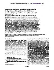
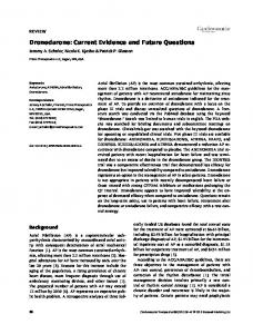
168 A. Monier et al. Fig. 3. A. Sample sites for Pacific Ocean metagenomes and satellite inferred chlorophyll a concentrations (mg m-3) over a period spanning the cruise (14 September–8 November 2007). Black indicates land, white missing data, pink symbols indicate sample sites. B. Phosphate (blue) and nitrate (yellow) concentrations as well as in vivo fluorescence (green) at sample collection sites.
from BpV-1 that were present in the host version. How these deletions affect function is still unclear; deletions are seen in this region in some eukaryotic taxa as well, although involving fewer amino acids. Most of the eukaryotic viral genomes analysed herein also encoded the gene phoH (Table 1). Although not a transporter this is relevant because phoH is found in the PHO (phosphate) regulon of E. coli and other bacteria, although its precise role is unclear (see discussions in Martiny et al., 2006; Sullivan et al., 2010). We identified phoH homologues in all available Mamiellales-virus genomes (OtV-1, OtV-2, OtV-5, OlV-1, BpV1, BpV-2 and MpV-1) but not EhV-86 (Table 1). Unlike the phoH genes in cyanobacteria and cyanophages (present in both hosts and viruses) or pstS and pho4 genes, which were found in all sequenced hosts, phoH genes appeared to be present only in the O. tauri and E. huxleyi genomes, based on BLASTp and tBLASTn similarity searches against available proteomes and genomes. In addition, the O. tauri-encoded version did not appear to be closely related to versions in viruses infecting Ostreococcus.
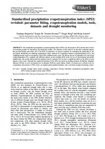
PHO4 superfamily members in the natural environment We scanned metagenomes from three Pacific Ocean environments (Fig. 3), coastal (high PO4), mesotrophic (moderate PO4) and oligotrophic (moderate PO4 and low nitrogen) for members of the PHO4 superfamily (Table S2a) as well as Global Ocean Sampling (GOS) data (Rusch et al., 2007) using a HMM (Fig. S3, Table S2b). The detected PHO4 sequences were assigned to branches on a fixed maximum-likelihood reference tree built from the overall Pfam alignment (Fig. 4). A large fraction of environmental sequences were assigned to two groups containing Gammaproteobacteria. One of these groups contained sequences from cultured Shewanella strains and some cyanobacterial gene sequences (Arthrospira and Synechococcus WH5701) branched in the same region of the tree. WH5701 is a halotolerant strain and some environmental sequences assigned to this group might be from the nitrogen fixing cyanobacterium Crocosphaera. The Crocosphaera gene model containing the PHO4 domain (ZP_00517140) appeared to be missing a section of the conserved C-terminal domain that is part of
© 2011 Society for Applied Microbiology and Blackwell Publishing Ltd, Environmental Microbiology, 14, 162–176
Phosphate and commonalities in marine viral-host exchanges
169
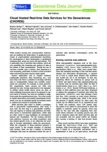
Fig. 4. Metagenomic sequences assigned to branches on a maximum-likelihood tree built from the PHO4 Pfam alignment. Squares represent (yellow) GOS sequences and (fuchsia) metagenomic sequences containing the PHO4 Pfam model (see Experimental procedures) from our Pacific Ocean transect (Fig. 3). Two hundred and forty and 2751 putative PHO4 sequences were detected in the Pacific metagenomes and GOS data respectively using our HMM. Due to the number of sequences in the Pfam alignment and computational time needed, bootstrap analysis was not performed for the maximum-likelihood reference tree.
the PHO4 Pfam model and was therefore not included in the tree, but had high similarity to the WH5701 version (ZP_01085811; 69% identity at the amino-acid level). Prochlorococcus and Synechococcus do not appear to encode genes containing the PHO4 Pfam domain. Environmental sequences were assigned to other bacterial groups as well (Fig. 4). The PHO4 Pfam model is composed of both high- and some low-affinity transporters and affinities could not be inferred based on placement in the overall Pfam analysis (Fig. 4). Although there appear to be numerous bacterial pho4 homologues in environmental community DNA (Fig. 4), if bacteriophages exchange this gene with their hosts, as eukaryotic viruses appear to, then any number might belong to phage rather than bacteria. Indeed, the flanking region of a Sanger sequenced shotgun clone from the Pacific Ocean deep chlorophyll maximum (DCM, 86 m) metagenome, recognized by our PHO4 superfamily HMM, indicated it was phage derived. In our phylogenetic analysis of this clone, the pho4-encoding ORF branched in an unsupported position with several bacterial taxa, close to the marine alphaproteobacterium
strain HIMB59 (Fig. 5). In contrast, the flanking ORF in this cloned sequence encoded a hypothetical protein so far only seen in cyanophages. Three hundred and nineteen environmental sequences were assigned to branches within the region of the PHO4 superfamily tree containing cultured eukaryotic algae, the Bathycoccus targeted metagenome pho4 gene sequence and the viral versions discussed above (Fig. 4). These environmental sequences were then assigned placements on the refined-eukaryotic pho4 tree (Fig. 2), which included more positions than the superfamily tree. The majority of eukaryotic environmental sequences grouped with prasinophytes, stramenopiles, haptophytes or corresponding viruses. A large number of those assigned to the Mamiellales (Bathycoccus/Ostreococcus) clade appeared to be from viral DNA templates (i.e. those assigned to OlV-1/ OtV-2 or BpV-1) rather than host-derived templates. For the haptophyte lineage, environmental sequences were not placed with E. huxleyi or its virus EhV-86, but rather branched in a position basal to the E. huxleyi-EhV-86 node, suggesting they may have originated from other
© 2011 Society for Applied Microbiology and Blackwell Publishing Ltd, Environmental Microbiology, 14, 162–176
170 A. Monier et al.
Fig. 5. Architecture of a cloned Pacific Ocean metagenome sequence and phylogeny of detected ORFs. This cloned sequence (1139 nt, JF837193) was retrieved from the DCM at Pacific Ocean Station 67–155. Maximum-likelihood phylogenetic trees were reconstructed with phyML using JTT matrix and 100 bootstrap replicates. Homologous sequences were retrieved by BLASTp searches against NCBI-NR and Moore marine microbial genomics databases. Note that taxon sampling for the hypothetical protein phylogeny is influenced by the fact that most sequenced marine phage genomes are from cyanophages.
prymnesiophyte taxa, which include widespread uncultured marine groups (Worden and Not, 2008; Liu et al., 2009; Shi et al., 2009; Cuvelier et al., 2010), or potentially their viruses. Sequenced genomes are not available for Pelagomonas (or Pelagomonas-infecting viruses); therefore, a pho4 gene from this taxon (assuming it contains the pho4 gene) was not included in our analysis. Several Pelagomonas, but not Aureococcus, SSU rDNA sequences were present in clone libraries (data not shown) from the Pacific sites (Table 2). Hence, environmental pho4 sequences assigned to the Aureococcus node were likely from its more oceanic relative Pelagomonas. The analysis showed that eukaryotic algae and viruses inhabiting both PO4 replete (Fig. 3) and deplete (Figs. 1 and S3, and some cultured taxon isolation sites) environments contained the putatively high-affinity inorganic PO4 transporter gene (Fig. 2). Given the high degree of homology between host and respective viral pho4 sequences, unambiguous assignment to either host- or infecting virus branches was not always possible, particularly for shortmetagenomic reads with limited phylogenetic information. The fact that our Pacific Ocean metagenome sequences came from larger size fractions (0.1 to ⱕ 0.8, 0.8 to ⱕ 3.0 and 3.0 to ⱕ 20 mm) as opposed to sampling protocols that
target the viral size fraction (e.g. tangential flow filtration for < 0.1 mm sized particles), indicates that the virally derived environmental sequences were present in infected hosts, especially for the two largest size fractions we sequenced. The proportion of viruses in nature that encode the pho4 gene is still not known. Broader implications In marine environments, P-availability is linked to primary production levels and shifts in phytoplankton community composition. PO4-uptake via different molecular mechanisms mitigates P-starvation and is important for survival in oligotrophic waters (e.g. Dyhrman et al., 2006b; Coleman and Chisholm, 2010). The distribution of the ‘green’-like pho4 homologues across green algae, haptophytes and some stramenopiles, indicates this putatively high-affinity transporter is important to physiological success. Together with previously hypothesized pstS HGT from cyanobacteria to cyanophages (Sullivan et al., 2010), the host-virus exchanges hypothesized herein show unexpected commonalities in the retention of PO4uptake related genes in viral genomes from both viruses that infect eukaryotic hosts and bacteriophages.
© 2011 Society for Applied Microbiology and Blackwell Publishing Ltd, Environmental Microbiology, 14, 162–176
Phosphate and commonalities in marine viral-host exchanges Experimentally, E. huxleyi viral lysis rates are reportedly higher in P-replete than P-deplete conditions (Bratbak et al., 1993). Similar observations were reported for Synechococcus grown in P-deplete conditions, in which they appear to be infected and lysed at significantly lower rates by the bacteriophage S-PM2, than in P-replete conditions (Wilson et al., 1996). The presence or absence of PO4uptake related genes in the genome of the infecting virus might mechanistically underpin these observations. For example, the S-PM2 genome does not appear to encode pstS (Mann et al., 2005), consistent with its low lysis rates in P-deplete conditions. Viral nutrient requirements and the ability of a virus, or lack thereof, to increase host nutrient uptake under limiting conditions could explain many such results. Moreover, presence of PO4-uptake related genes in a significant fraction of sequenced viral genomes, but not known nitrogen-uptake related genes, supports the hypothesis that viruses are more susceptible to PO4limitation than nitrogen limitation due to a high nucleic acid to protein ratio. The prevalence of pho4 in marine viral genomes indicates it may alleviate bottom-up control of viruses by PO4-limitation, allowing them to propagate even under low PO4 conditions. This in turn could influence phytoplankton top-down controls, enhancing the ability of eukaryotic viruses and bacteriophages alike to replicate and induce host mortality. Thus, expression and manipulation of PO4-uptake related genes presumably influence both the fitness and demise of multiple marine microbial taxa. Our data reveal evolutionary pressures felt by marine viruses and highlight the complex influences of nutrient bioavailability on microbial interactions and dynamics.
Experimental procedures Identification of transporter genes in virus genomes Transporter protein sequences and corresponding annotations were retrieved from TransportDB (Ren et al., 2007). To avoid biases resulting from different gene prediction methods we predicted ORFs for all the eukaryotic viral genomes investigated (Table 1). Viral genome sequences were downloaded from GenBank, and stop-stop ORFs with a minimum size of 60-amino-acid residues were retrieved. A combination of BLASTx and BLASTp searches against TransportDB were performed and BLAST hits having an e-value less than 0.001 were considered for further analysis. Results were controlled using BLASTp searches against NR and searches against Pfam-A using the hmmscan module of HMMer 3 (Eddy, 2009).
Environmental sample collection and contextual analyses Samples were collected using a rosette equipped with Niskin bottles, a CTD and fluorometer on two research expeditions, one aboard the R/V Seward Johnson (July 2006) and the
171
other on R/V Western Flyer (October 2007). The first cruise was in the tropical Atlantic Ocean and the sample sequenced here was collected on 12 July 2006 at Station 13 (SJ0609) from the deep chlorophyll maximum (Fig. 1, Table 2). One L of water was filtered by gravity onto a 0.45 mm pore size Supor filter until reduced (concentrated) to a volume of 10 ml, this concentrated sample was then used for flow sorting as below. The second cruise was performed in the eastern North Pacific and samples collected at three sites (Table 2). In both cases samples for phosphate and nitrate analysis were collected at multiple depths from the surface to the base of the euphotic zone. For DNA for traditional bulk metagenomic sequencing, approximately 200 l of water were filtered first through a 20 mm mesh, and then sequentially through 293 mm diameter, 3 mm pore size filters (Pall Sciences Versapor-3000T), a 0.8 mm pore size filters (Pall Sciences Supor-800) and finally a 0.1 mm pore size filters (Pall Sciences Supor-100). Surface ocean chlorophyll concentrations (Figs 1 and 3; Fig. S3) were derived from the MODIS instrument carried on NASA’s Aqua satellite (http:// oceancolor.gsfc.nasa.gov).
Metagenome templates and construction Processing for materials used to construct the targeted metagenome (sorted population) and the traditional metagenomes (size fractionated filters) was different. For the former, a 454-FLX and Sanger blended metagenomic assembly was generated for Bathycoccus. The analysed cells were first sorted by flow cytometry based on distinctive scatter and chlorophyll signals (Fig. S1) using an InFlux cell sorter (BD), directly after water collection. To avoid contamination, the instrument was cleaned extensively. Sheath fluid was 0.2 mm filtered, autoclaved PBS made in 18.2 MegaOhm H2O and 750 ml were run through the instrument after cleaning (immediately before sorting). Contamination control materials were from left and right test deflections run for 1 min (sample flow rate ~25 ml min-1). All sort droplets were immediately frozen cryogenically. Upon return to land the population sort was resorted to enhance purity levels (Fig. S1). The sample and controls were then amplified by MDA and the products debranched and precipitated prior to sequencing according to methods in Cuvelier and colleagues (2010). Universal 16S rRNA gene primers were used to verify the absence of product in controls (indicating the instrument provided no prokaryotic contamination). A combination of Sanger shotgun sequencing and 454 pyrosequencing was then performed on the DNA as detailed in Cuvelier and colleagues (2010). For Sanger sequencing, 3 Kbp shotgun libraries were constructed using debranched MDA products and end-sequencing yielded 71 328 reads (totalling 52.83 Mbp). Pyrosequencing was also performed on debranched MDA products using the Genome Sequencer FLX System (454 Life Sciences) according to the manufacturer protocol and resulted in 118 Mbp of sequence. Pyrosequence reads were assembled using the 454 Newbler assembler version 1.1.02.15 and the consensus sequence shredded into 1 Kbp shreds with 100 bp overlaps. The 454 shred data were assembled with Sanger sequences using the lucy trimmer, to remove vector and low-quality sequence, and the Paracel Genome Assembler (lucy
© 2011 Society for Applied Microbiology and Blackwell Publishing Ltd, Environmental Microbiology, 14, 162–176
172 A. Monier et al. version 1.19p, PGA version 2.6.2). The assembly process generated 1929 contigs containing 9 859 508 bp. To assess purity of the Bathycoccus sort we constructed 18S rDNA clone libraries using universal 18S rDNA primers (Moon-van der Staay et al., 2000). The resulting 741 sequences were analysed using Mothur v1.17 (Schloss et al., 2009). Sequences were first aligned against the SILVA 18S rRNA gene reference alignment (Pruesse et al., 2007). After end-trimming, clustering at 99% sequence identity resulted in six clusters and representatives of these were used for phylogenetic analysis. The maximum-likelihood tree was constructed using phyML v2.4 (Guindon and Gascuel, 2003) using an alignment where all gap-containing positions were removed and the TrNG substitution model was selected by AIC criterion (MrAIC script, distributed by J.A.A. Nylander, Uppsala University), and 100 bootstrap replicates. Because mitochondrial 16S rDNA databases are limited, BLASTn was performed against two databases, NCBI reference genomes and NCBI non-redundant nucleotide database (NT) using the 16S rDNA clones generated herein as query sequences. In order to further insure that only scaffolds originating from Bathycoccus were analysed, ORFs spanning at least 180 nucleotides (between two stop codons) were retrieved using EMBOSS GetORF (Rice et al., 2000). The resulting putative ORFs were used as BLASTp queries (Altschul et al., 1997) against the NCBI non-redundant database (NR-DB). Taxonomic information for each of the best BLASTp hit (E-value cutoff: 1e-3) was retrieved using the NCBI taxonomy database and assigned to the corresponding ORF. A scaffold was classified as Bathycoccus-like if more than half of its predicted ORFs had their best BLASTp hits (E-value cutoff: 1e-3) against a protein sequence originating from any of the five available prasinophyte genomes (Derelle et al., 2006; Palenik et al., 2007; Worden et al., 2009), including the publically available but unpublished genome from Ostreococcus RCC809. That is, at least three ORFs had to be detected on the scaffold, and two or more of these had to have best BLASTp hits to prasinophytes in order to be included in subsequent analyses. Additionally, scaffolds smaller than 7 kb were discarded. Of 10 Mb total assembly, scaffolds composing 7.1 Mb fit the criteria of more than half the ORFs being prasinophyte-like (with a minimum of three predicted ORFs), and among these 5.1 Mb were scaffolds of 7 kb or more (183 scaffolds). For each Pacific Ocean traditional metagenome, three different size fractions were extracted and sequenced independently. The nominal size fractions were 0.1 to < 0.8 mm, 0.8 to < 3 mm and 3 to < 20 mm. DNA was extracted from a fragment of each 293 mm filter using a sucrose protocol as described in Cuvelier and colleagues (2010). These materials were then divided and sequenced using the 454-FLX platform and Sanger sequencing.
PHO4 homologue searches The PHO4 model alignment from Pfam (PF01384) was retrieved and used to build a hidden Markov (HMM) model based on amino-acid sequences using hmmbuild [part of HMMer 3 (Eddy, 2009)]. This HMM was then used with hmmsearch (also part of HMMer 3) to detect PHO4 homologues not yet integrated in the PF01384 model alignment.
Searches were conducted against microbial eukaryotic proteomes not yet published or released in NR-DB and Uniprot (Apweiler et al., 2010) but publicly available (i.e. E. huxleyi, A. anophagefferens, Ostreococcus sp. RCC809) and NR-DB, allowing the detection of E. siliculosus (Cock et al., 2010), Chlorella sp. NC64A and Volvox carteri f. nagariensis homologues. The detected putative PHO4 sequences were then manually validated using BLASTp searches against NR-DB. It should be noted that the gene name pho4 has also been used to refer to genes that are not directly related to PO4 transport. Environmental PHO4 sequences were identified from GOS [ORFs were retrieved from CAMERA (Seshadri et al., 2007)] and CN207 [after stop-stop ORF predictions with a minimum size of 40 amino-acid residues, as implemented by ORF_finder, part of the RAMMCAP analysis pipeline (Li, 2009)] datasets using the PF01384 HMM enriched with other detected PHO4 sequences (added to the model alignment with hmmalign) using hmmsearch with an E-value threshold of 1e-10 and gathering cutoff. The percentage of pho4 genes detected in our 454-FLX reads was lower than from GOS samples (Sanger sequences) or some of our Sanger libraries (Table S2). Frequency differences for PHO4 superfamily members in different samples were not compared since they could be a function of (i) the two data types (Sanger and 454-FLX) used or (ii) the fact that short reads encode less information for successful domain detection. Furthermore, differences in nominal pore sizes, samples and relative depth of sequencing compound difficulties in interpreting frequency results.
Phylogenetic analyses The overall PHO4 superfamily phylogenetic tree reconstruction (Fig. 4) was based on a non-redundant Pfam model alignment PF01384 with the addition of newly identified PHO4 amino-acid sequences (see above paragraph), which were aligned using hmmalign (HMMer 3). To reduce the number of taxa in the alignment prior to phylogenetic analysis representative sequences were selected from a 95% similarity clustering using Uclust (Edgar, 2010). In addition, short sequences (i.e. smaller than 200-amino-acid residues) were removed from the multiple sequence alignment. The final alignment was composed of 954 protein sequences. The maximum-likelihood PHO4 tree was reconstructed using RAxML 7.0.4 (Stamatakis, 2006), an estimate gamma shape parameter, a WAG matrix and empirical amino-acid frequencies. Statistical support was not computed for this reference tree due to the computational time needed with the large number of sequences and positions in the Pfam alignment, as well as the use of maximum-likelihood methods. From the overall PHO4 superfamily protein tree, a region of the tree where most eukaryotic algal PHO4 protein sequences branched was identified and corresponding eukaryotic sequences were extracted from the alignment. This allowed us to construct an alignment that retained more informative positions than the entire Pfam alignment. Sequences were aligned using CLUSTAL W2 and the alignment was curated manually. A few additional sequences were added using T_Coffee v. 8.91 (Notredame et al., 2000). The majority of gap-containing sites were removed prior to
© 2011 Society for Applied Microbiology and Blackwell Publishing Ltd, Environmental Microbiology, 14, 162–176
Phosphate and commonalities in marine viral-host exchanges phylogenetic analysis. This alignment served for a maximumlikelihood phylogenetic reconstruction using phyML v. 3 (Guindon and Gascuel, 2003) using Jones-Taylor-Thornton (JTT) matrix and 100 bootstrap replicates. Homologues to the hypothetical protein and PHO4 superfamily protein sequences encoded on a potential marine bacteriophage metagenomic Sanger-sequenced shotgun clone were retrieved using a combination of BLASTp and tBLASTn searches against NR-DB and NCBI reference genomes and genomes of the Moore Marine Microbial Genomics Initiative. Sequences were aligned using T_Coffee v.8.91. Resulting multiple sequence alignments were cleaned using Gblocks (Castresana, 2000) allowing 50% of gapped-positions and was manually inspected using seaview 4 (Gouy et al., 2010). Phylogenetic reconstructions were conducted using phyML v. 3 with JTT matrix and 100 bootstrap replicates.
Placement of metagenomic PHO4 superfamily sequences PHO4 superfamily members detected in traditional metagenomes from the Pacific (as well as in GOS data) using our HMM search were then assigned to branches or nodes on the PHO4 protein family tree using pplacer (Matsen et al., 2010). A WAG matrix was used and Bayesian posterior probabilities computed to establish statistical support of the placements. The resulting tree with mapped environmental sequences was visualized and edited with Dendroscope (Huson et al., 2007) and taxonomic information for each of the tree node was retrieved through UniProtKB identifiers (Apweiler et al., 2010). Environmental sequences assigned to the eukaryotic/ algal region of the superfamily tree (Fig. 4) were extracted and used for a second round of phylogenetic placement (with same parameters as the initial run) on the manually curated eukaryotic PHO4 phylogenetic tree. Only environmental PHO4 sequences assigned placements that retained statistical support (i.e. P ⱖ 0.75) were considered further.
Nucleotide sequence accession numbers Traditional metagenome Sanger sequences from this study have been deposited in GenBank under Accession Numbers JF837193-JF837211 and 454-FLX sequences are in the CAMERA database (https://portal.camera.calit2.net/ gridsphere/gridsphere). The targeted Bathycoccus metagenome was deposited in NCBI genomes under accession AFUW00000000.
Acknowledgements We thank the captain and crews of the R/V Seward Johnson and R/V Western Flyer as well as J Zehr and J Montoya for cruise space on SJ0609. We are deeply grateful to A Engman, E Demir-Hilton, ML Cuvelier and MP Simmons for providing invaluable assistance with cruise sampling, H Wilcox for performing traditional metagenomic extractions as well as T Ishoey, T Woyke and SG Tringe for help with the population metagenome. C Suttle and MB Sullivan provided helpful discussions. H Moreau and E Derelle kindly confirmed
173
Bathycoccus genome characteristics reported herein by comparison with a genome project for Bathycoccus. This work was supported by the David and Lucille Packard Foundation, NSF OCE-083672 and a Gordon and Betty Moore Foundation (GBMF) Young Investigator award to AZW; a DOE Community Sequencing Program award to AZW and JAE; and a GBMF Grant 1668 to AZW, EVA and GW.
References Altschul, S.F., Madden, T.L., Schaffer, A.A., Zhang, J., Zhang, Z., Miller, W., and Lipman, D.J. (1997) Gapped BLAST and PSI-BLAST: a new generation of protein database search programs. Nucleic Acids Res 25: 3389–3402. Andersen, R.A. (2004) Biology and systematics of heterokont and haptophyte algae. Am J Bot 91: 1508–1522. Apweiler, R., Martin M.J., O’Donovan, C., Magrane, M., Alam-Faruque, Y., Antunes, R., et al. (2010) The Universal Protein Resource (UniProt) in 2010. Nucleic Acids Res 38: D142–D148. Armbrust, E.V., Berges, J.A., Bowler, C., Green, B.R., Martinez, D., Putnam, N.H., et al. (2004) The genome of the diatom Thalassiosira pseudonana: ecology, evolution, and metabolism. Science 306: 79–86. Azam, F., Fenchel, T., Field, J.G., Gray, J.S., Meyer-Reil, L.A., and Thingstad, F. (1983) The ecological role of watercolumn microbes in the sea. Mar Ecol Prog Ser 10: 257– 263. Bowler, C., Allen, A.E., Badger, J.H., Grimwood, J., Jabbari, K., Kuo, A., et al. (2008) The Phaeodactylum genome reveals the evolutionary history of diatom genomes. Nature 456: 239–244. Bratbak, G., Egge, J.K., and Heldal, M. (1993) Viral mortality of the marine alga Emiliania huxleyi (Haptophyceae) and termination of algal blooms. Mar Ecol Prog Ser 93: 39–48. Castresana, J. (2000) Selection of conserved blocks from multiple alignments for their use in phylogenetic analysis. Mol Biol Evol 17: 540–552. Chen, J., Cassar, S.C., Zhang, D., and Gopalakrishnan, M. (2005) A novel potassium channel encoded by Ectocarpus siliculosus virus. Biochem Biophys Res Commun 326: 887–893. Chung, C.C., Hwang, S.P., and Chang, J. (2003) Identification of a high-affinity phosphate transporter gene in a prasinophyte alga, Tetraselmis chui, and its expression under nutrient limitation. Appl Environ Microbiol 69: 754–759. Cock, J.M., Sterck, L., Rouze, P., Scornet, D., Allen, A.E., Amoutzias, G., et al. (2010) The Ectocarpus genome and the independent evolution of multicellularity in the brown algae. Nature 465: 617–621. Coleman, M.L., and Chisholm, S.W. (2010) Ecosystemspecific selection pressures revealed through comparative population genomics. Proc Natl Acad Sci USA 107: 18634– 18639. Colson, P., and Raoult, D. (2010) Gene repertoire of amoebaassociated giant viruses. Intervirology 53: 330–343. Cuvelier, M.L., Allen, A.E., Monier, A., McCrow, J.P., Messié, M., Tringe, S.G., et al. (2010) Targeted metagenomics and ecology of globally important uncultured eukaryotic phytoplankton. Proc Natl Acad Sci USA 107: 14679–14684.
© 2011 Society for Applied Microbiology and Blackwell Publishing Ltd, Environmental Microbiology, 14, 162–176
174 A. Monier et al. Demir-Hilton, E., Sudek, S., Cuvelier, M.L., Gentemann, C., Zehr, J.P., and Worden, A.Z. (2011) Global distribution patterns of distinct clades of the photosynthetic picoeukaryote Ostreococcus. ISME J 5: 1095–1107. Derelle, E., Ferraz, C., Rombauts, S., Rouze, P., Worden, A.Z., Robbens, S., et al. (2006) From the Cover: genome analysis of the smallest free-living eukaryote Ostreococcus tauri unveils many unique features. Proc Natl Acad Sci USA 103: 11647–11652. Derelle, E., Ferraz, C., Escande, M.L., Eychenie, S., Cooke, R., Piganeau, G., et al. (2008) Life-cycle and genome of OtV5, a large DNA virus of the pelagic marine unicellular green alga Ostreococcus tauri. PLoS ONE 3: e2250. Diaz, M., Esteban, A., Fernandez-Abalos, J.M., and Santamaria, R.I. (2005) The high-affinity phosphate-binding protein PstS is accumulated under high fructose concentrations and mutation of the corresponding gene affects differentiation in Streptomyces lividans. Microbiology 151: 2583–2592. Dyhrman, S.T., Haley, S.T., Birkeland, S.R., Wurch, L.L., Cipriano, M.J., and McArthur, A.G. (2006a) Long serial analysis of gene expression for gene discovery and transcriptome profiling in the widespread marine coccolithophore Emiliania huxleyi. Appl Environ Microbiol 72: 252– 260. Dyhrman, S.T., Chappell, P.D., Haley, S.T., Moffett, J.W., Orchard, E.D., Waterbury, J.B., and Webb, E.A. (2006b) Phosphonate utilization by the globally important marine diazotroph Trichodesmium. Nature 439: 68–71. Eddy, S.R. (2009) A new generation of homology search tools based on probabilistic inference. Genome Inform 23: 205–211. Edgar, R.C. (2010) Search and clustering orders of magnitude faster than BLAST. Bioinformatics 26: 2460–2461. Field, C.B., Behrenfeld, M.J., Randerson, J.T., and Falkowski, P. (1998) Primary production of the biosphere: integrating terrestrial and oceanic components. Science 281: 237–240. Foulon, E., Not, F., Jalabert, F., Cariou, T., Massana, R., and Simon, N. (2008) Ecological niche partitioning in the picoplanktonic green alga Micromonas pusilla: evidence from environmental surveys using phylogenetic probes. Environ Microbiol 10: 2433–2443. Gobler, C.J., Berry, D.L., Dyhrman, S.T., Wilhelm, S.W., Salamov, A., Lobanov, A.V. et al. (2011) Niche of harmful alga Aureococcus anophagefferens revealed through ecogenomics. Proc Natl Acad Sci USA 108: 4352– 4357. Gouy, M., Guindon, S., and Gascuel, O. (2010) SeaView version 4: a multiplatform graphical user interface for sequence alignment and phylogenetic tree building. Mol Biol Evol 27: 221–224. Guillou, L., Eikrem, W., Chretiennot-Dinet, M., Le Gall, F., Massana, R., Romari, K., et al. (2004) Diversity of picoplanktonic prasinophytes assessed by direct nuclear SSU rDNA sequencing of environmental samples and novel isolates retrieved from oceanic and coastal marine ecosystems. Protist 155: 193–214. Guindon, S., and Gascuel, O. (2003) A simple, fast, and accurate algorithm to estimate large phylogenies by maximum likelihood. Syst Biol 52: 696–704.
Hampl, V., Hug, L., Leigh, J.W., Dacks, J.B., Lang, B.F., Simpson, A.G., and Roger, A.J. (2009) Phylogenomic analyses support the monophyly of Excavata and resolve relationships among eukaryotic ‘supergroups’. Proc Natl Acad Sci USA 106: 3859–3864. Huson, D.H., Richter, D.C., Rausch, C., Dezulian, T., Franz, M., and Rupp, R. (2007) Dendroscope: an interactive viewer for large phylogenetic trees. BMC Bioinformatics 8: 460. Keeling, P.J., Burger, G., Durnford, D.G., Lang, B.F., Lee, R.W., Pearlman, R.E., et al. (2005) The tree of eukaryotes. Trends Ecol Evol 20: 670–676. Li, W. (2009) Analysis and comparison of very large metagenomes with fast clustering and functional annotation. BMC Bioinformatics 10: 359. Liu, H., Probert, I., Uitz, J., Claustre, H., Aris-Brosou, S., Frada, M., et al. (2009) Extreme diversity in noncalcifying haptophytes explains a major pigment paradox in open oceans. Proc Natl Acad Sci USA 106: 12803– 12808. McDonald, S.M., Plant, J.N., and Worden, A.Z. (2010) The mixed lineage nature of nitrogen transport and assimilation in marine eukaryotic phytoplankton: a case study of Micromonas. Mol Biol Evol 27: 2268–2283. Mann, N.H., Clokie, M.R., Millard, A., Cook, A., Wilson, W.H., Wheatley, P.J., et al. (2005) The genome of S-PM2, a ‘photosynthetic’ T4-type bacteriophage that infects marine Synechococcus strains. J Bacteriol 187: 3188–3200. Martiny, A.C., Coleman, M.L., and Chisholm, S.W. (2006) Phosphate acquisition genes in Prochlorococcus ecotypes: evidence for genome-wide adaptation. Proc Natl Acad Sci USA 103: 12552–12557. Matsen, F.A., Kodner, R.B., and Armbrust, E.V. (2010) pplacer: linear time maximum-likelihood and Bayesian phylogenetic placement of sequences onto a fixed reference tree. BMC Bioinformatics 11: 538. Monier, A., Claverie, J.M., and Ogata, H. (2007) Horizontal gene transfer and nucleotide compositional anomaly in large DNA viruses. BMC Genomics 8: 456. Monier, A., Pagarete, A., de Vargas, C., Allen, M.J., Read, B., Claverie, J.M., and Ogata, H. (2009) Horizontal gene transfer of an entire metabolic pathway between a eukaryotic alga and its DNA virus. Genome Res 19: 1441– 1449. Moon-van der Staay, S., van der Staay, G., Guillou, L., Vaulot, D., Claustre, H., and Medlin, L. (2000) Abundance and diversity of prymnesiophytes in the picoplankton community from the equatorial Pacific Ocean inferred from 18S rDNA sequences. Limnol Oceanogr 45: 98–109. Moran, N.A., and Jarvik, T. (2010) Lateral transfer of genes from fungi underlies carotenoid production in aphids. Science 328: 624–627. Moreau, H., Piganeau, G., Desdevises, Y., Cooke, R., Derelle, E., and Grimsley, N. (2010) Marine prasinovirus genomes show low evolutionary divergence and acquisition of protein metabolism genes by horizontal gene transfer. J Virol 84: 12555–12563. Moustafa, A., Beszteri, B., Maier, U.G., Bowler, C., Valentin, K., and Bhattacharya, D. (2009) Genomic footprints of a cryptic plastid endosymbiosis in diatoms. Science 324: 1724–1726.
© 2011 Society for Applied Microbiology and Blackwell Publishing Ltd, Environmental Microbiology, 14, 162–176
Phosphate and commonalities in marine viral-host exchanges Not, F., Massana, R., Latasa, M., Marie, D., Colson, C., Eikrem, W., et al. (2005) Late summer community composition and abundance of photosynthetic picoeukaryotes in Norwegian and Barents Seas. Limnol Oceanogr 50: 1677– 1686. Notredame, C., Higgins, D.G., and Heringa, J. (2000) T-Coffee: a novel method for fast and accurate multiple sequence alignment. J Mol Biol 302: 205–217. Palenik, B., Grimwood, J., Aerts, A., Rouze, P., Salamov, A., Putnam, N., et al. (2007) The tiny eukaryote Ostreococcus provides genomic insights into the paradox of plankton speciation. Proc Natl Acad Sci USA 104: 7705– 7710. Plugge, B., Becker, B., and Wolf, A.H. (1999) Several genes in Chlorella virus strain CVG-1 encode putative virion components. J Gen Virol 80: 1067–1072. Pruesse, E., Quast, C., Knittel, K., Fuchs, B.M., Ludwig, W., Peplies, J., and Glockner, F.O. (2007) SILVA: a comprehensive online resource for quality checked and aligned ribosomal RNA sequence data compatible with ARB. Nucleic Acids Res 35: 7188–7196. Ren, Q., Chen, K., and Paulsen, I.T. (2007) TransportDB: a comprehensive database resource for cytoplasmic membrane transport systems and outer membrane channels. Nucleic Acids Res 35: D274–D279. Rice, P., Longden, I., and Bleasby, A. (2000) EMBOSS: the European Molecular Biology Open Software Suite. Trends Genet 16: 276–277. Rohwer, F., and Thurber, R.V. (2009) Viruses manipulate the marine environment. Nature 459: 207–212. Rusch, D.B., Halpern, A.L., Sutton, G., Heidelberg, K.B., Williamson, S., Yooseph, S., et al. (2007) The Sorcerer II Global Ocean Sampling Expedition: Northwest Atlantic through Eastern Tropical Pacific. PLoS Biol 5: e77. Scanlan, D.J., Ostrowski, M., Mazard, S., Dufresne, A., Garczarek, L., Hess, W.R., et al. (2009) Ecological genomics of marine picocyanobacteria. Microbiol Mol Biol Rev 73: 249– 299. Schloss, P.D., Westcott, S.L., Ryabin, T., Hall, J.R., Hartmann, M., Hollister, E.B., et al. (2009) Introducing mothur: open-source, platform-independent, community-supported software for describing and comparing microbial communities. Appl Environ Microbiol 75: 7537–7541. Seshadri, R., Kravitz, S.A., Smarr, L., Gilna, P., and Frazier, M. (2007) CAMERA: a community resource for metagenomics. PLoS Biol 5: e75. Sharon, I., Alperovitch, A., Rohwer, F., Haynes, M., Glaser, F., Atamna-Ismaeel, N., et al. (2009) Photosystem I gene cassettes are present in marine virus genomes. Nature 461: 258–262. Shi, X.L., Marie, D., Jardillier, L., Scanlan, D.J., and Vaulot, D. (2009) Groups without cultured representatives dominate eukaryotic picophytoplankton in the oligotrophic South East Pacific Ocean. PLoS ONE 4: e7657. Stamatakis, A. (2006) RAxML-VI-HPC: maximum likelihoodbased phylogenetic analyses with thousands of taxa and mixed models. Bioinformatics 22: 2688–2690. Sullivan, M.B., Coleman, M.L., Weigele, P., Rohwer, F., and Chisholm, S.W. (2005) Three Prochlorococcus cyanophage genomes: signature features and ecological interpretations. PLoS Biol 3: 790–806.
175
Sullivan, M.B., Lindell, D., Lee, J.A., Thompson, L.R., Bielawski, J.P., and Chisholm, S.W. (2006) Prevalence and evolution of core photosystem II genes in marine cyanobacterial viruses and their hosts. PLoS Biol 4: e234. Sullivan, M.B., Huang, K.H., Ignacio-Espinoza, J.C., Berlin, A.M., Kelly, L., Weigele, P.R., et al. (2010) Genomic analysis of oceanic cyanobacterial myoviruses compared with T4-like myoviruses from diverse hosts and environments. Environ Microbiol 12: 3035–3056. Vardi, A., Van Mooy, B.A., Fredricks, H.F., Popendorf, K.J., Ossolinski, J.E., Haramaty, L., and Bidle, K.D. (2009) Viral glycosphingolipids induce lytic infection and cell death in marine phytoplankton. Science 326: 861–865. Weynberg, K.D., Allen, M.J., Ashelford, K., Scanlan, D.J., and Wilson, W.H. (2009) From small hosts come big viruses: the complete genome of a second Ostreococcus tauri virus, OtV-1. Environ Microbiol 11: 2821–2839. Weynberg, K.D., Allen, M.J., Gilg, I.C., Scanlan, D.J., and Wilson, W.H. (2011) Genome sequence of Ostreococcus tauri virus OtV-2 enlightens the role of picoeukaryote niche separation in the ocean. J Virol doi:10.1128/JVI.02131-10. Wilson, W.H., Carr, N.G., and Mann, N.H. (1996) The effect of phosphate status on the kinetics of cyanophage infection in the oceanic cyanobacterium Synechococcus sp. WH7803. J Phycol 32: 506–516. Wilson, W.H., Schroeder, D.C., Allen, M.J., Holden, M.T., Parkhill, J., Barrell, B.G., et al. (2005) Complete genome sequence and lytic phase transcription profile of a Coccolithovirus. Science 309: 1090–1092. Worden, A.Z. (2006) Picoeukaryote diversity in coastal waters of the Pacific Ocean. Aquat Microb Ecol 43: 165– 175. Worden, A.Z., and Not, F. (2008) Ecology and diversity of picoeukaryotes. In Microbial Ecology of the Oceans. Kirchman, D.L. (ed.). Hoboken, USA: Wiley, p. 594. Worden, A.Z., Lee, J.H., Mock, T., Rouze, P., Simmons, M.P., Aerts, A.L., et al. (2009) Green evolution and dynamic adaptations revealed by genomes of the marine picoeukaryotes Micromonas. Science 324: 268–272. Wurch, L.L., Haley, S.T., Orchard, E.D., Gobler, C.J., and Dyhrman, S.T. (2011) Nutrient-regulated transcriptional responses in the brown tide-forming alga Aureococcus anophagefferens. Environ Microbiol 13: 468–481.
Supporting information Additional Supporting Information may be found in the online version of this article: Fig. S1. Flow cytometry histograms of the pre-concentrated tropical Atlantic sample that was sorted. The arrows indicate the target population. Top panels are the sample as it appeared at sea during sorting. The right hand panel shows Synechococcus cells (R1), which have been gated out from the left hand panel. The bottom panel shows the sorted population as thawed and rerun on land where it was resorted for purity. R1 again shows the position where Synechococcus would be expected. Note cytometer set up is never identical between runs (even if strived for) hence position of populations in top and bottom panels is slightly different.
© 2011 Society for Applied Microbiology and Blackwell Publishing Ltd, Environmental Microbiology, 14, 162–176
176 A. Monier et al. Fig. S2. Analysis of an 18S rDNA clone library from the sorted, MDA amplified population template. All of the 741 successfully sequenced 18S rDNA clones were phylogenetically placed with Bathycoccus. Prior to this maximum-likelihood analysis (PhyML) sequences were clustered at 99% identity (sequences were not manually curated) and a single representative of each of the six resulting clusters used in the alignment and tree. Eight hundred and thirty homologous positions were analysed after gap removal with 100 boot straps. After discarding gapped positions and ambiguous positions in the alignment, differences between Bathyoccus sequences were so few that it resulted in the observed polytomy in this reconstruction. Fig. S3. Aqua MODIS chlorophyll concentration 1 February 2004–30 September 2005, the period spanning collection of sequenced GOS samples. Grey indicates land, white missing data, yellow circles indicate sites at which PHO4 was detected and black circles indicate those where no PHO4 were detected although the metagenome from the site was analysed. Table S1. BLASTn results of cluster representatives from the population sort 16S rDNA clone library. The sequences appear to be distantly related to those in both NCBI environmental nucleotide (env_nt) and reference genomic sequence (ref_genomes) databases and most related to uncultured bacteria from the nucleotide non-redundant database (nt). Note clone library sequence assemblies were not manually curated.
Table S2a. Number of metagenome sequences and number of identified PHO4 sequences. Those under Total Pho4 were placed on the PHO4 tree (after retrieval using the HMM), but do not necessarily have statistical support for their respective placements. Those under Euk/Virus heading could result from hosts or their respective viruses, while those in the viral column were unambiguously assigned to viruses. For both these categories only sequences retaining placement support (with probability ⱖ 0.75) are included. Total nonredundant ORFs are based on six-frame translation and minimum length of 40 and 60 amino acids for 454-FLX and Sanger respectively. Breakdown of read numbers is given for all sequencing technologies in Table S4. Table S2b. Summary of PHO4 detected in the Global Ocean Sampling predicted protein datasets as deposited in CAMERA. All PHO4 were identified using the HMM and those in the columns Euk/Virus or Virus also had supported positions in the phylogenetic analysis (with probability ⱖ 0.75). Sequences in Euk/Virus could result from hosts or their respective viruses, while those in the viral column were unambiguously assigned to viruses. Total reads does not reflected the number of predicted proteins and the number was not easily retrievable. Please note: Wiley-Blackwell are not responsible for the content or functionality of any supporting materials supplied by the authors. Any queries (other than missing material) should be directed to the corresponding author for the article.
© 2011 Society for Applied Microbiology and Blackwell Publishing Ltd, Environmental Microbiology, 14, 162–176