Biochimica et Biophysica Acta 1833 (2013) 494–502
Contents lists available at SciVerse ScienceDirect
Biochimica et Biophysica Acta journal homepage: www.elsevier.com/locate/bbamcr
Review
Biogenesis of mitochondrial carrier proteins: Molecular mechanisms of import into mitochondria Alessandra Ferramosca, Vincenzo Zara ⁎ Department of Environmental and Biological Sciences and Technologies, University of Salento, Lecce, Italy
a r t i c l e
i n f o
Article history: Received 2 October 2012 Received in revised form 18 November 2012 Accepted 19 November 2012 Available online 29 November 2012 Keywords: Mitochondria biogenesis Protein import Mitochondrial carrier Precursor protein TOM complex TIM complex
a b s t r a c t Mitochondrial metabolite carriers are hydrophobic proteins which catalyze the flux of several charged or hydrophilic substrates across the inner membrane of mitochondria. These proteins, like most mitochondrial proteins, are nuclear encoded and after their synthesis in the cytosol are transported into the inner mitochondrial membrane. Most metabolite carriers, differently from other nuclear encoded mitochondrial proteins, are synthesized without a cleavable presequence and contain several, poorly characterized, internal targeting signals. However, an interesting aspect is the presence of a positively charged N-terminal presequence in a limited number of mitochondrial metabolite carriers. Over the last few years the molecular mechanisms of import of metabolite carrier proteins into mitochondria have been thoroughly investigated. This review summarizes the present knowledge and discusses recent advances on the import and sorting of mitochondrial metabolite carriers. © 2012 Elsevier B.V. All rights reserved.
1. Introduction Mitochondria play a fundamental role in eukaryotic cells because they are primarily involved in energy production with oxidative mechanisms. These organelles can be divided into four different subcompartments, the outer membrane, the intermembrane space, the inner membrane and the matrix. The total number of proteins present in these subcompartments is quite high and is generally estimated to be around 1000. Mitochondria contain their own genome, which, however, encodes only a handful of mitochondrial proteins. Therefore, most of the mitochondrial proteins, being encoded by nuclear DNA, are synthesized on cytosolic polysomes and are then transported into mitochondria. This process is known as mitochondrial protein import and is part of the more general process of intracellular trafficking of proteins. The latter also includes the transport of several proteins into other cellular organelles such as peroxisomes, endoplasmic reticulum, nucleus and chloroplasts. The transport of proteins from their site of synthesis to the final destination into these organelles is a complicated process and requires the help of distinct import machineries of proteinaceous nature. In the case of mitochondria, a specialized protein import machinery has been identified and carefully characterized over the last few decades [1–11].
⁎ Corresponding author at: Department of Environmental and Biological Sciences and Technologies, University of Salento, I-73100 Lecce, Italy. Tel.: +39 0832 298705; fax: +39 0832 298626. E-mail address:
[email protected] (V. Zara). 0167-4889/$ – see front matter © 2012 Elsevier B.V. All rights reserved. http://dx.doi.org/10.1016/j.bbamcr.2012.11.014
The newly synthesized mitochondrial proteins, precursor proteins, are generally characterized by an amino-terminal extension or presequence which contains targeting information to mitochondria. The presequences consist of positively charged, hydroxylated and hydrophobic amino acid residues that are organized as an amphipatic α-helix [12]. The helix therefore shows two faces, one hydrophobic and the other hydrophilic, and possesses a net positive charge. This organization of the mitochondrial presequence is important in view of the specific interaction that it establishes with the receptors on the outer mitochondrial membrane. The presequence is cleaved off after or during the import of preproteins into mitochondria by specific processing peptidases [13]. A second group of precursor proteins lacks the amino-terminal extension but contains internal targeting sequences. The metabolite carrier family [14,15] exemplifies this group of proteins with internal targeting signals. Metabolite carriers play a central role in mitochondrial bioenergetics, because they connect intramitochondrial and cytosolic reactions, exchanging specific charged substrates across the lipid bilayer of the inner membrane [14–16]. All precursor proteins, with and without presequence, are generally imported into mitochondria post-translationally, even though a co-translational mechanism can also occur [17]. The protein import machinery of mitochondria is composed of three main translocation complexes, named TOM, TIM23 and TIM22. The TOM translocation complex is localized in the outer mitochondrial membrane, whereas the other two are present in the inner membrane. The outer membrane TOM translocase is the main entry point of all precursor proteins directed into mitochondria. The outer mitochondrial membrane additionally contains the sorting and assembly machinery
A. Ferramosca, V. Zara / Biochimica et Biophysica Acta 1833 (2013) 494–502
(SAM complex, also termed TOB), which is required for the biogenesis of β-barrel proteins into the outer membrane [18], but these proteins are first imported through TOM prior to relocation to the SAM complex. TIM23 and TIM22 represent two distinct inner membrane TIM translocases. TIM23 is responsible for the import of the presequencecontaining preproteins, while TIM22 facilitates the transport of the precursor proteins bearing internal targeting signals. A further machinery named MIA (mitochondrial intermembrane space assembly machinery) is located in the intermembrane space of mitochondria and mediates oxidative protein transport and folding [19]. Here, we report the present knowledge and the most recent achievements regarding the biogenesis of mitochondrial metabolite carriers. 2. Characteristics of the internal targeting signals The metabolite carriers of the inner mitochondrial membrane, with a few exceptions, represent a good example of proteins that do not carry an amino-terminal cleavable presequence. These proteins are largely hydrophobic and are made up of three homologous segments, each containing about 100 amino acid residues [14]. Each segment is characterized by two transmembrane α-helices connected by a large hydrophilic loop (Fig. 1). This repetitive structure, characteristic of the mitochondrial carriers, along with the lack of a cleavable presequence, has led to the hypothesis that they may contain multiple internal targeting signals. This has been investigated by several authors [20–24] who were able to demonstrate the import into mitochondria of truncated forms of the ADP/ATP carrier (AAC), the most representative member of this protein family. It is still unclear, however, how many internal targeting signals the AAC contains, and whether they are equivalent during the transport of this hydrophobic protein into the inner mitochondrial membrane. The mitochondrial carriers possess a conserved amino acid sequence motif, PX(D/E)XX(K/R), known as carrier signature (CS). This motif is found at the C-terminal end of the first helix of each module, in proximity to the matrix-exposed loop [25,26] and is repeated one to three times in the polypeptide chain (Fig. 1). The CS motif is possibly involved in specific interactions with the components of the mitochondrial protein import machinery and could play a role in the biogenesis of the carrier proteins [27,28]. In fact, a recent study [29] showed that the carrier signature of the yeast dicarboxylate carrier (DIC) [30] is able to facilitate the translocation of the precursor protein across the mitochondrial outer membrane. According to this observation, the transmembrane proline within the AAC signature sequences seems to play a role in the mitochondrial ADP/ATP carrier biogenesis [31]. However, the carrier signature does not seem to be essential for the biogenesis of this class of mitochondrial proteins. The third CS-containing, carboxy-terminal segment of the AAC is not equivalent to the other two in the targeting ability to mitochondria [24]. This last portion was indeed responsible for the membrane potential-guided insertion of the carrier protein into the inner mitochondrial membrane. It has also been demonstrated that the three segments of this carrier protein cooperate in the recruitment of dimers of Tom70 [32]. This receptor, localized on the outer mitochondrial membrane, is specifically involved in the recognition of members of the metabolite carrier family. Furthermore, the three segments of the AAC were also required for the import of the protein into mitochondria in a partially folded conformation [32]. It is thus well established that the mitochondrial import machinery is able to efficiently recognize a network of interactions generated among differently located amino acid residues in the carrier proteins. However, only little is known about the distinct molecular interactions that determine the individual steps of the import pathway [33]. Interestingly, only a few members of the mitochondrial carrier family possess a cleavable presequence. They are the mammalian
495
Fig 1. Schematic structure of a carrier protein inserted into the inner mitochondrial membrane. Regions containing carrier signature (CS) are colored in gray. The overall carrier architecture consists of six transmembrane helices (H1–H6), connected by three loops on the matrix side and two loops on the intermembrane space. Matrix loops contain short amphipatic helices (h1–2, h3–4 and h5–6).
phosphate (PiC) [34] and citrate (CIC) carriers [35–37] and the AAC of higher plants [38]. The plant AAC exhibits a presequence of 76 amino acid residues with a net positive charge of + 2, the bovine PiC a presequence of 49 amino acids with a positive charge of + 3, the rat and eel CIC a presequence of 13 and 20 amino acids, respectively, with a net positive charge of + 2. It is clearly evident that the length of presequences is extremely different among these members of the carrier family, even though they all show a net positive charge. Furthermore, the AAC of other organisms, such as mammals and yeast, is devoid of presequence, as well as the phosphate and the citrate carriers in yeast and plants. In addition, it must be considered that these presequence-containing mitochondrial carriers are homologous to other members of the carrier family. In fact, they show the typical tripartite structure, which is the main characteristic of the mitochondrial metabolite carriers. It is therefore plausible that also the presequence-carrying protein carriers possess internal targeting information to mitochondria [36,37,39]. The role of cleavable presequences of mitochondrial metabolite carriers has therefore been investigated in detail. First, the import of the bovine heart PiC has been studied in isolated mitochondria from mammals and yeast [40]. This protein was imported into mitochondria in a membrane potential-dependent manner and the presequence was cleaved off after or during the insertion of the carrier into the inner membrane. Very interestingly, the mature protein, devoid of the 49-amino acid residue presequence, was also imported into mitochondria and correctly assembled in the inner membrane as the authentic carrier protein [41]. The import efficiency of the mature PiC into mitochondria was about 50% of that shown by the presequence-containing preprotein. The presequence of the PiC fused to the cytosolic dihydrofolate reductase was not able to target this fusion protein to mitochondria very efficiently. These results led to the conclusion that the bovine heart PiC possesses internal targeting information to mitochondria, as in the case of other members of the carrier family. With further experiments it was demonstrated that the presequence of the mammalian PiC plays a role in determining the specificity of import and in increasing the efficiency of protein import into mammalian mitochondria [41]. In agreement with these results, the very long presequence of the plant AAC was unnecessary for the targeting and import of this carrier into mitochondria [42]. The presequence of rat and eel CIC was also dispensable both for targeting to mitochondria and insertion into the inner membrane [36,37]. Interestingly, it was found that the CIC presequence was important to avoid aggregation of the newly synthesized polypeptide chain, thereby keeping the precursor protein soluble in the cytosol. This presequence was also able to influence the folding state of the precursor protein in the cytosol prior to import into mitochondria. Notably, the chaperoning effect of the presequence was completely retained if the positive charges were exchanged with negative charges [37].
496
A. Ferramosca, V. Zara / Biochimica et Biophysica Acta 1833 (2013) 494–502
On the basis of these results it can be concluded that the presequences of mitochondrial proteins do not necessarily act as mediators of targeting. Accordingly, examples are known in which amino-terminal extension prevents or retards the folding of the mature part of mitochondrial preproteins [43]. 3. Cytosolic transport of carrier precursors to mitochondria As mentioned, the newly synthesized preproteins are imported into mitochondria post-translationally. The unfolded hydrophobic mitochondrial precursor proteins are kept soluble by specialized chaperone proteins [44,45]. The most investigated chaperones are the cytosolic Hsp70 of Saccharomyces cerevisiae and Neurospora crassa [46] and Hsp90, which often works in parallel with Hsp70 [47]. The cytosolic Hsp70 does not seem to possess specificity for the organelle where the preprotein is targeted [48–50]. Two different portions are present in the cytosolic Hsp70, an amino-terminal ATPase domain and a peptide-binding domain. The first corresponds to the highly conserved 44 kDa amino-terminal domain which binds to ATP or ADP, while the second is the adjacent domain of 18 kDa which is less conserved and binds to unfolded proteins. It has been found that the ATP-bound form of the cytosolic Hsp70 binds to precursor proteins weakly. On the contrary, the ADP-bound form of this chaperone interacts with the precursor proteins strongly [51,52]. Hsp70 catalyzes a cyclic hydrolysis of ATP to ADP and this results in rapid peptide binding and release. Mammalian cells utilize both Hsp90 as well as Hsc70 for the transfer of precursor proteins from the ribosome to the TOM complex [53]. Hsc70 is the homologous protein present in mammalian reticulocyte lysate [54,55]. The role of presequences in the context of chaperone-mediated targeting has been recently investigated [56]. It is known that Hsc70 and Hsp90 bind different types of substrate proteins: Hsc70 preferentially binds hydrophobic sequences with an extended conformation, while Hsp90 is thought to interact with the surface of partially folded proteins [57,58]. How these modes of interaction apply to carrier protein presequences remains undefined. The amino-terminal presequences and mature segments of the PiC and CIC show a specific pattern of interaction with the cytosolic chaperones Hsc70 and Hsp90 [56]. In particular, the deletion of the PiC presequence reduced the binding of the protein to Hsc70 but not to Hsp90. On the other hand, deletion of the presequence from the CIC had little effect on the contribution to import of either Hsc70 or Hsp90. These results suggest that the presequences of PiC and CIC improve import competence by different mechanisms. Indeed, PiC presequence provides a binding site for a particular chaperone, Hsc70, whereas CIC presequence reduces the aggregation of the polypeptide independent of any external chaperone activity [56]. Hsp/Hsc70 and Hsp90 interact with a wide range of co-chaperone proteins, which connect additional specific chaperoning functions or biochemical activities to chaperone complexes [59]. Therefore, the early steps of carrier biogenesis are still under investigation and the role of presequences in the context of chaperone-mediated targeting needs further investigation. 4. Translocation of carrier proteins across the outer membrane Tom20 and Tom70 are the main receptors for protein import localized on the outer mitochondrial membrane. Both receptors are anchored to the lipid bilayer with their N-terminal portions, thereby protruding in the cytosol with the hydrophilic carboxy-terminal domains. Both Tom20 and Tom70 loosely interact with the GIP (general insertion pore) complex [60,61], which represents the import channel of the outer mitochondrial membrane. The deletion of both genes encoding these proteins is lethal for the yeast S. cerevisiae, thus indicating that the presence of at least one of these receptors is necessary
to ensure cell viability [62]. These two receptors, in fact, show some overlapping specificities in recognition and binding of mitochondrial precursor proteins. The receptor Tom70, which is present in the outer mitochondrial membrane as a homodimer [32], plays a fundamental role in binding and import of mitochondrial carriers that have internal targeting signals [63–65]. The binding of Tom70 to mitochondrial carrier proteins is specific and is not influenced by the addition of mitochondrial presequences. A core domain of 25 kDa, able to recognize hydrophobic proteins containing internal targeting information, has been identified in the central part of the Tom70 sequence [66]. The mitochondrial carriers contain several internal binding sequences, which are able to interact with Tom70 [67,68]. These amino acid sequences are both polar and non-polar, thus excluding the possibility that only the positively charged sequences possess targeting ability to mitochondria. In addition, a network of interactions among various amino acid residues, localized in different positions in the polypeptide chain of the carriers, is responsible for the binding of these proteins to Tom70. As stated before, the targeting information to mitochondria of the metabolite carriers does not seem to reside in a specific consensus amino acid sequence. Tom70 may have an additional role in ensuring the solubility of the presequence-containing precursor proteins [69]. In addition, Tom70 provides a docking site for cytosolic multi-chaperone complexes that contain precursor protein and the co-chaperones DNAJA1 and DNAJA2 [53,70]. Tom70 contains 11 tetratricopeptide repeat motifs or TPR [71], which are important for dynamic protein–protein interactions. In fact, it has been demonstrated that these TPR motifs are involved in the interaction of Tom70 with cytosolic Hsp70 and Hsp90 [53]. In particular, the consensus EEVD motif at the C-terminus of the chaperones was found to be absolutely necessary for the interaction with the TPR clamp domain in the N-terminal sub-domain of yeast Tom70 [53,72]. Tom20 functions with Tom70 in the recognition of presequences. Recently, a direct and functional interaction between Tom20 and TPR clamp domain of Tom70 via a conserved C-terminal DDVE motif has been reported [73]. This suggests a novel model of chaperone displacement mechanism, in which Tom20 binds Tom70 to facilitate precursor protein release from the chaperones by competition. The carrier presequences are not optimized for strong binding to import receptors, but for an equilibrium that allows both specific binding and rapid release [39]. The precursor proteins released from Tom70/Tom20 are delivered to the GIP. These receptors and the minor component Tom71 [74] are peripherally associated to the core GIP complex (Fig. 2). The GIP has a size of about 400 kDa, when analyzed under non-denaturing conditions, and includes various proteins of different molecular masses. These are the channel-forming protein Tom40 [75,76], the receptor protein Tom22 [77–80] and the small proteins Tom5 [81], Tom6 [82,83], Tom7 [84], involved in complex stability and dynamics. The GIP complex is made up of two-three channels and each of them includes two Tom40 molecules [85]. The fact that the GIP complex possesses at least two different pores is intriguing. This has led to the hypothesis [4] that such an arrangement of the GIP complex probably allows the import of multiple domains of polytopic membrane proteins at the same time. In this context it is worth emphasizing that Tom22 plays an important role in stabilizing and in controlling the gate of the channels [86]. Little is known about regulation of TOM translocases. Very recently it has been proposed that biogenesis and function of the TOM complex are regulated by cytosolic protein kinases, which are able to control mitochondrial protein homeostasis [87]. In particular, the carrier import receptor Tom70 is phosphorylated by protein kinase A (PKA) under fermentable conditions in yeast and this leads to the inhibition of its receptor activity and consequently to a decreased import of carrier proteins into mitochondria.
A. Ferramosca, V. Zara / Biochimica et Biophysica Acta 1833 (2013) 494–502
497
Fig. 2. The translocase of the outer mitochondrial membrane (TOM complex). Receptors are shown in yellow, GIP components are shown in blue.
5. Translocation of carrier precursors across the intermembrane space The preproteins containing a presequence are directly transported from the outer to the inner membrane of mitochondria at the level of the contact sites. This is possible because there is a direct interaction, even if transient, between the outer (TOM) and the inner membrane (TIM23) translocation complexes. Less evidence exists for such a mechanism in the case of the hydrophobic carrier proteins, which have to cross the hydrophilic environment of the intermembrane space prior to reaching their final location. In fact, no interaction has been described between the TOM and the TIM22 complex, the latter being the inner membrane translocase specialized in the import of the members of the carrier family. A series of small proteins, identified in the intermembrane space of yeast mitochondria, facilitates the transport of the hydrophobic proteins across this hydrophilic subcompartment. These proteins include Tim8, Tim9, Tim10, Tim12 and Tim13 [27,88–91]. Tim9, Tim10 and Tim12 are essential for the viability of yeast, whereas Tim8 and Tim13 are not. These small Tim proteins function as chaperones guiding the precursor preproteins from the outer to the inner mitochondrial membrane. In addition, they not only direct carrier proteins to the inner mitochondrial membrane, but also bind to β-barrel proteins or substrates for the SAM pathway coming out of the Tom40 channel [92]. Mammalian homologues have also been identified [90,93,94] and are designated Tim8a (or DDP1), Tim8b (or DDP2), Tim9, Tim10a, Tim10b and Tim13. The discovery of these small Tim chaperones both in yeast and in mammals has led to a new interest in the study of the biogenesis of mitochondrial metabolite carriers, which represent the main hydrophobic proteins interacting with the small Tims. Interestingly, mutations in the gene encoding the human homologue of Tim8 or DDP1 cause a serious X-linked neurodegenerative pathology called human deafness dystonia syndrome or Mohr–Tranebjaerg syndrome [90]. A direct interaction between the metabolite carriers and the small Tim chaperones has been demonstrated with cross-linking experiments [27,88,89,95]. In the mitochondrial intermembrane space the hydrophobic segments of the carrier proteins can be prone to off-pathway misfolding reactions becoming import-incompetent. The interaction between the small Tims and the metabolite carriers is highly specific and involves particular regions in both types of proteins [27]. In the case of the small Tims these regions include the two typical C(X)3C motifs which form disulfide-bonded helical hairpin structures [96,97]. The metabolite carriers bind to the small Tims with their hydrophobic portions and not with the CS motif. Whereas the carrier signature is localized near the large hydrophilic loops protruding into the mitochondrial matrix, the hydrophobic regions are stably embedded in the inner mitochondrial membrane in the fully imported carrier protein. The three proteins Tim9, Tim10 and Tim12 are located in different positions in the mitochondrial intermembrane space. In fact, Tim9 and Tim10 are mainly involved in the formation of a complex of about 70 kDa that is soluble in the intermembrane space, whereas Tim12 is present on the outer surface of the inner mitochondrial membrane at
the level of the TIM22 complex. The soluble complex of 70 kDa is a hexamer containing three molecules of Tim9 and three molecules of Tim10. Small amounts of Tim9 and Tim10 were also found at the level of the inner membrane TIM22 complex. The Tim9–Tim10 complex resembles a six-bladed α-helical propeller, which binds to hydrophobic patches in carrier precursors emerging from the outlet of the Tom40 channel [98]. A similar structure and binding mechanism was described for the homologous Tim8–Tim13 complex, which binds to a part of the substrates for the TIM22 or SAM pathway at the outlet of the Tom40 channel [99]. However, the molecular mechanisms of interaction of the Tim9–Tim10 complex with precursor proteins are not completely understood, although it has been proposed that Tim10 is involved in substrate binding whereas Tim9 acts in complex stabilization [100–102]. Recently, a possible functional role of Tim9 in facilitating the translocation of precursor substrates into the intermembrane space has been proposed [103]. Interestingly, different organization and functions have been proposed for the small mammalian Tims [104]. In fact, two hetero-oligomeric complexes of about 70 kDa and 450 kDa have been identified in human mitochondria. It has been proposed that there are two forms of the 70-kDa complex, each of them composed of different subunits: one complex contains Tim9–Tim10a whereas the second contains Tim9–Tim10a–Tim10b and is part of a higher molecular weight complex of 450 kDa. This large complex identified in mammalian mitochondria does not contain Tim22 differently from the results obtained in yeast where all of Tim9–10–12 is found in a stable complex of 300 kDa together with Tim22. In addition, differently from yeast, both human complexes are tightly associated with the inner membrane, even if the strength of the association of the Tim9–10a complex with the inner membrane appears to be weaker. Moreover, Tim10b might be the functional homolog of yeast Tim12 since it is exclusively found in the 450 kDa complex and not in the 70-kDa complexes. However, further studies are still required to elucidate the substrate specificity of the mammalian small Tim proteins and the mechanism by which these proteins bind substrates. In this context, researchers are trying new approaches to characterize protein translocation in mammalian mitochondria, since alterations of protein translocation pathways are involved in human diseases. Recently, the identification of a small molecule inhibitor of carrier proteins translocation has been reported [105]. This novel approach may be useful for characterizing protein translocation and for facilitating mechanistic studies in mammalian mitochondria. 6. Translocation of carrier precursors across the inner membrane The TOM and TIM complexes present in the outer and in the inner membrane, respectively, show distinct properties. The inner membrane channels are more strictly regulated with respect to the TOM channel in order to prevent the leakage of ions across this membrane. In fact, while the outer mitochondrial membrane is freely permeable to small molecules, the inner one constitutes a barrier that is impermeable to almost all molecules, even to protons. The polypeptide chains crossing the inner membrane must therefore adapt strictly to the import channels in order to avoid the loss of small species across the lipid bilayer
498
A. Ferramosca, V. Zara / Biochimica et Biophysica Acta 1833 (2013) 494–502
[106]. Furthermore, the diameter of the inner membrane import channels is smaller than that of the GIP present in the outer membrane. The channels of the inner membrane are also characterized by a certain degree of flexibility. The TIM23 complex is responsible for the import of presequencecontaining proteins and, in order to accomplish this function, it requires the presence of both the membrane potential across the inner mitochondrial membrane and the ATP hydrolysis in the matrix. The TIM22 complex facilitates the transport of precursor proteins with internal targeting signals and only requires the membrane potential. This complex is located in the inner mitochondrial membrane, as well as the TIM23 complex, and is made up of Tim22 [107], Tim54 [108] and Tim18 [109,110] (Fig. 3). Very recently, Sdh3 has been identified as a further subunit of the carrier translocase TIM22 [111]. This subunit, firmly integrated in the inner membrane, is involved in electron transfer in the mitochondrial complex II (succinate dehydrogenase). Surprisingly, genetic and biochemical analysis revealed that Sdh3 is also a genuine component of the TIM22 complex, interacting with Tim18 during assembly of the carrier translocase. While Tim22, Tim54, Tim18 and Sdh3 are integral components of the inner mitochondrial membrane and represent the core of this import channel, Tim9, Tim10 and Tim12 are only loosely associated with the outer surface of the inner membrane at the level of the TIM22 complex. Tim22 constitutes a further channel in the inner membrane that is responsible for the import of hydrophobic proteins into mitochondria. The metabolite carriers of the inner membrane are the main proteins that use this translocation channel. Tim22 has been overexpressed and reconstituted into artificial membranes where it formed a channel specific for cations [112]. This channel was activated by both membrane potential and the internal targeting signals present in the metabolite carrier proteins. The TIM22 channel showed a diameter of about 16 Å when analyzed by electron microscopy [113]. With these experiments and the electrophysiology data it was concluded that the translocase contained two pores. They were not functionally independent, but cooperated during the import of hydrophobic proteins. In particular, it has been proposed that during the translocation of a precursor protein there is the opening of one pore, while the second undergoes closure. This suggests a strict coordination in the function of the twin-pore translocase. The function of the other components of this complex, i.e. Tim18, Tim54 and Sdh3, is less known if compared to Tim22. It has been proposed that Tim54, which exposes a large domain to the intermembrane space, probably serves as a docking point for the Tim9–Tim10–Tim12 complex; Tim18 is involved in the assembly of the TIM22 complex [114]; Sdh3 seems to be required for the biogenesis of Tim18 and Tim22 [111]. Mammalian homologues of the yeast proteins constituting the TIM22 complex have been described [115]. Very recently it has been hypotesized that the presence of Oxa1, a protein insertase of the mitochondrial inner membrane, is required for an efficient import of carrier proteins into mitochondria [116]. Nevertheless, the molecular mechanisms underlying this specific function of Oxa1 are not known, although it has been proposed that it promotes the folding of newly imported carrier proteins in the inner membrane [116].
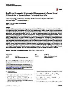
7. General import pathway of the metabolite carriers into mitochondria We can therefore summarize the various steps of import of mitochondrial metabolite carriers from their site of synthesis in the cytosol to their final destination in the inner mitochondrial membrane [117–120]. In particular, five different stages of import have been described for the AAC, the most studied member of this family (Fig. 4). Following synthesis in the cytosol (stage I), the chaperone-bound AAC is targeted to mitochondria thanks to the presence of internal targeting signals. In this stage Hsp70/Hsp90 docks onto Tom70 and the precursor proteins are transferred to Tom70, which may prevent precursor aggregation. On the surface of mitochondria, the multiple import signals within carrier precursors are recognized by several Tom70 molecules in a cooperative manner (stage II). The AAC is then translocated from the outer membrane into the intermembrane space (stage III). However, when the membrane potential is dissipated by the addition of valinomycin (and potassium ions) to mitochondria the AAC is translocated only partially (stage IIIa). In this condition the precursor protein accumulates in the Tom40 channel thus remaining accessible for externally added proteases. In stage IIIa the carboxy-terminal part of the AAC appears to bind tightly to the TOM complex, while the remaining domains are already bound by the soluble 70 kDa complex composed of Tim9 and Tim10 [24]. On the contrary, in energized mitochondria, the AAC is released from the TOM complex and immediately binds to the TIM22 complex of the inner membrane (stage IIIb). After that, the AAC integrates into the inner membrane at the level of the TIM22 complex (stage IV). A role for Oxa1 in the TIM22-mediated protein biogenesis was recently proposed [116]. This stage is eventually followed by the dimerization of this hydrophobic carrier in the lipid bilayer of the inner membrane [36,37,120,121] where the protein acquires its final and functional conformation (stage V). In this context, it is worth underlying that mitochondrial carriers may form oligomeric structures in the inner membrane, which are necessary to mediate metabolite transport, as recently found in the case of the pyruvate carrier [122,123]. The AAC is therefore transported from the outer to the inner membrane of mitochondria with the help of the soluble Tim9–Tim10 complex in the intermembrane space and of the TIM22 complex in the inner membrane. In spite of the fact that a direct link between the TOM and the TIM22 complexes has never been found, it has been proposed that the transport of the AAC may occur at the level of translocation contact sites [22]. Subsequent studies [124] carried out with the dicarboxylate carrier, another member of the metabolite carrier family [30], revealed a partially different mechanism of import. The dicarboxylate carrier was completely translocated across the outer mitochondrial membrane in the absence of the membrane potential, thereby becoming resistant to externally added proteinase K. This carrier protein therefore accumulated as a soluble intermediate in the intermembrane space of de-energized mitochondria. This new translocation intermediate, never found before with the AAC or other members of the carrier family, was named stage III* of import. On the basis of these specific properties, the dicarboxylate carrier represents a good
Fig. 3. The translocase of the inner mitochondrial membrane for hydrophobic proteins carrying internal targeting signals (TIM22 complex).
A. Ferramosca, V. Zara / Biochimica et Biophysica Acta 1833 (2013) 494–502
499
Fig. 4. Different stages characterizing the import of the newly synthesized ADP/ATP carrier (AAC) into mitochondria. For schematic reasons, in the outer and inner membranes of mitochondria only the TOM and the TIM22 complexes are reported. Furthermore, the cytosolic chaperones bound to the AAC precursor are shown in orange. Following synthesis in the cytosol (stage I), the chaperone-bound AAC is targeted to mitochondria thanks to the presence of internal targeting signals. Multiple molecules of the receptor Tom70 specifically recognize the carrier protein on the mitochondrial surface (stage II). AAC is then translocated from the outer membrane into the intermembrane space (stage III). However, when the mitochondrial membrane potential (Δψ) is dissipated, the AAC is translocated only partially (stage IIIa). On the contrary, in energized mitochondria, AAC is released from the TOM complex and immediately binds to the TIM22 complex of the inner membrane (stage IIIb). Stage IV is represented by the membrane potential-dependent insertion of the hydrophobic protein in the inner membrane through the Tim22 channel and Oxa1. Finally, in stage V, the dimerization of the ADP/ATP carrier in its functional location occurs.
candidate for exploring the role played by the small Tims in the intermembrane space of mitochondria. In accordance with these results, the possible presence of soluble translocation intermediates of the metabolite carriers in the intermembrane space has also been proposed in other studies [96,125]. 8. Perspectives Many components of the mitochondrial protein import machinery were identified over the past 20 years. Since then, investigations seemed to turn to more detailed studies on the mechanism of protein transport. On the basis of the previously mentioned data, also the biogenesis of some hydrophobic carrier proteins has been investigated in detail, thereby obtaining a large amount of new information. Nevertheless, several important questions remain unanswered. One aspect of carrier biogenesis which merits further investigation is the precise role played by the positively charged N-terminal presequence in a subset of mitochondrial carriers. Since the vast majority of studies on the biogenesis of mitochondrial carrier proteins have focused exclusively on the characterization of AAC of yeast, only little is known about the presequences and the biogenesis of other carrier proteins. It has recently emerged that carrier presequences have multiple and distinct roles during the biogenesis of this class of proteins. Furthermore, it has been found that some of these functions are mediated by specific interactions of carrier presequences both with cytosolic
chaperones and/or mitochondrial membrane receptors. However, more experiments are needed in order to reach a unifying vision of the significance and of the molecular mechanisms of action of these carrier presequences. Moreover, it is generally assumed that hydrophobic interactions play an important role in driving the translocation of carrier proteins across the mitochondrial outer membrane. On the other hand, it is also clear that charged residues participate in the import mechanism, both at the outer and the inner mitochondrial membrane. Further investigations are therefore required to determine the individual residues that are involved, and to elucidate the function of these residues in translocation and membrane insertion of the carrier proteins. Another aspect is the folding state of the carrier proteins during their translocation into mitochondria. Recent studies suggest that, unlike presequence-containing preproteins, different regions of the mature polypeptide chain of the metabolite carriers are required for targeting and translocation into mitochondria. In this way, the complex network of interactions generated among differently located residues in the metabolite carriers is efficiently recognized by the protein import machinery of mitochondria. However, the exact nature of this structural arrangement is still unclear and further studies are necessary in order to clarify what happens during carrier translocation in the intermembrane space. Future efforts will be directed in these and possibly other directions. This also in view of the fact that a defective carrier biogenesis has profound pathological implications because of the possible imbalance of mitochondrial bioenergetics.
500
A. Ferramosca, V. Zara / Biochimica et Biophysica Acta 1833 (2013) 494–502
References [1] W. Neupert, Protein import into mitochondria, Annu. Rev. Biochem. 66 (1997) 863–917. [2] S.A. Paschen, W. Neupert, Protein import into mitochondria, IUBMB Life 52 (2001) 101–112. [3] N. Pfanner, N. Wiedemann, Mitochondrial protein import: two membranes, three translocases, Curr. Opin. Cell Biol. 14 (2002) 400–411. [4] K.N. Truscott, K. Brandner, N. Pfanner, Mechanisms of protein import into mitochondria, Curr. Biol. 13 (2003) R326–R337. [5] N. Wiedemann, A.E. Frazier, N. Pfanner, The protein import machinery of mitochondria, J. Biol. Chem. 279 (2004) 14473–14476. [6] M.J. Baker, A.E. Frazier, J.M. Gulbis, M.T. Ryan, Mitochondrial protein-import machinery: correlating structure with function, Trends Cell Biol. 17 (2007) 456–464. [7] W. Neupert, J.M. Herrmann, Translocation of proteins into mitochondria, Annu. Rev. Biochem. 76 (2007) 723–749. [8] N. Bolender, A. Sickmann, R. Wagner, C. Meisinger, N. Pfanner, Multiple pathways for sorting mitochondrial precursor proteins, EMBO Rep. 9 (2008) 42–49. [9] A. Chacinska, C.M. Koehler, D. Milenkovic, T. Lithgow, N. Pfanner, Importing mitochondrial proteins: machineries and mechanisms, Cell 138 (2009) 628–644. [10] O. Schmidt, N. Pfanner, C. Meisinger, Mitochondrial protein import: from proteomics to functional mechanisms, Nat. Rev. Mol. Cell Biol. 11 (2010) 655–667. [11] T. Becker, L. Böttinger, N. Pfanner, Mitochondrial protein import: from transport pathways to an integrated network, Trends Biochem. Sci. 37 (2012) 85–91. [12] G. von Heijne, J. Steppuhn, R.G. Herrmann, Domain structure of mitochondrial and chloroplast targeting peptides, Eur. J. Biochem. 180 (1989) 535–545. [13] O. Gakh, P. Cavadini, G. Isaya, Mitochondrial processing peptidases, Biochim. Biophys. Acta 1592 (2002) 63–77. [14] F. Palmieri, The mitochondrial transporter family (SLC25): physiological and pathological implications, Pflugers Arch. 447 (2004) 689–709. [15] F. Palmieri, C.L. Pierri, Mitochondrial metabolite transport, Essays Biochem. 47 (2010) 37–52. [16] F. Palmieri, F. Bisaccia, L. Capobianco, V. Iacobazzi, C. Indiveri, V. Zara, Structural and functional properties of mitochondrial anion carriers, Biochim. Biophys. Acta 1018 (1990) 147–150. [17] T. Beddoe, T. Lithgow, Delivery of nascent polypeptides to the mitochondrial surface, Biochim. Biophys. Acta 1592 (2002) 35–39. [18] N. Wiedemann, V. Kozjak, A. Chacinska, B. Schönfisch, S. Rospert, M.T. Ryan, N. Pfanner, C. Meisinger, Machinery for protein sorting and assembly in the mitochondrial outer membrane, Nature 424 (2003) 565–571. [19] N. Mesecke, N. Terziyska, C. Kozany, F. Baumann, W. Neupert, K. Hell, J.M. Herrmann, A disulfide relay system in the intermembrane space of mitochondria that mediates protein import, Cell 121 (2005) 1059–1069. [20] G.S. Adrian, M.T. McCammon, D.L. Montgomery, M.G. Douglas, Sequences required for delivery and localization of the ADP/ATP translocator to the mitochondrial inner membrane, Mol. Cell. Biol. 6 (1986) 626–634. [21] N. Pfanner, P. Hoeben, M. Tropschug, W. Neupert, The carboxyl-terminal two-thirds of the ADP/ATP carrier polypeptide contains sufficient information to direct translocation into mitochondria, J. Biol. Chem. 262 (1987) 14851–14854. [22] C. Smagula, M.G. Douglas, Mitochondrial import of the ADP/ATP carrier protein in Saccharomyces cerevisiae. Sequences required for receptor binding and membrane translocation, J. Biol. Chem. 263 (1988) 6783–6790. [23] C.S. Smagula, M.G. Douglas, ADP/ATP carrier of Saccharomyces cerevisiae contains a mitochondrial import signal between aminoacids 72 and 111, J. Cell. Biochem. 36 (1988) 323–327. [24] M. Endres, W. Neupert, M. Brunner, Transport of the ADP/ATP carrier of mitochondria from the TOM complex to the TIM22.54 complex, EMBO J. 18 (1999) 3214–3221. [25] D.R. Nelson, C.M. Felix, J.M. Swanson, Highly conserved charged-pair networks in the mitochondrial carrier family, J. Mol. Biol. 277 (1998) 285–308. [26] H. Nury, C. Dahout-Gonzalez, V. Trezeguet, G.J. Lauquin, G. Brandolin, E. Pebay-Peyroula, Relations between structure and function of the mitochondrial ADP/ATP carrier, Annu. Rev. Biochem. 75 (2006) 713–741. [27] C. Sirrenberg, M. Endres, H. Fölsch, R.A. Stuart, W. Neupert, M. Brunner, Carrier protein import into mitochondria mediated by the intermembrane proteins Tim10/Mrs11 and Tim12/Mrs5, Nature 391 (1998) 912–915. [28] M.F. Bauer, S. Hofmann, W. Neupert, M. Brunner, Protein translocation into mitochondria: the role of TIM complexes, Trends Cell Biol. 10 (2000) 25–31. [29] V. Zara, A. Ferramosca, L. Capobianco, K.M. Baltz, O. Randel, J. Rassow, F. Palmieri, P. Papatheodorou, Biogenesis of yeast dicarboxylate carrier: the carrier signature facilitates translocation across the mitochondrial outer membrane, J. Cell Sci. 120 (2007) 4099–4106. [30] L. Palmieri, A. Vozza, A. Hönlinger, K. Dietmeier, A. Palmisano, V. Zara, F. Palmieri, The mitochondrial dicarboxylate carrier is essential for the growth of Saccharomyces cerevisiae on ethanol or acetate as the sole carbon source, Mol. Microbiol. 31 (1999) 569–577. [31] M. Babot, C. Blancard, L. Pelosi, G.J. Lauquin, V. Trézéguet, The transmembrane prolines of the mitochondrial ADP/ATP carrier are involved in nucleotide binding and transport and its biogenesis, J. Biol. Chem. 287 (2012) 10368–10378. [32] N. Wiedemann, N. Pfanner, M.T. Ryan, The three modules of ADP/ATP carrier cooperate in receptor recruitment and translocation into mitochondria, EMBO J. 20 (2001) 951–960.
[33] P. Rehling, K. Brandner, N. Pfanner, Mitochondrial import and the twin-pore translocase, Nat. Rev. Mol. Cell Biol. 5 (2004) 519–530. [34] M.J. Runswick, S.J. Powell, P. Nyren, J.E. Walker, Sequence of the bovine mitochondrial phosphate carrier protein: structural relationship to ADP/ATP translocase and the brown fat mitochondria uncoupling protein, EMBO J. 6 (1987) 1367–1373. [35] R.S. Kaplan, J.A. Mayor, D.O. Wood, The mitochondrial tricarboxylate transport protein. cDNA cloning, primary structure, and comparison with other mitochondrial transport proteins, J. Biol. Chem. 268 (1993) 13682–13690. [36] V. Zara, A. Ferramosca, I. Palmisano, F. Palmieri, J. Rassow, Biogenesis of rat mitochondrial citrate carrier (CIC): the N-terminal presequence facilitates the solubility of the preprotein but does not act as a targeting signal, J. Mol. Biol. 325 (2003) 399–408. [37] V. Zara, V. Dolce, L. Capobianco, A. Ferramosca, P. Papatheodorou, J. Rassow, F. Palmieri, Biogenesis of eel liver citrate carrier (CIC): negative charges can substitute for positive charges in the presequence, J. Mol. Biol. 365 (2007) 958–967. [38] M. Emmermann, H.P. Braun, U.K. Schmitz, The ADP/ATP translocator from potato has a long amino-terminal extension, Curr. Genet. 20 (1991) 405–410. [39] V. Zara, A. Ferramosca, P. Papatheodorou, F. Palmieri, J. Rassow, Import of rat mitochondrial citrate carrier (CIC) at increasing salt concentrations promotes presequence binding to import receptor Tom20 and inhibits membrane translocation, J. Cell Sci. 118 (2005) 3985–3995. [40] V. Zara, J. Rassow, E. Wachter, M. Tropschug, F. Palmieri, W. Neupert, N. Pfanner, Biogenesis of the mitochondrial phosphate carrier, Eur. J. Biochem. 198 (1991) 405–410. [41] V. Zara, F. Palmieri, K. Mahlke, N. Pfanner, The cleavable presequence is not essential for import and assembly of the phosphate carrier of mammalian mitochondria but enhances the specificity and efficiency of import, J. Biol. Chem. 267 (1992) 12077–12081. [42] T. Mozo, K. Fischer, U.I. Flügge, U.K. Schmitz, The N-terminal extension of the ADP/ATP translocator is not involved in targeting to plant mitochondria in vivo, Plant J. 7 (1995) 1015–1020. [43] P. Hájek, J.Y. Koh, L. Jones, D.M. Bedwell, The amino-terminus of the F1 ATP-ase beta-subunit precursor functions as an intramolecular chaperone to facilitate mitochondrial protein import, Mol. Cell. Biol. 17 (1997) 7169–7177. [44] J. Rassow, N. Pfanner, Molecular chaperones and intracellular protein translocation, Rev. Phys. Biochem. Pharmacol. 126 (1995) 199–264. [45] V.R. Agashe, F.U. Hartl, Roles of molecular chaperones in cytoplasmic protein folding, Semin. Cell Dev. Biol. 11 (2000) 15–25. [46] J.C. Young, J.M. Barral, F.U. Hartl, More than folding: localized functions of cytosolic chaperones, Trends Biochem. Sci. 28 (2003) 541–547. [47] J.C. Young, I. Moarefi, F.U. Hartl, Hsp90: a specialized but essential protein-folding tool, J. Cell Biol. 154 (2001) 267–273. [48] R.J. Deshaies, B.D. Koch, M. Werner-Washburne, E.A. Craig, R. Schekman, A subfamily of stress protein facilitates translocation of secretory and mitochondrial precursor polypeptides, Nature 332 (1988) 800–805. [49] H. Murakami, D. Pain, G. Blobel, 70 kD heat shock-related protein is one of at least two cytosolic factors stimulating protein import into mitochondria, J. Cell Biol. 107 (1988) 2051–2057. [50] F.U. Hartl, Molecular chaperones in cellular protein folding, Nature 381 (1996) 571–579. [51] D. Schmid, A. Baici, H. Gehring, P. Christen, Kinetics of molecular chaperone action, Science 263 (1994) 971–973. [52] H. Theyssen, H.P. Schuster, L. Packschies, B. Bukau, J. Reinstein, The second step of ATP binding to DnaK induces peptide release, J. Mol. Biol. 263 (1996) 657–670. [53] J.C. Young, N.J. Hoogenraad, F.U. Hartl, Molecular chaperones Hsp90 and Hsp70 deliver preproteins to the mitochondrial import receptor Tom70, Cell 112 (2003) 41–50. [54] W.P. Sheffield, G.C. Shore, S.K. Randall, Mitochondrial precursor protein. Effects of 70-kilodalton heat shock protein on polypeptide folding, aggregation, and import competence, J. Biol. Chem. 265 (1990) 11069–11076. [55] K. Terada, K. Ohtsuka, N. Imamoto, Y. Yoneda, M. Mori, Role of heat shock cognate 70 protein in import of ornithine transcarbamylase precursor into mammalian mitochondria, Mol. Cell. Biol. 15 (1995) 3708–3713. [56] V. Zara, A. Ferramosca, P. Robitaille-Foucher, F. Palmieri, J.C. Young, Mitochondrial carrier protein biogenesis: role of the chaperones Hsc70 and Hsp90, Biochem. J. 419 (2009) 369–375. [57] B. Bukau, J. Weissman, A. Horwich, Molecular chaperones and protein quality control, Cell 125 (2006) 443–451. [58] L.H. Pearl, C. Prodromou, Structure and mechanism of the Hsp90 molecular chaperone machinery, Annu. Rev. Biochem. 75 (2006) 271–294. [59] J.C. Young, Mechanisms of the Hsp70 chaperone system, Biochem. Cell Biol. 88 (2010) 291–300. [60] P.J. Dekker, M.T. Ryan, J. Brix, H. Müller, A. Hönlinger, N. Pfanner, Preprotein translocase of the outer mitochondrial membrane: molecular dissection and assembly of the general import pore complex, Mol. Cell. Biol. 18 (1998) 6515–6524. [61] K.P. Künkele, S. Heins, M. Dembowski, F.E. Nargang, R. Benz, R. Thieffry, J. Walz, R. Lill, S. Nussberger, W. Neupert, The preprotein translocation channel of the outer membrane of mitochondria, Cell 93 (1998) 1009–1019. [62] L. Ramage, T. Junne, K. Hahne, T. Lithgow, G. Schatz, Functional cooperation of mitochondrial protein import receptors in yeast, EMBO J. 12 (1993) 4115–4123. [63] T. Söllner, R. Pfaller, G. Griffiths, N. Pfanner, W. Neupert, A mitochondrial import receptor for the ADP/ATP carrier, Cell 62 (1990) 107–115. [64] H.F. Steger, T. Söllner, M. Kiebler, K.A. Dietmeier, R. Pfaller, K.S. Trulzsch, M. Tropschug, W. Neupert, N. Pfanner, Import of ADP/ATP carrier into mitochondria: two receptors act in parallel, J. Cell Biol. 111 (1990) 2353–2363.
A. Ferramosca, V. Zara / Biochimica et Biophysica Acta 1833 (2013) 494–502 [65] V. Hines, A. Brandt, G. Griffiths, H. Horstmann, H. Brutsch, G. Schatz, Protein import into yeast mitochondria is accelerated by the outer membrane protein MAS70, EMBO J. 9 (1990) 3191–3200. [66] J. Brix, G.A. Ziegler, K. Dietmeier, J. Schneider-Mergener, G.E. Schulz, N. Pfanner, The mitochondrial import receptor Tom70: identification of a 25 kDa core domain with a specific binding site for preproteins, J. Mol. Biol. 303 (2000) 479–488. [67] J. Brix, K. Dietmeier, N. Pfanner, Differential recognition of preproteins by the purified cytosolic domains of the mitochondrial import receptors Tom20, Tom22 and Tom70, J. Biol. Chem. 272 (1997) 20730–20735. [68] J. Brix, S. Rüdiger, B. Bukau, J. Schneider-Mergener, N. Pfanner, Distribution of binding sequences for the mitochondrial import receptors Tom20, Tom22 and Tom70 in a presequence-carrying preprotein and a non-cleavable preprotein, J. Biol. Chem. 274 (1999) 16522–16530. [69] H. Yamamoto, K. Fukui, H. Takahashi, S. Kitamura, T. Shiota, K. Terao, M. Uchida, M. Esaki, S. Nishikawa, T. Yoshihisa, K. Yamano, T. Endo, Roles of Tom70 in import of presequence-containing mitochondrial proteins, J. Biol. Chem. 284 (2009) 31635–31646. [70] M.K. Bhangoo, S. Tzankov, A.C. Fan, K. Dejgaard, D.Y. Thomas, J.C. Young, Multiple 40-kDa heat-shock protein chaperones function in Tom70-dependent mitochondrial import, Mol. Biol. Cell 18 (2007) 3414–3428. [71] Y. Wu, B. Sha, Crystal structure of yeast mitochondrial outer membrane translocon member Tom70p, Nat. Struct. Mol. Biol. 13 (2006) 589–593. [72] A.C. Fan, M.K. Bhangoo, J.C. Young, Hsp90 functions in the targeting and outer membrane translocation steps of Tom70-mediated mitochondrial import, J. Biol. Chem. 281 (2006) 33313–33324. [73] A.C. Fan, G. Kozlov, A. Hoegl, R.C. Marcellus, M.J. Wong, K. Gehring, J.C. Young, Interaction between the human mitochondrial import receptors Tom20 and Tom70 in vitro suggests a chaperone displacement mechanism, J. Biol. Chem. 286 (2011) 32208–32219. [74] J. Schlossmann, R. Lill, W. Neupert, D.A. Court, Tom71, a novel homologue of the mitochondrial preprotein receptor Tom70, J. Biol. Chem. 271 (1996) 17890–17895. [75] D. Vestweber, J. Brunner, A. Baker, G. Schatz, A 42K outer-membrane protein is a component of the yeast mitochondrial protein import site, Nature 341 (1989) 205–209. [76] K.P. Baker, A. Schaniel, D. Vestweber, G. Schatz, A yeast mitochondrial outer membrane protein essential for protein import and cell viability, Nature 348 (1990) 605–609. [77] M. Kiebler, P. Keil, H. Schneider, I.J. van der Klei, N. Pfanner, W. Neupert, The mitochondrial receptor complex: a central role of MOM22 in mediating pre-protein transfer from receptors to the general insertion pore, Cell 74 (1993) 483–492. [78] T. Lithgow, T. Junne, K. Suda, S. Gratzer, G. Schatz, The mitochondrial outer membrane protein Mas22p is essential for protein import and viability of yeast, Proc. Natl. Acad. Sci. U. S. A. 91 (1994) 11973–11977. [79] A. Hönlinger, M. Kübrich, M. Moczko, F. Gartner, L. Mallet, F. Busserau, C. Eckerskorn, F. Lottspeich, K. Dietmeier, M. Jacquet, N. Pfanner, The mitochondrial receptor complex: Mom22 is essential for cell viability and directly interacts with preproteins, Mol. Cell. Biol. 15 (1995) 3382–3389. [80] A. Mayer, F.E. Nargang, W. Neupert, R. Lill, MOM22 is a receptor for mitochondrial targeting sequences and cooperates with MOM19, EMBO J. 14 (1995) 4204–4211. [81] K. Dietmeier, A. Hönlinger, U. Bömer, P.J. Dekker, C. Eckerskorn, F. Lottspeich, M. Kübrich, N. Pfanner, Tom5 functionally links mitochondrial preprotein receptors to the general import pore, Nature 388 (1997) 195–200. [82] C.K. Kassenbrock, W. Cao, M.G. Douglas, Genetic and biochemical characterization of ISP6, a small mitochondrial outer membrane protein associated with the protein translocation complex, EMBO J. 12 (1993) 3023–3034. [83] A. Alconada, M. Kübrich, M. Moczko, A. Hönlinger, N. Pfanner, The mitochondrial receptor complex: the small subunit Mom8b/Isp6 supports association of receptors with the general insertion pore and transfer of preproteins, Mol. Cell. Biol. 15 (1995) 6196–6205. [84] A. Hönlinger, U. Bömer, A. Alconada, C. Eckerskorn, F. Lottspeich, K. Dietmeier, N. Pfanner, Tom7 modulates the dynamics of the mitocondrial outer membrane translocase and plays a pathway-related role in protein import, EMBO J. 15 (1996) 2125–2137. [85] C. Meisinger, M.T. Ryan, K. Hill, K. Model, J.H. Lim, A. Sickmann, H. Müller, H.E. Meyer, R. Wagner, N. Pfanner, Protein import channel of the outer mitochondrial membrane: a highly stable Tom40-Tom22 core structure differentially interacts with preproteins, small tom proteins, and import receptors, Mol. Cell. Biol. 21 (2001) 2337–2348. [86] S. van Wilpe, M.T. Ryan, K. Hill, A.C. Maarse, C. Meisinger, J. Brix, P.J. Dekker, M. Moczko, R. Wagner, M. Meijer, B. Guiard, A. Hönlinger, N. Pfanner, Tom22 is a multifunctional organizer of the mitochondrial preprotein translocase, Nature 401 (1999) 485–489. [87] O. Schmidt, A.B. Harbauer, S. Rao, B. Eyrich, R.P. Zahedi, D. Stojanovski, B. Schönfisch, B. Guiard, A. Sickmann, N. Pfanner, C. Meisinger, Regulation of mitochondrial protein import by cytosolic kinases, Cell 144 (2011) 227–239. [88] C.M. Koehler, E. Jarosch, K. Tokatlidis, K. Schmid, R.J. Schweyen, G. Schatz, Import of mitochondrial carriers mediated by essential proteins of the intermembrane space, Science 279 (1998) 369–373. [89] C.M. Koehler, S. Merchant, W. Oppliger, K. Schmid, E. Jarosch, L. Dolfini, T. Junne, G. Schatz, K. Tokatlidis, Tim9p, an essential partner subunit of Tim10p for the import of mitochondrial carrier proteins, EMBO J. 17 (1998) 6477–6486. [90] C. Koehler, D. Leuenberger, S. Merchant, A. Renold, T. Junne, G. Schatz, Human deafness dystonia syndrome is a mitochondrial disease, Proc. Natl. Acad. Sci. U. S. A. 96 (1999) 2141–2146.
501
[91] A. Adam, M. Endres, C. Sirrenberg, F. Lottspeich, W. Neupert, M. Brunner, Tim9, a new component of the TIM22.54 translocase in mitochondria, EMBO J. 18 (1999) 313–319. [92] N. Wiedemann, K.N. Truscott, S. Pfannschmidt, B. Guiard, C. Meisinger, N. Pfanner, Biogenesis of the protein import channel Tom40 of the mitochondrial outer membrane: intermembrane space components are involved in an early stage of the assembly pathway, J. Biol. Chem. 279 (2004) 18188–18194. [93] M.F. Bauer, K. Gempel, A.S. Reichert, G.A. Rappold, P. Lichtner, K.D. Gerbitz, W. Neupert, M. Brunner, S. Hoffman, Genetic and structural characterization of the human mitochondrial inner membrane translocase, J. Mol. Biol. 289 (1999) 69–82. [94] U. Rothbauer, S. Hofmann, N. Mühlenbein, S.A. Paschen, K.D. Gerbitz, W. Neupert, M. Brunner, M.F. Bauer, Role of deafness dystonia peptide 1 (DDP1) in import of human Tim23 into the inner membrane of mitochondria, J. Biol. Chem. 276 (2001) 37327–37334. [95] M.T. Ryan, H. Müller, N. Pfanner, Functional staging of ADP/ATP carrier translocation across the outer mitochondrial membrane, J. Biol. Chem. 274 (1999) 20619–20627. [96] S.P. Curran, D. Leuenberger, W. Oppliger, C. Koehler, The Tim9p–Tim10p complex binds to the transmembrane domains of the ADP/ATP carrier, EMBO J. 21 (2002) 942–953. [97] C.M. Koehler, The small Tim proteins and the twin Cx3C motif, Trends Biochem. Sci. 29 (2004) 1–4. [98] C.T. Webb, M.A. Gorman, M. Lazarou, M.T. Ryan, J.M. Gulbis, Crystal structure of the mitochondrial chaperone TIM9.10 reveals a six-bladed alpha-propeller, Mol. Cell 21 (2006) 123–133. [99] K.N. Beverly, M.R. Sawaya, E. Schmid, C.M. Koehler, The Tim8–Tim13 complex has multiple substrate binding sites and binds cooperatively to Tim23, J. Mol. Biol. 382 (2008) 1144–1156. [100] M.A. Vergnolle, C. Baud, A.P. Golovanov, F. Alcock, P. Luciano, L.Y. Lian, K. Tokatlidis, Distinct domains of small Tims involved in subunit interaction and substrate recognition, J. Mol. Biol. 351 (2005) 839–849. [101] M.A. Vergnolle, F.H. Alcock, N. Petrakis, K. Tokatlidis, Mutation of conserved charged residues in mitochondrial TIM10 subunits precludes TIM10 complex assembly, but does not abolish growth of yeast cells, J. Mol. Biol. 371 (2007) 1315–1324. [102] I.E. Gentle, A.J. Perry, F.H. Alcock, V.A. Likić, P. Dolezal, E.T. Ng, A.W. Purcell, M. McConnville, T. Naderer, A.L. Chanez, F. Charrière, C. Aschinger, A. Schneider, K. Tokatlidis, T. Lithgow, Conserved motifs reveal details of ancestry and structure in the small TIM chaperones of the mitochondrial intermembrane space, Mol. Biol. Evol. 24 (2007) 1149–1160. [103] M.J. Baker, C.T. Webb, D.A. Stroud, C.S. Palmer, A.E. Frazier, B. Guiard, A. Chacinska, J.M. Gulbis, M.T. Ryan, Structural and functional requirements for activity of the Tim9–Tim10 complex in mitochondrial protein import, Mol. Biol. Cell 20 (2009) 769–779. [104] N. Mühlenbein, S. Hofmann, U. Rothbauer, M.F. Bauer, Organization and function of the small tim complexes acting along the import pathway of metabolite carriers into mammalian mitochondria, J. Biol. Chem. 279 (2004) 13540–13546. [105] S.A. Hasson, R. Damoiseaux, J.D. Glavin, D.V. Dabir, S.S. Walker, C.M. Koehler, Substrate specificity of the TIM22 mitochondrial import pathway revealed with small molecule inhibitor of protein translocation, Proc. Natl. Acad. Sci. U. S. A. 107 (2010) 9578–9583. [106] M.P. Schwartz, A. Matouschek, The dimensions of the protein import channels in the outer and inner mitochondrial membranes, Proc. Natl. Acad. Sci. U. S. A. 96 (1999) 3086–13090. [107] C. Sirrenberg, M.F. Bauer, B. Guiard, W. Neupert, M. Brunner, Import of carrier proteins into the mitochondrial inner membrane mediated by Tim22, Nature 384 (1996) 582–585. [108] O. Kerscher, J. Holder, M. Srinivasan, R.S. Leung, R.E. Jensen, The Tim54p–Tim22p complex mediates insertion of proteins into the mitochondrial inner membrane, J. Cell Biol. 139 (1997) 1663–1675. [109] O. Kerscher, N.B. Sepuri, R.E. Jensen, Tim18p is a new component of the Tim54p– Tim22p translocon in the mitochondrial inner membrane, Mol. Biol. Cell 11 (2000) 103–116. [110] C.M. Koehler, M.P. Murphy, N.A. Bally, D. Leuenberger, W. Oppliger, L. Dolfini, T. Junne, G. Schatz, E. Or, Tim18p, a new subunit of the TIM22 complex that mediates insertion of imported proteins into the yeast mitochondrial inner membrane, Mol. Cell. Biol. 20 (2000) 1187–1193. [111] N. Gebert, M. Gebert, S. Oeljeklaus, K. von der Malsburg, D.A. Stroud, B. Kulawiak, C. Wirth, R.P. Zahedi, P. Dolezal, S. Wiese, O. Simon, A. Schulze-Specking, K.N. Truscott, A. Sickmann, P. Rehling, B. Guiard, C. Hunte, B. Warscheid, M. van der Laan, N. Pfanner, N. Wiedemann, Dual function of Sdh3 in the respiratory chain and TIM22 protein translocase of the mitochondrial inner membrane, Mol. Cell 44 (2011) 811–818. [112] P. Kovermann, K.N. Truscott, B. Guiard, P. Rehling, N.B. Sepuri, H. Müller, R.E. Jensen, R. Wagner, N. Pfanner, Tim22, the essential core of the mitochondrial protein insertion complex forms a voltage-activated and signal-gated channel, Mol. Cell 9 (2002) 363–373. [113] P. Rehling, K. Model, K. Brandner, P. Kovermann, A. Sickmann, H.E. Meyer, W. Kuhlbrandt, R. Wagner, K.N. Truscott, N. Pfanner, Protein insertion into the mitochondrial inner membrane by a twin-pore translocase, Science 299 (2003) 1747–1751. [114] K. Wagner, N. Gebert, B. Guiard, K. Brandner, K.N. Truscott, N. Wiedemann, N. Pfanner, P. Rehling, The assembly pathway of the mitochondrial carrier translocase involves four preprotein translocases, Mol. Cell. Biol. 28 (2008) 4251–4260.
502
A. Ferramosca, V. Zara / Biochimica et Biophysica Acta 1833 (2013) 494–502
[115] M.F. Bauer, U. Rothbauer, N. Mühlenbein, R.J.H. Smith, K.D. Gerbitz, W. Neupert, M. Brunner, S. Hofmann, The mitochondrial Tim22 preprotein translocase is highly conserved throughout the eukaryotic kingdom, FEBS Lett. 464 (1999) 41–47. [116] M. Hildenbeutel, M. Theis, M. Geier, I. Haferkamp, H.E. Neuhaus, J.M. Herrmann, M. Ott, The membrane insertase oxa1 is required for efficient import of carrier proteins into mitochondria, J. Mol. Biol. 423 (2012) 590–599. [117] N. Pfanner, W. Neupert, Distinct steps in the import of ADP/ATP carrier into mitochondria, J. Biol. Chem. 262 (1987) 7528–7536. [118] K.N. Truscott, N. Pfanner, Import of carrier proteins into mitochondria, Biol. Chem. 380 (1999) 1151–1156. [119] N. Pfanner, A. Geissler, Versatility of the mitochondrial protein import machinery, Nat. Rev. Mol. Cell Biol. 2 (2001) 339–349. [120] A. Palmisano, V. Zara, A. Hönlinger, A. Vozza, P.J. Dekker, N. Pfanner, F. Palmieri, Targeting and assembly of the oxoglutarate carrier: general principles for biogenesis of carrier proteins of the mitochondrial inner membrane, Biochem. J. 333 (1998) 151–158.
[121] L. Capobianco, A. Ferramosca, V. Zara, The mitochondrial tricarboxylate carrier of silver eel: dimeric structure and cytosolic exposure of both N- and C-termini, J. Protein Chem. 21 (2002) 515–521. [122] S. Herzig, E. Raemy, S. Montessuit, J.L. Veuthey, N. Zamboni, B. Westermann, E.R. Kunji, J.C. Martinou, Identification and functional expression of the mitochondrial pyruvate carrier, Science 337 (2012) 93–96. [123] D.K. Bricker, E.B. Taylor, J.C. Schell, T. Orsak, A. Boutron, Y.C. Chen, J.E. Cox, C.M. Cardon, J.G. Van Vranken, N. Dephoure, C. Redin, S. Boudina, S.P. Gygi, M. Brivet, C.S. Thummel, J. Rutter, A mitochondrial pyruvate carrier required for pyruvate uptake in yeast, Drosophila, and humans, Science 337 (2012) 96–100. [124] V. Zara, I. Palmisano, J. Rassow, F. Palmieri, Biogenesis of the dicarboxylate carrier (DIC): translocation across the mitochondrial outer membrane and subsequent release from the TOM channel are membrane potential-independent, J. Mol. Biol. 310 (2001) 965–971. [125] C.M. Koehler, S. Merchant, G. Schatz, How membrane proteins travel across the mitochondrial intermembrane space, Trends Biochem. Sci. 24 (1999) 428–432.