African Journal of Biotechnology Vol. 7 (25), pp. 4765-4773, 29 December, 2008 Available online at http://www.academicjournals.org/AJB DOI: 10.5897/AJB08.038 ISSN 1684–5315 © 2008 Academic Journals
Review
Biological control of Microcystis dominated harmful algal blooms R. Jabulani Gumbo1, 2*, Gina Ross1 and E. Thomas Cloete1 1
Department of Microbiology and Plant Pathology, University of Pretoria, Hillcrest, Pretoria, ZA0002, South Africa. Department of Hydrology and Water Resources, University of Venda, P/Bag x5050, Thohoyandou, 0950, South Africa.
2
Accepted 24 October, 2008
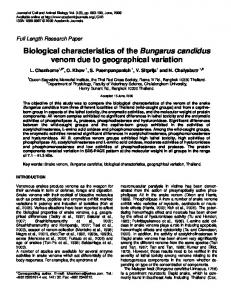
Freshwater resources are now threatened by the presence and increase of harmful algal blooms (HAB) all over the world. The HABs are sometimes a direct result of anthropogenic pollution entering water bodies, such as partially treated nutrient-rich effluents and the leaching of fertilisers and animal wastes. The impact of HABs on aquatic ecosystems and water resources, as well as their human health implications are well documented. Countermeasures have been proposed and implemented to manage HABs with varying levels of success. The use of copper algicides, though effective in managing HABs, often results in negative impacts such as copper toxicity and release of microcystins into surrounding water after cyanobacterial lysis. Biological control of HABs presents a possible solution. Predatory bacteria that have been isolated as potential biological control agents include members of the Bacteroides-Cytophaga-Flavobacterium, ranging from Bacillus spp. to Flexibacter spp., Cytophaga and Myxobacteria. Various mechanisms of predation have been proposed, including; physical contact between prey and predator, release of extracellular substances, entrapment of prey by the predator followed by antibiosis and endoparasitism or ectoparasitism of the host by the predator. Despite an increasing amount of work being done in this field, research is usually limited to laboratory cultures; assessment of microbial control agents is seldom extrapolated to field conditions. Key words: Biological control, Microcystis aeruginosa, harmful algal blooms, predatory bacteria. INTRODUCTION Harmful algal blooms (HABs) are a regular occurrence in eutrophic water bodies during spring, winter and summer and are on the increase world wide (Shilo, 1970; Zohary, 1987; Rashidan and Bird, 2001; Choi et al., 2005) including South Africa (Harding and Paxton, 2001; Harding et al., 2004). Cyanobacteria, mostly dominated by Microcystis, thrive in eutrophic waters producing blooms (Figure 1) rich in toxins and metabolites that reduces water quality with adverse effects on lake ecology, livestock, human water supply and recreational amenities (Sigee et al., 1999; Nakamura et al., 2003b). The freshwater cyanobacterial species that are often implicated with microcystin toxicity are: Microcystis, Anabaena, Oscillatoria and Nostoc. Nodularin toxicity is caused by
*Corresponding author. E-mail:
[email protected],
[email protected]. Tel: +27 15 962 8563. Fax: +27 15 962 4749.
a marine cyanobacterium called Nodularia spumigena (Rapala et al., 1994; Cronberg et al., 2003). The toxins are harmful to humans, fish, birds and other animals. Illness and death may occur after oral ingestion of cyanobacterial cells, or by contact with water that harbours the toxin-releasing strains of cyanobacteria. Animal deaths may also occur following bioaccumulation of cyanobacterial toxins (Richard et al., 1983). In many parts of the world, microcystins are a major concern to drinking water providers from a health and economic perspective. Microcystins have been linked to liver damage, which prompted the World Health Organization (WHO) to adopt a provisional guideline value for microcystins-LR (L for leucine and R for arginine) of 1.0 µg.l-1 drinking water (WHO, 1998; Hoeger et al., 2004). Earlier, Ueno et al. (1996) proposed a more stringent guideline value of 0.01 µgl-1, based on a possible correlation of primary liver cancer in certain locations in China and consumption of contaminated water containing microcystins (Oberholster et al., 2004). In Australia, the
4766
Afr. J. Biotechnol.
Figure 1. Harmful algal blooms scums visible in Hartbeespoort dam, South Africa (June, 2005).
potable water standard for microcystins was set at 1.3 µgl-1 (NHMRZ/ARMCANZ, 2001). Conventional water treatment processes are capable of removing intact cyanobacterial cells from raw water if operated in conjunction with dissolved air flotation. Ruptured or damaged cells may release intracellular toxins into the surrounding water, necessitating the use of expensive removal processes such as activated carbon and/or oxidative ozone and chlorine (Haider et al., 2003). Although cyanobacteria are normally removed during the production of potable water, consumers may be exposed to sub-lethal dosages of cyanobacterial toxins derived from contaminated dams and reservoirs (Lam et al., 1995). In Australia, elevated concentrations of microcystins were linked epidemiologically to an outbreak of human hepatoenteritis (Falconer et al., 1983). Microcystins are generally very stable compounds, are resistant to chemical breakdown and are persistent in natural waters for weeks to several months (Sivonen and Jones, 1999). However, the toxins are susceptible to breakdown by aquatic bacteria found naturally in rivers and reservoirs. Bourne et al. (1996) isolated a bacterial species identified as Spingomonas capable of degrading microcystin-LR and RR but not the closely related cyclic pentapeptide nodularin. The bacterium was reported to utilize the toxin as its sole carbon and nitrogen source. The bacterial degradation process may result in approxi-
mately 90 per cent removal of microcystin in a period of 2 to 10 days. Of major interest is what role these bacteria play in cyanobacterial cell lysis. Microcystin-degrading bacteria and predatory bacteria that are antagonistic towards cyanobacteria have been isolated from algal blooms. A strategy involving predatory bacteria may provide an environmental friendly solution to HABs. The development of a successful biological control strategy includes factors such as the predatorprey ratio and the mechanism of cyanobacterial lysis. This review considers the biological control of HAB as the primary approach to controlling HABs with emphasis on the use of bacteria. CURRENT USE OF ALGICIDES HARMFUL ALGAL BLOOMS
IN
MANAGING
Mechanical and physico-chemical methods have been devised in attempts to manage cyanobacterial blooms, with limited success. The most direct control method involves the use of chemical treatments such as algicides, including copper, Reglone A (diquat, 1,1ethylene-2, 2-dipyridilium dibromide), potassium permanganate, chlorine and Simazine (2-chloro-4,6bis(ethylamino)-s-triazine (Lam et al., 1995). Copper sulphate or organo-copper compounds have been used
Gumbo et al.
4767
successfully to control harmful algal blooms in raw water supplies intended for human consumption (Lam et al., 1995). These chemicals induced cyanobacterial cell lysis, followed by the release of toxins into surrounding waters. An appropriate waiting period has to follow these treatments to allow for the degradation of the toxins (WHO, 1999). These algicides are toxic to other aquatic microorganisms, may accumulate in the sediment at harmful concentrations and may cause long-term damage to the lake ecology (Mason, 1996). However, there is an increasing need to reduce the use of chemicals for environmental and safety reasons. Thus, the development of non-chemical control measures such as biological control is of great importance to the management of HABs. Other water treatment chemicals ® such as Phoslock (Greenop and Robb, 2001; Robb et al., 2003), alum and lime (within pH 6 - 10) controlled cyanobacteria blooms through nutrient precipitation and cell coagulation, and did not cause a significant increase in extracellular toxins (Lam et al., 1995). The major limitation for daily use of these chemical substances was their prohibitive cost.
or organisms. Secord (2003) has given an excellent treatise of this phenomenon with real world case studies with regards to the management of nuisance pests. Biocontrol of cyanobacteria, like other control measures for nuisance organisms, is often viewed with caution. This may be attributed to the experiences of plant pathologists who observed the destruction of important crops such as chestnut blight in the United States and potato blight in Ireland after the accidental release of pathogens (Atlas and Bartha, 1998). Further readings are recommended to obtain precise details of high profile cases of successful and catastrophic failures of biocontrol in the last century (Secord, 2003). Viral pathogens would be ideal as biocontrol agents as they are target selective and specific for nuisance cyanobacteria. However, bacterial agents are considered more suitable than viruses as biocontrol agents because bacteria can survive on alternate food sources during non-bloom periods and the possibility of mutation within the host is not problematic, as bacterial predation is not reliant on unique attachment receptors (Rashidan and Bird, 2001).
BIOLOGICAL BLOOMS
ALGAL
The use of microorganisms to control cyanobacterial blooms
One alternative approach to the control of algal blooms involves the use of biological control (biocontrol) agents. Predatory bacteria that are indigenous to the lake environment have been isolated from algal blooms. Daft et al. (1985) proposed the following seven attributes that defined a good predatory bacterial agent: adaptability to variations in physical conditions; ability to search for or trap prey; capacity and ability to multiply; prey consumption; ability to survive low prey densities (switch or adapt to other food sources); wide host range and ability to respond to changes in the host. In addition to these, this work suggests an eighth attribute, that is, the predatory bacteria should be indigenous to the particular water environment, thus providing an environmentally friendly solution. This is in agreement with Sigee et al. (1999), who suggested that the microbial antagonists must be indigenous species of that particular lake environment, having not undergone any gene modification or enhancement. The practice of introduction of foreign microbial agents has raised some concern with regards to environmental safety due to the so-called host specificity paradigm involving host switching (HS) and host range expansion (HRE) (Secord, 2003). The foreign microbial agents are naturally reproductive and may exploit the opportunities that are available in the new environment by shifting their host affinities to other host species (set of species) and/or add another target species other than the original target. The change in direction of the microbial antagonist is difficult to anticipate, and there is the possibility that the organisms may affect other economically important crops
In the natural environment, there are predatory microorganisms that are antagonistic towards particular nuisance organisms (weeds, cyanobacteria) thus providing a natural means of controlling these nuisance organisms. Microbial agents antagonistic to algae (bacteria, fungi, virus and protozoa) have been isolated from harmful algal blooms (Shilo, 1970; Burnham et al., 1981; Ashton and Robarts, 1987; Yamamoto et al., 1998; Sigee et al., 1999; Walker and Higginbotham, 2000; Rashidan and Bird, 2001; Nakamura et al., 2003a; Choi et al., 2005). These microbial agents may play a major role in the prevention, regulation and termination of harmful algal blooms. Such microbial populations are called microbial herbicides (Atlas and Bartha, 1998). The biological control of cyanobacteria provides a potential control measure to reduce the population of nuisance algal blooms to manageable levels. In many cases these bacterial agents are species- or genus-specific (Rashidan and Bird, 2001), while others attack a variety of cyanobacteria (Daft et al., 1975). The predatory bacteria are classified as members of the Bacteroides-CytophagaFlavobacterium, ranging from Bacillus spp. to Flexibacter spp., Cytophaga and Myxobacteria (Table 1). Choi et al. (2005) isolated the bacterium, Streptomyces neyagawaensis, which had a Microcystis-killing ability, from the sediment of a eutrophic lake in Korea. Under natural conditions, Cytophaga spp. were implicated in the demise of marine red tides caused by the flagellate Chatonella spp. in the Seto Inland Sea of Japan. Bacillus cereus N14 was isolated by Nakamura et al. (2003a) from a eutrophic lake in Japan and caused lysis of the
CONTROL
OF
HARMFUL
4768
Afr. J. Biotechnol.
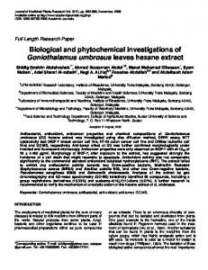
Table 1. Lysis of cyanobacteria by different bacterial pathogens.
Mechanism of cell lysis 1
2
Contact
Entrapment
3
Endoparasitism Ectoparasitism Not specified Not specified Not specified 4
Predatory bacteria S. neyagawaensis Bacillus cereus Cytophaga F. flexilis, F. sancti M. fulvus BGO2 M. xanthus PCO2 Bdellovibrio-like bacteria B. bacteriovorus Xanthomonas Saprospira albida Bacillus spp
Major host Cyanobacteria Microcystis Microcystis Microcystis Oscillatoria williamsii Phormidium luridum Phormidium luridum Microcystis aeruginosa Phormidium luridum Anabaena, Oscillatoria Microcystis aeruginosa Anabaena variabilis
Extra cellular Substances Not identified Not identified Not identified Identified Not identified Not identified Not identified Not identified Not identified Not identified Not identified
Predator- Prey ratio Not specified 1:1 Not specified Not specified 1:6 x 10⁷ 1:10 Not specified 1:1 Not specified Not specified Not specified
Flask shaking conditions Not specified Not specified Not specified Not specified 100 rpm 100 rpm Not specified Shaker Shake flasks Not specified Not specified
Reference Choi et al. (2005) Nakamura et al. (2003a) Rashidan and Bird (2001) Sallal (1994) Burnham et al. (1984) Burnham et al. (1981) Caiola and Pellegrini (1984) Burnham et al. (1976) Walker and Higginbotham (2000) Ashton and Robarts (1987) Wright and Thompson (1985)
1
Contact = Initial physical contact between bacteria and cyanobacteria is established and leads to bacterial secretion of extracellular substances causing damage to cyanobacterial cell walls. Final result is cell lysis and death. 2 Entrapment = Bacteria surround the cyanobacterial cell in ‘wolf-like pack’; establish physical contact with the cyanobacteria, bacterial secretion of extracellular substances that cause damage to cyanobacterial cell wall. Final result is cell lysis and death. 3 Endoparasitism = Bacteria penetrate the cyanobacterial cytoplasm, multiply inside cell using cyanobacterial nutrients. Final result is cell lysis and death. 4 Ectoparasitism = Bacteria do penetrate the cyanobacterial cytoplasm, associate closely with prey, deriving nutritional benefits that lead to prey death by starvation.
cyanobacteria Microcystis aeruginosa and M. viridis. The bacterium Saprospira albida, isolated from Hartbeespoort Dam, lysed Microcystis aeruginosa (Ashton and Robarts, 1987). Caiola and Pellegrini (1984) showed cells of Microcystis aeruginosa that were infected and lysed by Bdellovibrio-like bacteria in bloom containing water samples from Lake Varse, Italy. Blakeman and Fokkerna (1982) observed that naturally occurring, resident microorganisms adapt to survive and grow in a specific habitat. If these organisms were effective antagonists against a pathogen, they would be preferred for biocontrol purposes. Organisms from other habitats, which may be equally antagonistic to the pathogen, would be less likely to survive, and consequently would have to be reapplied more
frequently. The same would be true in other habitats, such as where antagonists are used to control cyanobacterial blooms. Therefore, augmentative biocontrol (deliberately enhancing the predator population through culturing in the laboratory) with resident predatory organisms is attractive as it offers certain advantages, such as being highly specific to the target organism, with no destruction of other organisms and no direct chemical pollution that might affect humans (Sigee et al. 1999). However, there are disadvantages, which include the limited destruction of the target organism, limited survival of the microbial agent or its removal by other organisms, problems of large scale production, storage and application, as well as reluctance to apply microbial agents in a field environment.
Predator-prey ratio If predatory microbial agents are present in the natural ecosystem, why then are the harmful algal blooms so persistent in nature? According to Fraleigh and Burnham (1988), the predator population in a lake could not survive and increase to a threshold density while depending on lake inorganic nutrients alone, but also required algal carbon. The predator bacteria population increased during the algal bloom period, partly due to availability of algal carbon. The control of the host prey was dependent on a threshold density exceeding 1 x 10⁷ cells.ml-1 in order to initiate cyanobacterial lysis. Rashidan and Bird (2001) isolated Cytophaga bacteria from a temperate lake in Quebec, Cana-
Gumbo et al.
da. The bacteria were capable of lysing bloom-forming cyanobacteria. The population of Cytophaga strain C1 correlated well with the abundance of Anabaena in the natural lake environment. The bacterial population was at its peak when the cyanobacterial population was at its lowest. Daft et al. (1971) isolated four bacterial pathogens of cyanobacteria of which three (CP-1, CP-2 and CP-3) were from a wastewater treatment plant (Forfar sewage works, Scotland) and the fourth (CP-4) was from a lysed Oscillatoria bloom (Lake Windermere, England). Under laboratory conditions, these bacterial pathogens were able to lyse Anabaena flos-aquae, A. circinalis, Aphanizomenon flos-aquae and Microcystis aeruginosa. The bacterium CP-1 was found to be the most effective and underwent trials with field samples in an enclosed mesocosm, and a predator-prey ratio of approximately 5 -1 10 cells.ml was needed to cause rapid lysis of Microcystis. Nakamura et al. (2003a) found that a predator-prey ratio of 1:1 was needed for Bacillus cereus to lyse a Microcystis culture. Burnham et al. (1981, 1984) isolated Myxococcus xanthus strain PCO2 and Myxococcus fulvus strain BGO2 and BGO3 from grab samples obtained from roadside ditches draining agricultural fields in Ohio, USA. The myxococcal strains effectively lysed agitated aqueous populations of Phormidium luridium and derived nutritional benefits from the cyanobacteria. M. fulvus strain BG02, at an initial predator density of 0.5 cells.ml-1, was capable of lysing a Phormidium population of 3 x 10⁷ cells.ml-1, a predator-prey ratio of 1:6 x 10⁷. Phormidium luridum was lysed by Myxococcus xanthus PCO2 when the predator-prey ratio exceeded 1:10. Phormidium luridum was also lysed by Bdellovibrio bacteriovorus, at a predator-prey ratio of 1:1 (Burnham et al.,1976). It is clear that the predator-prey ratio needed for cyanobacterial lysis is an important parameter to consider when using predatory organisms for biocontrol purposes. This ratio differs between species of prey and predator, and therefore needs to be determined for each relationship specifically. In a natural environment, it appears that the prey and predator are usually in contact with one another, but that the population of the predator is always lower. To be successful, the predator should preferably be able to colonize the cyanobacterial bloom, and multiply to numbers above the critical predator-prey ratio. Thus augmentative biocontrol may provide a means to increase the predator population to above the threshold needed to induce large-scale cyanobacterial lysis (Daft et al., 1973; Rashidan and Bird, 2001). Mechanisms of cyanobacterial lysis The mechanism of cyanobacterial lysis following exposure to a bacterial agent is poorly understood. Various mechanisms have been elucidated, including antibiosis, production of lytic enzymes, parasitism and competitive exclusion (Table 1). Cyanobacterial lysis by bacteria is
4769
caused by: contact lysis (Nakamura et al., 2003a; Choi et al., 2005); production of lytic enzymes or extracellular products (Burnham et al., 1981; Nakamura et al., 2003a); antibiosis after entrapment of the host (Daft et al., 1985; Sigee et al., 1999) and parasitism (Caiola and Pellegrini, 1984; Rashidan and Bird, 2001). Contact lysis The cyanobacterial cell wall resembles that of a Gramnegative bacterium, but is significantly thicker (Rapala et al., 2002). The cell wall consists of three or four outer layers between the plasma membrane (or plasmalemma) and the sheath (Holm-Hansen, 1968). The cell wall thickness may range from 10 to 20 nm and is coated with a relatively thick capsule of proteinaceous material (Skulberg et al., 1993). The outer membrane may be smooth or contain invaginations. It extends into the cell to form structures called mesosomes, which regulate substances entering and exiting the cell. In the cytoplasm, there are thylakoid membranes which are considered as sites for enzymatic reactions including photosynthesis, electron transport and ATP synthesis. The inner membrane consists of globular protein and mucopolymer molecules, with the mucopeptides being responsible for the additional structural strength of the cell. The cyanobacterial cell wall can be disrupted by the enzymatic actions of lysozyme and penicillin (HolmHansen, 1968). Burnham et al. (1984) examined the degradation of cyanobacteria by bacteria and pointed out that the peptidoglycan component of the cyanobacterial cell wall was the ‘weak link’ against predatory bacteria. Cyanobacterial lipopolycaccharides (LPS) differ to the LPS of other Gram-negative bacteria. They have a greater variety of long chain unsaturated fatty acids and hydroxy fatty acids with two or more double bonds, including the unusual fatty acid β-hydroxypalmitic acid which is found in the lipid A moiety. Other Gram-negative bacteria contain almost exclusively saturated and mono-unsaturated fatty acids with one double bond. Cyanobacterial LPS often lack ketodeoxyoctonate, a common LPS component of Gram-negative bacterial outer membranes, and contain only small amounts of bound phosphates when compared with other bacteria (Brock et al., 1994; Hoiczyk and Hansel, 2000). Contact between the predatory bacterium and the cyanobacterium is a pre-requisite for effective lysis to take place. Shilo (1970) and Daft et al. (1971) observed that during this contact, the predatory bacteria released lytic enzymes or extracellular substances that resulted in the dissolution of the cyanobacterial cell membrane. Agitation or turbulence disturbed this physical contact, and no cyanobacterial cell lysis was observed in the absence of contact. This indicated that the lysing enzyme was not excreted into the medium. Cyanobacterial lysis of Lysobacter by bacterium CP isolates again illustrated that
4770
Afr. J. Biotechnol.
contact was necessary for lysis (Daft et al., 1985; Rashidan and Bird, 2001). Although no extracellular lytic enzymes were produced by CP isolates, within 20 min after establishing contact with the cyanobacteria, the host cell was disrupted, presumably due to the transfer of enzymes across the adjacent cell walls. This type of predation involved the production of extracellular chemicals or enzymes by the prey during contact with the host. Daft et al. (1971) showed that extracellular products alone are insufficient for lysis to occur, and that the bacterial cells must be present. Bacteria caused lysis of Nostoc ellipsosporum by inhibiting algal metabolic activity (nitrogenase activity and photosynthesis). There was no evidence of extracellular enzymes but the enzymes responsible for causing cyanobacterial cell lysis appeared to be on the bacterial surface, provided that there was contact between the organisms. Myxobacter lysis of vegetative cells of Nostoc ellipsosporum was observed whereas heterocysts were unaffected. As the cell walls of heterocysts contain cellulose and those of vegetatative cells do not, this suggested that the bacteria were unable to degrade cellulose. Adams and Duggan (1999) again demonstrated the greater resistance of heterocysts and akinetes to predatory bacteria when compared with vegetative cells. This might be attributed to the thicker heterocyst cell wall. During the differentiation of a vegetative cell into a heterocyst, major structural and biochemical changes occurred that affected nitrogen fixation. The cell wall was thickened by the decomposition of three extra layers external to the normal cell structure. The inner layer consisted of glycolipid; the center layer was a homogeneous layer consisting of polysaccharide, and the outer layer was a fibrous layer. The culture supernatant of B. cereus was effective in the lysis of M. aeruginosa and M. viridis (Nakamura et al., 2003a). Based on microscopic observation, the B. cereus cells were observed to attach to the surface of the cyanobacteria cell thereby inducing cell aggregation. The extracellular substances that were released lysed the cyanobacterial cell wall, leaving the chlorophyll a intact. The extracellular substances effectively lysed the cyanobacterial cells within 24 h under alkaline conditions, which are most prevalent during a bloom. The unidentified extracellular substances were non proteinaceous, hydrophilic, heat stable and had a molecular weight of less than 2 kDa. The studies of Choi et al. (2005) showed that the unidentified anti-algal substances originated in the bacterial periplasm and were secreted when the bacterium, S. neyagawaensis, was in physical contact with M. aeruginosa. Although the growth of M. aeruginosa was suppressed, there was no increase in bacterial biomass. Antibiosis after entrapment of host Burnham et al. (1981, 1984) indicated that the entrapment of cyanobacteria and release of enzymes, possibly
antibiotics appeared to be an efficient system for cyanobacterial cell lysis. The predatory bacteria Myxococcus xanthus PCO2 and M. fulvus BGO2 were capable of inducing lysis of both agar- and liquid-grown cultures of the filamentous cyanobacterium Phormidium luridum, var. olivacea. The predatory bacteria caused rapid cyanobacterial lysis in agitated liquid grown cultures of Phormidium, which indicated that a mechanism other than the contact lysis was operating. It appeared that Myxococcus formed colonial spherules, which entrapped the cyanobacteria prey in a ‘wolflike manner’. The formation of these spherules was dependent on the number of myxococci per ml in an aqueous environment. It took about an hour to form mature spherules with 107 myxococci per ml, followed by rapid lysis of 107 Phormidium cells per ml (a predatorprey ratio of 1:1). The cyanobacterial prey cultures were inoculated with myxococci (predator-prey ratios of 1:10 and 1:100) and were lysed within 48 h. The earliest sign of cyanobacteria degradation was shown by light microscopy and involved the separation of a trichome into shorter filaments and single cells. The motile nature of Myxococcus, gradually moved the filamentous cyanobacteria to the centre of the core of the spherule. Once the cyanobacteria reached the core, there was physical contact between the predator and prey leading to the release of enzymes that acted on the cyanobacterial cell wall. Transmission electron microscopy studies showed that the Phormidium skeletal remains lacked the peptidoglycan layer. Myxococcus strains appeared to be effective predators, especially M. fulvus BGO2, which lysed a Phormidium culture with a density of 107 cells per ml, reducing it to 103 in 2 days (Fraleigh and Burnham, 1988). The standard reference strain M. xanthus ATCC 25232 caused very little cyanobacteria lysis. Myxococcus strains lysed cyanobacteria cells of Phormidium growing in an agitated autotrophic aqueous environment. This is important for biological control of cyanobacteria. In nature, the aqueous environment is never ‘still’ but in continuous flux, causing mixing of water columns and layers. Parasitism There are few published reports on Bdellovibrio (Burnham et al., 1976) and Bdellovibrio-like bacteria (Wilkinson, 1979; Caiola and Pellegrini, 1984) that caused cyanobacteria lysis. In a separate but unrelated study, Burnham et al. (1968) demonstrated that Bdellovibrio bacteriovorus penetrated a Gram-negative Escherichia coli, causing its lysis and death. Burnham et al. (1968) demonstrated the endoparasitic behaviour of B. bacteriovorus. The Bdellovibrio’s actively and violently stroke the host, E. coli, with the end of the cell opposite the sheathed flagellum. During this initial period of irreversible attachment to host, Bdellovibrio commenced a grating motion which lasted for several minutes as
Gumbo et al.
observed by phase contrast microscopy. During attachment the Bdellovibrio developed unique receptors that bound tightly to the host. Attempts to separate the Bdellovibrio and hosts using violent shaking or vortex mixing at maximum speed had no visible effect. The Bdellovibrio continued to push into the host cytoplasm space while the host was constricting in an attempt to prevent entrance by the predator. At the penetration pore, there was no visible damage to the host cell wall. Once inside the prey, Bdellovibrio commenced to inactivate host metabolism and feed off its nutrients (Yair et al., 2003). The exhaustion of cytoplasm contents triggered the Bdellovibrio to undergo multiple fission replications to produce progeny called bdelloplast. The bdelloplast, now flagellated, emerged after breaking the prey cell wall leaving behind ghost prey remnants. The bacterium B. bacteriovorus behaved as an ectoparasite. When the bacterium was added to an aqueous culture of Phormidium luridum it caused lysis of the cyanobacteria (Burnham et al., 1976). The mechanism of cyanobacterial lysis was not endoparasitic as expected; an extracellular substance was released that dissolved the cyanobacteria cell wall. The bacterium was then able to gain nutrients from the cyanobacterium. FIELD APPLICATION OF BIOLOGICAL CONTROL AGENTS Although there are non-indigenous bacterial agents that have been isolated and characterised, it appears that the studies on application of biocontrol agents are rather limited. Most of the studies have been limited to lysis of laboratory-cultured cyanobacteria. Before application of bacterial biocontrol agents to freshwater systems, information must be available on: the anti-algal activity against target alga, the effects of bacteria on other organisms in the freshwater ecosystem and the prediction of the algal dynamics after removal of target alga (Choi et al., 2005). Another aspect of importance is agitation. Shilo (1970) and Daft et al. (1971) found that cyanobacterial lysis was ineffective if there was agitation, especially where contact lysis was involved. Under natural conditions, rapid mixing may favour the proliferation of cyanobacteria and discourage attachment of predatory bacteria. During a field trial performed by Wilkinson (1979) and Caiola and Pellegrini (1984) a Bdellovibrio-like bacterium caused lysis of Neofibularia irata, Jaspis stellifera and Microcystis cells respectively. The bdelloplast were localised within the cell wall and cyanobacteria cytoplasm membrane. The infecting bacterium was similar in size and appearance to previously described Bdellovibrio’s. These observations, though not replicated under controlled laboratory conditions, indicated the possibility of endoparasitism of the cyanobacteria by Bdellovibrio-like bacteria. The Bdellovibrio-like bacteria are an attractive biocontrol agent because they penetrate the host cells
4771
specifically, exhaust host cell contents and replicate within to form bdelloplasts, which attack further cyanobacteria cells. Nakamura et al. (2003b) immobilised Bacillus cereus N-14 in floating biodegradable plastic carriers, at a cell concentration of 3 x 10⁷ cells/ml per 1 g-dry weight of starch-carrier float. This was used as an effective in situ control of natural floating Microcystis blooms, eliminating 99% of floating cyanobacteria in 4 days. The bacteria utilized the starch as a nutrient source and amino acids were derived from the lysis of Microcystis. The floating carrier enabled immobilized bacteria to be directed to floating cyanobacteria blooms. Asaeda et al. (2001) installed two vertical curtains having depths that covered the epilimnion thickness of Terauchi dam in Japan. The purpose of the curtains was to curtail the nutrient supply from nutrient rich inflows to the downstream epilimnion of the reservoir. There was a marked reduction in cyanobacterial blooms downstream from the curtain in spring and summer. The curtain prevented the direct intrusion of nutrients into the downstream zone. Epilimnion cyanobacteria concentrations were higher in the upstream zones. Thus, within the upstream zone the cyanobacteria consumed large amounts of the inflow nutrients, reducing the nutrient supply to the downstream zone of the reservoir. Thus floating curtains such as these may be used to segregate Microcystis algal blooms, minimising turbulence and allow for the introduction of Nakamura et al. (2003b) floating carrier. This would allow the introduction of microbial antagonists, and afford the predator ample time to attach to the prey and initiate the lytic process. CONCLUSION There are increasing demands to reduce the use of chemicals such as copper sulphate or organo-copper compounds for HAB management for environmental and safety reasons. Thus, the development of a non-chemical control measure such as biocontrol is of great importance. Predatory bacteria are the more potent biological control agent when compared with viral pathogens. These bacterial agents have been isolated from a variety of sources such as the terminal stages of harmful algal blooms. In some studies, bacteria such as Sphingomonas species capable of biodegrading microcystins were isolated from eutrophic waters. Further research is required to evaluate whether these bacteria are antagonistic towards cyanobacteria. Ideally, a combination of strategies should be introduced; that is, combine predatory bacteria that directly lyse the cyanobacteria with microcystin degrading bacteria that then ‘mop up’ the released microcystins. Although the mechanisms of cyanobacterial lysis have been proposed, which include antibiosis, production of lytic enzymes, parasitism and competition for nutrients
4772
Afr. J. Biotechnol.
and space; it is often difficult to ascribe cyanobacterial lyses to one mechanism only. The predatory bacteria that induced cyanobacterial lysis appear to act in four major ways: contact lysis, production of lytic enzymes or extracellular products, antibiosis after entrapment of host and endoparasitism or ectoparasitism of host. Most of these studies were based on laboratory cultures, and need to be extended to field trials to determine which mechanisms may be applicable to large-scale applications. Bacterial antagonists have been isolated against a wide variety of cyanobacteria under laboratory conditions. However, care should be taken when extrapolating laboratory-based observations to field conditions. This requires an understanding of the mechanism of interaction between the cyanobacteria and predatory bacteria. Very little information is available on the successful use of predatory bacteria under natural conditions. The predator-prey ratio needed for cyanobacterial lysis is an important parameter to consider when using predatory organisms for biological control purposes. It is clear that the critical predator-prey ratio needs to be met or exceeded if successful cyanobacterial lysis is to occur. Augmentative biocontrol may provide a means to increase the predator population to above the threshold needed to induce large-scale cyanobacterial lysis. Further studies are required in the development of antialgal chemicals such as protease I that may cleave pentaglycine bridge in the cell wall of cyanobacteria. These anti-algal substances may be less toxic to the environment when compared to copper algicides. REFERENCES Adams DG, Duggan PS (1999). Transley Review No. 107 Heterocyst and Akinetes differentiation in cyanobacteria. New Phytol. 144: 3-33. Asaeda T, Pham HS, Nimal PDG, Manatunge J, Hocking GC (2001). Control of algal blooms in reservoirs with a curtain: a numerical analysis. Ecol. Eng. 16: 395-404. Ashton PJ, Robarts RD (1987). Apparent predation of Microcystis aeruginosa kutz emend elenkin by a saprospira-like bacterium in a hypertrophic lake (Hartbeespoort dam, South Africa). J. Limnol. Soc. S. Afr. 13: 44-47. Atlas RM, Bartha R (1998). Microbial ecology: fundamentals and th applications. 4 edition. Benjamin/Cummings Science Publishers, 2725 sand Hill Road, Menlo Park, California 94025. p. 698. Blakeman JP, Fokkerna NJ (1982). Potential for biological Control of plant diseases on the phylloplane. Ann. Rev. Phyopathol. 20: 167192. Bourne DG, Jones GJ, Blakeley RL, Jones A, Negri AP, Riddles P (1996). Enzymatic pathway for the bacterial degradation of the cyanobacterial peptide toxin microcystins LR. Appl. Environ. Microbiol. 62: 4086-4094. Brock TD, Madigan MT, Martinko JM, Parker J (1994). Biology of Microorganisms, 7th Edn., 909 pp. Prentice-Hall International, Englewood Cliffs, NJ. Burnham JC, Hashimoto T, Conti SF (1968). Electron microscopic observations on the penetration of Bdellovibrio bacteriovorus into Gram-negative bacterial hosts. J. Bacteriol. 96: 1366-1381. Burnham JC, Stetak T, Gregory L (1976). Extracellular lysis of the bluegreen alga Phormidium luridum by Bdellovibrio bacteriovorus. J. Phycol. 12: 306-313. Burnham JC, Collart SA, Highison BW (1981). Entrapment and lysis of
the cyanobacterium Phormidum luridum by aqueous colonies of Myxococcus xanthus PCO2. Arch. Microbiol. 129: 285-294. Burnham JC, Susan AC, Daft MJ (1984). Myxococcal predation of the cyanobacterium Phormidium luridum in aqueous environment. Arch. Microbiol. 137: 220-225. Caiola MG, Pellegrini S (1984). Lysis of Microcystis aeruginosa (Kutz) by Bdellovibrio-like bacteria. J. Phycol. 20: 471-475. Choi HJ, Kim BH, Kim JD, Han MS (2005). Streptomyces neyagawaensis as a control for the hazardous biomass of Microcystis aeruginosa (Cyanobacteria) in eutrophic freshwaters. Biol. Control. 33: 335-343. Cronberg G, Carpenter EJ, Carmichael WW (2003). Taxonomy of harmful cyanobacteria. In: Hallegraeff GM, Anderson DM, Cembella AD (eds.), Manual on Harmful Marine Microalgae. Unesco Publishing pp. 523-562. Daft MJ, Stewart WDP (1971). Bacterial pathogens of freshwater blue green algae. New Phytol. 70: 819-829. Daft MJ, McCord SB, Stewart WDP (1975). Ecological studies on algal lysing bacteria in fresh waters. Freshwater Biol. 5: 577-596. Daft MJ, Burnham JC, Yamamato Y (1985). Algal blooms: conesquences and potential cures. J. Appl. Bacteriol. Symposium Supplement. 175S-186S. Falconer IR, Beresford AM, Runnegar MTC (1983). Evidence of liver damage by toxin from a bloom of the blue green alga, Microcystis aeruginosa. Med. J. Austr. 1: 511-514. Fraleigh PC, Burnham JC (1988). Myxococcal predation on Cyanobacterial Populations: Nutrient Effects. Limnol. Oceanogr. 33: 476-483. Greenop B, Robb M (2001). Phosphorus in the Canning: 1999-2000 Phoslock™ trials. River Sci. 17: 1-7. Haider S, Naithani V, Viswanathan PN, Kakkar P (2003). Review Cyanobacterial toxins: a growing environmental concern. Chemosphere. 52: 1-21. Harding WR, Paxton B (2001). Cyanobacteria in South Africa: A Review. WRC Report No. TT 153/01, July 2001. Harding WR, Thornton JA, Steyn G, Panuska J, Morrison IR (2004). Hartbeespoort dam Remediation Project (Phase 1). Final Report (Volume 1). Project Number 58/2003. Completed October 2004. Department of Agriculture, Conservation, Environment and Tourism (DACET) of the Provincial Government of North West Province (NWPG), South Africa, p. 166. Hoeger SJ, Shaw G, Hitzfeld BC, Dietrich DR (2004). Occurrence and elimination of cyanobacterial toxins in two Australian drinking water treatment plants. Toxicon. 43: 639-649. Hoiczyk E, Hansel A (2000). Cyanobacterial cell walls: news from an unusual prokaryotic envelope. J. Bacteriol. 182: 1191-1199. Holm-Hasen O (1968). Ecology, physiology and biochemistry of bluegreen algae. Ann. Rev. Microbiol. 22: 47-70. Lam AKY, Prepas EE, David S, Hrudey SE (1995). Chemical control of heptatotoxic phytoplankton Blooms: Implications for human health. Water Res. 29: 184-554. rd Mason CF (1996). Biology of freshwater pollution, 3 edition. Longman, Essex. NHMRZ/ARMCANZ (2001). Australian drinking water guidelines, microorganism 3: toxic algae, Fact Sheets No. 17a-17d, National Health and Medical Research Council, Agriculture Resource Management Council of Australia and New Zealand, Canberra. Nakamura N, Nakano K, Sungira N, Matsumura M (2003a). A novel control process of cyanobacterial bloom using cyanobacteriolytic bacteria immobilized in floating biodegradable plastic carriers. Environ. Technol. 24: 1569-1576. Nakamura N, Nakano K, Sugiura N, Matsumura M (2003b). A novel cyanobacteriolytic bacterium, Bacillus cereus, Isolated from a Eutrophic Lake. J. Biosci. Bioeng. 95: 179-184. Oberholster PJ, Botha AM, Grobbelaar JU (2004). Review Microcystis aeruginosa: source of toxic microcystins in drinking water. Afr. J. Biotechnol. 3: 159-168. Rapala J, Lahti K, Niemelä SI, Sivonen K (1994). Biodegradability and adsorption on lake sediments of cyanobacterial hepatoxins and anatoxin-a. Lett. Appl. Microbiol. 19: 423-428. Rapala J, Lahti K, Räsänen LA, Anna-Liisa E, Niemelä SI, Sivonen K (2002). Endotoxins associated with cyanobacteria and their removal during drinking water treatment, Water Res. 36: 2627-2635.
Gumbo et al.
Rashidan KK, Bird DF (2001). Role of predatory bacteria in the termination of a cyanobacterial bloom. Microbial Ecol. 41: 97-105. Richard DS, Beatiie KA, Codd GA (1983). Toxicity of cyanobacterial blooms from Scottish freshwaters. Environ. Technol. Lett. 4: 377-382. Robb M, Greenop B, Goss Z, Douglas G, Adeney J (2003). Application of Phoslock an innovative phosphorus binding clay, to two Western Australian waterways: preliminary findings. Hydrobiologia. 494: 237243. Sallal AKJ (1994). Lysis of cyanobacteria with Flexibacter spp isolated from domestic sewage. Microbios. 77: 57-67. Secord D (2003). Biological control of marine invasive species: cautionary tales and land-based lesions. Biol. Invasions. 5: 117-131. Shilo M (1970). Lysis of Blue Green Algae by Myxobacter. J. Bacteriol. 104: 453-461. Sigee DC, Glenn R, Andrews MJ, Bellinger EG, Butler RD, Epton HAS, Hendry RD (1999). Biological control of cyanobacteria: principles and possibilities. Hydrobiologia In The Ecological Bases for Lake and Reservoir Management, Harper DM, Brierley, Ferguson AJD, Philips G (eds), Kluwer Academic Publishers, Netherlands. 395(396): 161172. Sivonen K, Jones G (1999). Cyanobacterial toxins. In: Chorus I, Bartram J (eds.), Toxic Cyanobacteria in water, E & FN Spon, London, pp. 41-111. Skulberg OM, Carmichael WW, Codd GA, Skulberg R (1993). Taxonomy of toxic Cyanophyceae (cyanobacteria). In: Falconer R. (Ed.). Algal toxins in seafood and drinking water. Academic Press Ltd., London, pp. 145-164. Ueno Y, Nagata S, Tsutsumi T, Hasegawa A, Watanabe MF, Park HD, Chen GC, Yu SZ (1996). Detection of microcystins, a blue green algal hepatotoxin, in drinking water sampled in Hauimen and Fusui, endemic areas of primary liver cancer in China, by highly sensitive immunoassay. Carcinogenesis. 17: 1317-1321.
4773
Walker HL, Higginbotham LR (2000). An aquatic bacterium that lyses cyanobacteria associated with off-flavor of channel catfish (Ictalurus punctatus). Biol. Contr. 18: 71-78. Wilkinson CR (1979). Bdellovibrio-Like Parasite of Cyanobacteria Symbiotic in Marine Sponges. Arch. Microbiol. 123: 101-103. World Health Organization (WHO) (1998). Guidelines for Drinking Water Quality. Second Edition, addendum to Volume 2, Health Criteria and Other Supporting Information, World Health Organization, Geneva, pp. 95-110. World Health Organization (WHO) (1999). Toxic cyanobacteria in water, Chorus I, Bartram J, (eds), E&FN Spon, Routledge, London. Wright SJL, Thompson RJ (1985). Bacillus volatiles antagonize cyanobacteria. FEMS Microbiol. Lett. 30: 263-267. Yair S, Yaacov D, Susan K, Jurkevitch E (2003). Small eats big: ecology and diversity of Bdellovibrio and like organism, and their dynamics in predator-prey interactions. Agronomie. 23: 433-439. Yamamoto Y, Kouchiwa T, Hodoki Y, Hotta K, Uchida H, Harada K (1998). Distribution and identification of actinomycetes lysing cyanobacteria in a eutrophic lake. J. Appl. Phycol. 10: 391-397. Zohary T (1987). On the ecology of hyperscum-forming Microcystis aeruginosa in a hypertrophic African lake. Unpublished PhD Thesis, University of Natal, Pietermaritzburg.