Anuário do Instituto de Geociências - UFRJ ISSN 0101-9759
Vol. 29 - 1 / 2006 p. 108-128
FORAMS 2006
Bleaching in Foraminifera with Algal Symbionts: Implications for Reef Monitoring and Risk Assessment Pamela Hallock1; D. E. Williams2; E. M. Fisher1 & S. K. Toler1 1
College of Marine Science, University of South Florida, 140 7th Ave. S., St. Petersburg, FL 33701, U.S.A.
[email protected] 2 RSMAS/CIMAS, 4600 Rickenbacker Causeway, Miami, Florida 33149, U.S.A.
Abstract Reef-dwelling larger foraminifers share key characteristics with reefbuilding corals: they are prolific producers of calcium carbonate, they are physiologically dependent upon algal endosymbionts, and representatives of both groups have suffered bleaching episodes in recent decades. Since 1991, bleaching has been observed in populations of Amphistegina in all subtropical oceans, with peak bleaching in 1992 and secondary peaks in 1998 and 2005. Amphistegina populations exhibiting chronic, intermediate-intensity bleaching characteristically show anomalously high incidences of shell breakage, shell deformities, evidence of predation, and microbial infestation. Asexual reproduction is profoundly affected; broods from partly bleached parents typically have fewer individuals, many of which are anomalous in shape and size. Key differences between bleaching in corals and Amphistegina are that corals typically bleach by expelling their symbionts, while Amphistegina bleach when damaged symbionts are digested, and that mass coral bleaching requires high light but correlates most consistently with elevated temperatures, while bleaching in Amphistegina is induced by light. Amphistegina are particularly sensitive to the shorter (300-490 nm) wavelengths of solar radiation, which have increased in intensity relative to longer visible wavelengths (>490-700 nm) in clear reef waters over the past 30 years as a consequence of stratospheric ozone depletion. Abundances and visual assessments of
FORAMS 2006 Bleaching in Foraminifera with Algal Symbionts: Implications for Reef Monitoring and Risk Assessment Pamela Hallock; D. E. Williams; E. M. Fisher & S. K. Toler
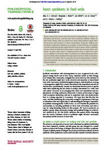
Amphistegina populations can be used as a low-cost risk-assessment tool. These protists are sensitive to environmental conditions over days to weeks, and provide a method to quickly distinguish between water quality (local) and photo-oxidative (global) stresses. Risk assessments based on the combined use of in situ measurements and low-cost indicators can provide resource managers with essential information to decide when more costly chemical or molecular procedures are needed to determine local sources of stress. Keywords: Foraminifera; coral; bleaching; symbiosis; UV; photoinhibition; population dynamics 1 Introduction Calcifying plant/animal symbioses, notably reef-building corals and larger benthic foraminifers, are unique components of tropical coastal ecosystems because they are not only important biological components, but also key producers of the geologic substratum (i.e., reef structure and sediments). Over the last 30 years, scientists have witnessed the decline of reef-building coral populations and communities, first locally, then over whole reef tracts and regions. Among the first victims were western Atlantic/Caribbean acroporids, which were decimated by white-band disease beginning in the 1970s (Gladfelter, 1982). Then came the first mass coral-bleaching events in 1983 and 1987 (Glynn, 1984; Williams & Bunckley Williams, 1990). By the late 1990’s, most scientists recognized that coral reefs were in decline worldwide (e.g., Dight & Scherl, 1997; Eakin et al., 1997; Risk, 1999), as the 1997-98 ENSO event was triggering mass bleaching of corals unprecedented in its global scale and intensity (e.g., Hoegh-Guldberg, 1999; Lough, 2000). 2 Discovery of Bleaching in Foraminifers Signs of bleaching are now commonly observed, at least during summer months, in members of the reef-dwelling, benthic foraminiferal genus Amphistegina. Bleaching is well documented in A. gibbosa d’Orbigny (Figure 1), the most common western Atlantic/Caribbean species, and in A. lessonii d’Orbigny, which is the most similar Indo-Pacific species. Bleaching also has been observed in A. lobifera Larsen and A. radiata (Fichtel & Moll). Although noted in laboratory experiments in the early 1980s (Hallock et al., 1986), bleaching was unknown in field populations until 1988, when a very small sample collected in the Bahamas during a post-bleaching coral survey revealed several A. gibbosa specimens that appeared “mottled” (Hallock et al., 1992). Healthy populations of A. gibbosa were observed in the Florida Keys, USA, as late as Anuário do Instituto de Geociências - UFRJ ISSN 0101-9759 - Vol. 29 - 1 / 2006 p. 108-128
109
FORAMS 2006 Bleaching in Foraminifera with Algal Symbionts: Implications for Reef Monitoring and Risk Assessment Pamela Hallock; D. E. Williams; E. M. Fisher & S. K. Toler
mid-May 1991. Early signs of bleaching were noted in late June 1991 during extremely calm local conditions (Hallock et al., 1992). By September 1991, approximately 80% of the adult A. gibbosa were partially to extensively bleached in Florida reef tract populations. By November, population densities had plummeted by 95%. The discovery of bleaching in Amphistegina populations prompted monitoring of Florida reef-tract populations, sampling of populations elsewhere as resources permitted, and laboratory observations and experiments. Sampling at Heron Island, Great Barrier Reef, in March 1992, confirmed that bleaching was occurring in A. lessonii and A. lobifera populations, and first revealed unusual shell breakage and repair (Hallock & Talge, 1993). Since 1992, bleaching has been observed in Amphistegina populations in all three oceans (Hallock, 2000).
Figure 1 Partly bleached, adult (>0.6 mm diameter) Amphistegina gibbosa. Scale bar 0.3 mm.
110
Anuário do Instituto de Geociências - UFRJ ISSN 0101-9759 - Vol. 29 - 1 / 2006 p. 108-128
FORAMS 2006 Bleaching in Foraminifera with Algal Symbionts: Implications for Reef Monitoring and Risk Assessment Pamela Hallock; D. E. Williams; E. M. Fisher & S. K. Toler
3 Bleaching in the Individual Amphistegina host naked diatom cells (i.e., cells lacking silica frustules) as endosymbionts (e.g., Lee et al., 1991). The calcite shell contains two kinds of cytoplasm, endoplasm and ectoplasm, which carry out distinct functions. The endoplasm, which is found within the shell in older chambers, contains the host nucleus or nucleii, a variety of kinds of organelles, lipid storage bodies, and the diatom endosymbionts. The latter are typically found just below the outer walls of the chambers (Talge & Hallock, 2003). The interior surface of the chamber wall is lined with pore cups, superficially resembling an egg crate (see, e.g., Toler & Hallock, 1998, Pl. 2); the diatom symbionts, though intracellular, characteristically are found one per pore cup along the periphery of the endoplasm (Talge & Hallock, 2003). The ectoplasm occurs primarily in the two most recently added chambers, from which it extends out of the shell as granuloreticulopodia, the streaming, anastomosing, microtubule-laden pseudopodia characteristic of the protoctistan Phylum Granuloreticulopodia (Lee et al., 2000). The functions of the ectoplasm include movement, food acquisition, and chamber formation. In addition to microtubles, organelles present in the ectoplasm include mitochondria and lysosomes (Travis & Bowser, 1991). Bleaching in Amphistegina, as in corals (e.g., Glynn, 1996), is caused by either loss of pigment in the diatoms or loss of the diatoms themselves (Talge & Hallock, 2003). Pigment loss can occur in response either to acute photo-inhibitory stress or to prolonged darkness. Loss of symbionts occurs under chronic or prolonged acute photo-inhibitory stress. Cytological studies indicates that the typical mechanism for symbiont loss is digestion by the host (Hallock et al., 1992; Talge & Hallock, 1995; 2003). Early stages of symbiont loss sometimes can be detected cytologically before color loss can be distinguished under a stereomicroscope (Talge & Hallock, 2003). However, if color loss is visible, i.e., white spots in the cytoplasm can be seen through the test wall (Hallock et al., 1992, Fig. 1.; Hallock et al., 1995, Pl. 1.1), damage will be evident cytologically (Hallock et al., 1992; Talge & Hallock, 1995). Color loss is seldom seen in A. gibbosa specimens smaller than 0.5 mm in diameter (Hallock et al., 1995). Early stages of bleaching (Hallock et al., 1992) are most commonly seen in intermediate-sized specimens, i.e., those between 0.5 and 0.8 mm in maximum diameter. Since September 1991, most specimens collected from Florida Keys sites whose diameters exceeded 0.8 mm exhibited some degree of bleaching; during summer months larger individuals have typically exhibited extensive loss of color (Williams et al., 1997; Williams, 2002). Anuário do Instituto de Geociências - UFRJ ISSN 0101-9759 - Vol. 29 - 1 / 2006 p. 108-128
111
FORAMS 2006 Bleaching in Foraminifera with Algal Symbionts: Implications for Reef Monitoring and Risk Assessment Pamela Hallock; D. E. Williams; E. M. Fisher & S. K. Toler
From a cytological perspective (Talge & Hallock, 1995; 2003), the earliest stages of bleaching include subtle damage to symbiont plastids and lysosome activity around the symbionts, followed by their subsequent breakdown. Autolysis of the endoplasm in the outer rows of chambers of the host cell is manifested as membrane deterioration, enlargement of vacuoles in the endoplasm, breakdown of organelles, and ultimately complete loss of cytoplasmic integrity. Deterioration does not appear to be reversible in chambers where autolysis occurs. However, field specimens collected in the late summer and autumn frequently are found with older chambers exhibiting extensive degradation, while recently added chambers appear normal in color, indicating uptake of healthy diatoms from the environment. An interesting paradox is that partially bleached specimens continue to add chambers. An explanation may be the division of function between the endoplasm and the ectoplasm (Talge & Hallock, 2003). Because the functions of the ectoplasm include movement, feeding and chamber addition, the ectoplasm contains mitochondria and lysosomes, so feeding provides the energy source for the ectoplasm (Travis & Bowser, 1991). We suspect that the ectoplasm is somehow “preprogrammed” to add chambers, even when the endoplasm cannot support them, explaining how bleaching-damaged individuals continue to increase in shell size. This is certainly a hypothesis that needs to be tested.
4 Bleaching in the Population Monthly monitoring of A. gibbosa populations in the Florida Keys began in May 1992, almost a year after the initial onset of bleaching. At that time, live individuals were very rare and what few could be found were at least partially bleached (Hallock et al., 1992). During the first two years, every field sampling revealed something unusual. Shell abnormalities such as distended protoconchs became relatively common; particularly striking were conjoined individuals (Hallock et al., 1995, Plate 1, Fig. 4) whose protoconchs had apparently failed to fully separate. Observing individuals in culture provided insights into anomalies in the newly collected field specimens. When partially bleached specimens tried to asexually reproduce, instead of producing broods of several hundred nearly identical offspring (see Harney et al., 1998, Plate 1.1), the results varied from production of several hundred tiny progenitor cells that failed to calcify, to production of a few misshapen offspring (see Harney et al., 1998, Plate 1.2). These reproductive failures indicated why the populations declined so 112
Anuário do Instituto de Geociências - UFRJ ISSN 0101-9759 - Vol. 29 - 1 / 2006 p. 108-128
FORAMS 2006 Bleaching in Foraminifera with Algal Symbionts: Implications for Reef Monitoring and Risk Assessment Pamela Hallock; D. E. Williams; E. M. Fisher & S. K. Toler
dramatically in fall 1991 and why they remained low through 1992, as well as why shell abnormalities became common in the field populations. Prior to the discovery of bleaching in Florida Keys populations in 1991, one of us (PH), had more than 20 years experience with field collections and laboratory-culture of Amphistegina spp. from both the Indo-Pacific and western Atlantic/Caribbean, including observations of thousands of asexual reproductions. Amphistegina appeared to conform to the classic foraminiferal life cycle of alternation of asexual and sexual generations (e.g., Lee et al., 1991). Asexual reproduction by multiple fission is the principal mechanism for increasing population density; sexual reproduction by gamete broadcasting has a much lower probability for success, even when population densities are high. In Florida reef-tract populations, asexual reproduction occurred most commonly in the spring, thereby rapidly increasing populations in the spring and summer. Sexual reproduction was more common in the autumn. Individuals grown to maturity in the laboratory from asexual broods prior to 1991 always produced gametes, never subsequent asexual broods. Yet specimens collected in September and November 1991, which successfully produced asexual broods in the laboratory, produced lineages of as many as four successive asexual generations (Harney et al., 1998). This dramatic discovery may indicate one reason why these foraminifers are so successful. When a population has been decimated by a stress, asexual reproduction can replicate surviving genotypes, increasing their populations and thereby providing the potential for later success of sexual reproduction. Bleaching stress also resulted in another shift in basic life-history strategy. Prior to 1991, asexual reproduction was seldom seen in individuals less than approximately 1 mm in maximum diameter (Hallock et al., 1986 and unpublished). However, as Hallock et al. (1992, Fig. 1) illustrated photographically and Hallock et al. (1995) and Williams et al. (1997) demonstrated numerically, after 1991 bleaching was seldom seen in smaller individuals, while specimens larger than 1 mm were typically extensively bleached. Hallock et al. (1995) and Talge et al. (1997) reported that, during summer months when bleaching was most prevalent, attempts at asexual reproduction were predominantly seen in specimens whose average diameter was less that 1 mm, at the same time proportions of non-viable or partially viable broods also increased. For example, in June through August 1993, fewer than 10 normal broods were produced of more than 50 asexual reproduction attempts observed in freshly collected specimens (Hallock et al., 1995); well over half of the parent specimens were smaller than 1 mm. A shift to earlier reproduction is a textbook response to increased mortality rates in adult size Anuário do Instituto de Geociências - UFRJ ISSN 0101-9759 - Vol. 29 - 1 / 2006 p. 108-128
113
FORAMS 2006 Bleaching in Foraminifera with Algal Symbionts: Implications for Reef Monitoring and Risk Assessment Pamela Hallock; D. E. Williams; E. M. Fisher & S. K. Toler
classes, particularly in suicide-reproducing (iteroparous) foraminifers (e.g., Hallock, 1985). Williams et al. (1997) reported on temporal trends in A. gibbosa populations sampled monthly on the Florida reef tract between 1992 and 1996. The highest incidences of bleaching occurred in May through July and were consistently lower in August and September, which are typically months when sea-surface temperatures peak in this region (Hallock et al., 1995; Williams, 2002). Over the 5 year study (Williams et al., 1997), the intensity of bleaching declined, as indicated by a decrease in the percentage of the adult population exhibiting bleaching during the summer maxima from 86% in 1992 to 60% in 1996. At the same time, proportions of juveniles in the population nearly doubled, indicating increased reproductive success as the stress diminished in intensity. Williams (2002) analyzed other population parameters including abundance, size- frequency distributions, mean individual size (diameter), percent juveniles, and percent of bleached adults, finding that all were influenced by bleaching. Bleaching resulted in suppression of reproduction and low numbers of juveniles in the populations; years with the highest incidences of bleaching were also the years with the lowest population densities and lowest proportions of juveniles. 5 Bleaching and Biotic Interactions Anomalous shell breakage was first noticed in field populations of Amphistegina lessonii from Heron Island in March 1992. During summer 1992 monitoring of Florida reef tract populations, shells of many specimens were remarkably fragile. When population densities began to recover in 1993, we began to record significant incidences of shell breakage in the field populations (Hallock & Talge, 1995, Pl. 1.2-3; Toler & Hallock, 1998, Pl.1.2-6). We also documented epiphytization and microbial infestation of the shells of live individuals (e.g., Hallock et al., 1995, Pl. 1.3; Toler & Hallock, 1998, Pl. 1.6) Hallock & Talge (1994) described a new species of foraminifer (Floresina amphiphaga Hallock and Talge) that was preying upon Amphistegina specimens. They also found that the F. amphiphaga were significantly more likely to be found on the intermediate-sized individuals (which tended to show early signs of bleaching), while being less likely to prey on healthy-appearing juveniles or extensively bleached adults. Shell breakage data for samples collected in the 1990s were compared with breakage in archived samples collected in the 1970s and 1980s (Hallock et al., 1995; Toler & Hallock, 114
Anuário do Instituto de Geociências - UFRJ ISSN 0101-9759 - Vol. 29 - 1 / 2006 p. 108-128
FORAMS 2006 Bleaching in Foraminifera with Algal Symbionts: Implications for Reef Monitoring and Risk Assessment Pamela Hallock; D. E. Williams; E. M. Fisher & S. K. Toler
1998). Incidences of broken shells, including evidence for breakage and repair, were variable in the samples from the 1990s, ranging from ~15% to >40%, which was consistently higher than 5-6 percent incidences of breakage in archived samples of A. gibbosa and A. lessonii specimens that had been collected live prior to 1991. Toler (2002) documented that breakage characteristics were consistent with predator-induced breakage reported in the literature. She also interpreted interannual trends in breakage, reporting that incidences of breakage were not as high during years that bleaching was most acute and widespread, and consequently when population densities were significantly reduced (1992-93). She found intermediate percentages of breakage during years when bleaching was less intense (1995-1997 and 1999). She found the highest incidences of breakage during 1994 and 1998, which were years when bleaching was very prevalent but not so intense that Amphistegina abundances were severely diminished. 6 Comparison with Bleaching in Corals Two important factors in global decline of coral reefs have been bleaching and disease. Mass coral-bleaching events occur in response to photo-oxidative stress that becomes acute when temperatures exceed normal maxima (e.g., Glynn, 1996; Warner et al., 1999; Downs et al., 2002). Reef-dwelling larger foraminifers share key characteristics with reef-building corals: both groups are prolific producers of calcium carbonate, both groups are physiologically dependent upon algal endosymbionts, and representatives of both groups have suffered bleaching episodes in recent decades. A key difference between bleaching in corals and foraminifers is that events of mass bleaching in corals have correlated most consistently with elevated sea-surface temperatures, while bleaching in Amphistegina is demonstrably associated with photoinhibitory stress (Hallock et al., 1995; Talge & Hallock, 2003; Williams & Hallock, 2004) and minimally influenced by temperature, at least up to month-long exposure to 32o C (Talge & Hallock, 2003). Thus, while corals that are susceptible to bleaching apparently live near their upper thermal thresholds (e.g., Glynn, 1996), Amphistegina thrive near their photoinhibitory thresholds and are particularly sensitive to shorter wavelengths of solar radiation. Recognizing the similarities and differences between these taxonomically very different symbiotic systems may facilitate understanding the dramatic increase in both bleaching and disease that has occurred in zooxanthellate corals over the past 30 years. For if chronic Anuário do Instituto de Geociências - UFRJ ISSN 0101-9759 - Vol. 29 - 1 / 2006 p. 108-128
115
FORAMS 2006 Bleaching in Foraminifera with Algal Symbionts: Implications for Reef Monitoring and Risk Assessment Pamela Hallock; D. E. Williams; E. M. Fisher & S. K. Toler
photoinhibitory stress has occurred in Florida reef-tract waters during the spring and summer over the past 13 years, as indicated by bleaching and breakage in Amphistegina populations, that stress has likely affected coral populations, possibly rendering them susceptible to bleaching when temperatures rise to acute levels and also increasing susceptibility to diseases. 7 Sources of Photo-inhibitory Stress Since Amphistegina populations worldwide have exhibited evidence for chronic photo-inhibitory stress since at least 1991, what could be the source of that stress? As noted earlier, the earliest signs of bleaching in Florida Keys populations were seen late June 1991 (Hallock et al., 1992) during extremely calm local conditions. As early as 1992, Hallock and co-workers postulated that the source of photo-inhibitory stress that induced bleaching in Amphistegina populations could be increased biologically damaging ultraviolet (UV-B) radiation reaching the Earth’s surface as a consequence of stratospheric-ozone depletion. Subsequent reports by a variety of researchers are consistent with the Hallock et al. (1992) hypothesis. By the 1990s, anthropogenic chlorofluorocarbons had reduced the critical stratospheric ozone layer by 10-15 percent at mid-latitudes, such that UV-B intensities previously only experienced near the summer solstice in the 1960s were (and continue to be) prevalent from April through August (Shick et al. 1996). Since UV-B reaching the Earth’s surface tends to increase by 2% for every 1% decline in stratospheric ozone (e.g., Shick et al., 1996; Moran & Sheldon, 2000), UV-B reaching the Florida reef tract has increased roughly 20-30% in spring to early summer as a result of ozone depletion. Global ozone depletion is further accelerated when anthropogenically produced chlorofluorocarbons interact with aerosols injected into the atmosphere by explosive volcanoes (e.g., Randel et al., 1995; Roscoe, 2001). Several major volcanic eruptions, notably Mexico’s El Chichõn in 1982, Colombia’s Nevada del Ruiz in late 1986, and especially Mt. Pinatubo in 1991, injected volcanic aerosols into the stratosphere that resulted in additional mid to low latitude ozone depletion by as much as 4% (Roscoe, 2001; Hallock et al., 2006). Shick et al. (1996) called the effects of the Mt. Pinatubo eruption a “natural experiment” in the possible damaging effects of ozone depletion on zooxanthellate corals. Because a mass bleaching event did not occur in the months following the Mt. Pinatubo eruption (as did occur following the El Chichõn and Nevada del Ruiz eruptions), Shick and colleagues dismissed ozone depletion as a factor in mass coral bleaching events.
116
Anuário do Instituto de Geociências - UFRJ ISSN 0101-9759 - Vol. 29 - 1 / 2006 p. 108-128
FORAMS 2006 Bleaching in Foraminifera with Algal Symbionts: Implications for Reef Monitoring and Risk Assessment Pamela Hallock; D. E. Williams; E. M. Fisher & S. K. Toler
However, Amphistegina in the Florida reef tract and elsewhere responded to that “natural experiment” by bleaching and, early in the event, exhibiting other maladies that were consistent with a mutagenic stressor (Hallock et al., 1995). The key to Amphistegina’s response as compared with that of zooxanthellate corals is that Amphistegina bleach in response to photo-oxidative stress induced by increased short-wavelength radiation, and likely to the increased ratio of short to longer wavelength radiation (Williams & Hallock, 2004). On the other hand, corals bleach in response to photo-oxidative stress, which can be induced by either high irradiance or by normal irradiance in the presence of elevated temperature (Lesser, 1996; 1997; Warner et al., 1999; Downs et al., 2002). Mt. Pinatubo put so much ash and aerosol into the stratosphere that climatic conditions cooled for several years following the eruption (e.g., Randel et al., 1995), so fewer reefs were thermally stressed (e.g., Hoegh-Guldberg, 1999). However, the possibility should not be dismissed that photo-oxidative stress, which induced bleaching and accelerated predation in Amphistegina beginning in 1991, was a factor in the dramatic increase in diseases reported in corals during the 1990s (e.g., Santavy & Peters, 1997; Goreau et al., 1998; Richardson, 1999). The quality and quantity of solar radiation reaching the seafloor in reef and coastal waters is a critical environmental parameter that has been modified by human activities. Stratospheric ozone depletion has not only increased the intensity of UV-B radiation reaching the Earth’s surface by as much 20% over the past several decades, but has also influenced how much shorter wavelength visible (400-500 nm) and UV (280-400 nm) is absorbed by seawater relative to how much penetrates to the sea floor. Pure water absorbs minimally at wavelengths below 500 nm; dissolved and particulate matter are primarily responsible for absorption and attenuation of the shorter wavelengths of light (e.g., Kirk, 1996). The relationship between colored dissolved organic matter (CDOM) in seawater and absorption of shorter wavelengths is negatively exponential, i.e., the shorter wavelengths are rapidly attenuated in waters containing any significant concentration of CDOM. However, the absorption of high energy radiation results in the simultaneous breakdown of CDOM, with a roughly three-fold multiplication factor (Moran & Sheldon, 2000). While an increase in rates of CDOM breakdown may be insignificant in turbid, CDOM-rich nearshore waters, accelerated CDOM breakdown may be a major reason why corals in clear, oceanic waters seem to be the most susceptible to bleaching and have succumbed to diseases such as white band (e.g., Gladfelter, 1972; Goreau et al., 1998) that are not directly related to pollution. For example, the 4% global reduction in stratospheric ozone following the Mt. Pinatubo eruption resulted in an approximately 8% increase in UV-B Anuário do Instituto de Geociências - UFRJ ISSN 0101-9759 - Vol. 29 - 1 / 2006 p. 108-128
117
FORAMS 2006 Bleaching in Foraminifera with Algal Symbionts: Implications for Reef Monitoring and Risk Assessment Pamela Hallock; D. E. Williams; E. M. Fisher & S. K. Toler
reaching the sea surface, which increased the rate of CDOM breakdown by as much as 24%, allowing substantially increased penetration of shorter wavelengths of solar radiation. Added to the estimated 10-15% longer-term ozone depletion prior to the eruption (Shick et al., 1996), the cumulative increase in the rate of CDOM breakdown in the 1990s on the order of 60-90%, as compared to rates prior to the 1970s. We postulate that the consequent increase in penetration of the higher energy solar radiation contributes to photo-oxidative stress in corals with zooxanthellae, predisposing corals to disease and to bleaching when sea-surface temperatures rise above normal. The compounding factors of ozone depletion, CDOM breakdown, and photo-oxidative stress in reef-building corals have significant management implications. First, these factors provide additional scientific justification for maintaining and strengthening international treaties regarding production of ozone-depleting chemicals. The second implication is directly applicable to local management; intact wetlands, coastal hammocks, mangroves and seagrass beds continuously produce tremendous volumes of humic substances, i.e., CDOM, which are carried into coastal waters and over the reefs by each tidal cycle, even during dry seasons or drought. Developed coastlines and uplands, by contrast, contribute minimal humic matter to coastal waters during dry weather, and pulses of turbid, sediment and nutrient-laden runoff during wet seasons and major storms. One consequence of coastline development is destabilization of the quality and quantity of solar radiation reaching the benthic community, with serious implications for photo-trophic organisms, particularly the animal-algal symbioses prevalent on coral reefs. Near intact coastlines, these organisms are protected from photo-oxidative stress by natural supplies of CDOM, whereas near developed coastlines, such organisms must contend with reduced light energy during turbidity events and higher, potentially damaging intensities solar radiation during dry weather. 8 Amphistegina as a Bioindicator of Local vs. Global Stress The discovery and intensive study of bleaching in the ubiquitous reefdwelling benthic foraminifers, Amphistegina spp., have provided a key to understanding how ozone depletion has contributed to the dramatic increase in photo-oxidative stress on coral reefs worldwide. Amphistegina, as single-celled protists, visually respond to acute photo-inhibition within hours to days and to chronic stress over several days to weeks. As a consequence, these foraminifers provide a bioindicator of damaging intensities of photo-oxidative stress on the reef that is independent of temperature (Hallock et al., 2003). When monitored in conjunction with existing global temperature networks, 118
Anuário do Instituto de Geociências - UFRJ ISSN 0101-9759 - Vol. 29 - 1 / 2006 p. 108-128
FORAMS 2006 Bleaching in Foraminifera with Algal Symbionts: Implications for Reef Monitoring and Risk Assessment Pamela Hallock; D. E. Williams; E. M. Fisher & S. K. Toler
Amphistegina spp. provide the potential for forecasting not only mass coralbleaching events but also elevated susceptibility to disease. Where coral-reef decline has occurred as visually indicated by degrading reef structure or historic records, or is occurring as indicated by coral mortality (Gomez et al., 1994; Edinger et al., 2000) or a disease outbreak (e.g., Santavy et al., 2001), a major challenge is to determine whether causes are local or global. A useful low cost tool can be the status of Amphistegina populations (Hallock-Muller, 1996; Hallock, 2000; Williams, 2002). Richardson et al. (2001) recommended assessing bleaching and disease in coral populations in late spring, when disease activity starts to increase, and late summer, when bleaching and disease tend to be highest. These are also the optimum times to assess Amphistegina populations to determine if water quality is favorable for calcifying symbioses (i.e., corals and larger foraminifers), based on abundance (Hallock-Muller, 1996; Hallock, 2000), or if photic stress is likely to induce susceptibility to disease or bleaching, based on size distribution, bleaching prevalence and intensity (Williams, 2002), or breakage prevalence (Toler, 2002). The following outline provides step by step instructions on how to utilize Amphistegina populations to indicate whether water quality supports calcifying symbioses and whether damaging photo-inhibitory stress is present in environment. A. Collecting: 1) Collect 3 to 5 samples using SCUBA or by snorkeling: a) Recommended depth range is 5 to 20 m. b) Recommended sampling strategy is stratified (i.e., targeting reefs of interest) random (i.e., collecting samples using a randomized search strategy) or haphazard (i.e., collecting available, suitable substrate with no a priori knowledge of the abundance or status of foraminifers that will be found). 2) Recommended time for sampling for latitudes above 20o north or south is between a month before the summer solstice and the autumnal equinox. Seasonality is not a factor at more equatorial latitudes. 3) A sample can be a piece of reef rubble, or in volcanic areas, a volcanic rock, that covers a bottom area of approximately 100 cm2, which is slightly larger than fist sized. If the substrate is hardbottom with abundant filamentous algae or macroalgae, a large fistful of algae can be substituted. Anuário do Instituto de Geociências - UFRJ ISSN 0101-9759 - Vol. 29 - 1 / 2006 p. 108-128
119
FORAMS 2006 Bleaching in Foraminifera with Algal Symbionts: Implications for Reef Monitoring and Risk Assessment Pamela Hallock; D. E. Williams; E. M. Fisher & S. K. Toler
4) Place each sample in its own labeled plastic bag and keep out of direct sunlight while transporting and processing. B. Sample processing: 1) Either on board the field vessel (if shade is available) or at the field laboratory, scrub each piece of rubble individually in a small pan of seawater using a small brush (e.g., toothbrush or small vegetable brush), thereby removing attached algae, sediment, and associated micro- and meiofauna. Algal samples should be shredded manually to release most of the attached foraminifers. 2) Set the rubble piece aside in its labeled bag for measurement and/or discard larger pieces of algae. When convenient, the rubble piece can be photographed against a gridded background or its outline traced to determine its approximate area of bottom cover. 3) Rinse and decant the resultant sediment-algae slurry until most of the easily suspended material (i.e., pieces of algae and muddy sediments) are rinsed away, leaving a clean-appearing sediment residue. 4) Processing to this point is often carried out at a site remote from the laboratory where the samples will be further processed. Thus, the sediment residue should be placed in a water-tight container for transport and protected from extremes of temperature and light. We use 0.5 l wide-mouth plastic jars and transport in insulated boxes. 5) At the laboratory site, disperse the sediment residue for each sample into an appropriately labeled 150 mm diameter gridded Petri dish, forming a very thin layer on the bottom; use multiple dishes if necessary. If there are substantial amounts of algae remaining in the sample, separate the algae into its own Petri dish to prevent possible degradation of the wholesample. 6) Place the sediment-residue samples either on a counter top where they are not exposed to direct sunlight or into an environmental chamber. As long as bright light and temperature extremes (e.g., ~+3oC) are avoided, strict temperature and light control is not critical. 7) Allow the samples to sit undisturbed for at least 24 hours. The Amphistegina are negatively geotaxic and will climb up through the sediment onto the sediment surface and sometimes up the walls of the dish. C. Sample and data analysis: 1) Examine the samples using a steromicroscope equipped with an ocular measuring device. Depending upon the overall goals of the project, the foraminifers can either be picked out using fine forceps or a fine artist’s brush, or directly evaluated. 120
Anuário do Instituto de Geociências - UFRJ ISSN 0101-9759 - Vol. 29 - 1 / 2006 p. 108-128
FORAMS 2006 Bleaching in Foraminifera with Algal Symbionts: Implications for Reef Monitoring and Risk Assessment Pamela Hallock; D. E. Williams; E. M. Fisher & S. K. Toler
2)
a) b) 3)
a) The basic requirements for using Amphistegina populations as bioindicators are to estimate abundance of live specimens and to determine the percentage of adult specimens that exhibit partial bleaching. Inexperienced observers may not be able to reliably distinguish bleached specimens from dead shells, but fully bleached specimens are typically rare and therefore seldom affect log-normalized abundance data. b) A hand-counting device with at least three assessment categories is useful, as minimum assessment categories are juveniles (4.5 indicate favorable hydrodynamic conditions and water quality. 2) Bleaching in adults (juveniles