BMC Structural Biology
BioMed Central
Open Access
Research article
Structural organization and interactions of transmembrane domains in tetraspanin proteins Oleg V Kovalenko1, Douglas G Metcalf2, William F DeGrado2 and Martin E Hemler*1,3 Address: 1Department of Cancer Immunology and AIDS, Dana-Farber Cancer Institute and Department of Pathology, Harvard Medical School, Boston, USA, 2Department of Biochemistry and Biophysics, School of Medicine, University of Pennsylvania, Philadelphia, USA and 3Dana-Farber Cancer Institute, D-1430, 44 Binney Street, Boston, MA 02115, USA Email: Oleg V Kovalenko -
[email protected]; Douglas G Metcalf -
[email protected]; William F DeGrado -
[email protected]; Martin E Hemler* -
[email protected] * Corresponding author
Published: 28 June 2005 BMC Structural Biology 2005, 5:11
doi:10.1186/1472-6807-5-11
Received: 29 March 2005 Accepted: 28 June 2005
This article is available from: http://www.biomedcentral.com/1472-6807/5/11 © 2005 Kovalenko et al; licensee BioMed Central Ltd. This is an Open Access article distributed under the terms of the Creative Commons Attribution License (http://creativecommons.org/licenses/by/2.0), which permits unrestricted use, distribution, and reproduction in any medium, provided the original work is properly cited.
Abstract Background: Proteins of the tetraspanin family contain four transmembrane domains (TM1-4) linked by two extracellular loops and a short intracellular loop, and have short intracellular N- and C-termini. While structure and function analysis of the larger extracellular loop has been performed, the organization and role of transmembrane domains have not been systematically assessed. Results: Among 28 human tetraspanin proteins, the TM1-3 sequences display a distinct heptad repeat motif (abcdefg)n. In TM1, position a is occupied by structurally conserved bulky residues and position d contains highly conserved Asn and Gly residues. In TM2, position a is occupied by conserved small residues (Gly/Ala/Thr), and position d has a conserved Gly and two bulky aliphatic residues. In TM3, three a positions of the heptad repeat are filled by two leucines and a glutamate/ glutamine residue, and two d positions are occupied by either Phe/Tyr or Val/Ile/Leu residues. No heptad motif is apparent in TM4 sequences. Mutations of conserved glycines in human CD9 (Gly25 and Gly32 in TM1; Gly67 and Gly74 in TM2) caused aggregation of mutant proteins inside the cell. Modeling of the TM1-TM2 interface in CD9, using a novel algorithm, predicts tight packing of conserved bulky residues against conserved Gly residues along the two helices. The homodimeric interface of CD9 was mapped, by disulfide cross-linking of single-cysteine mutants, to the vicinity of residues Leu14 and Phe17 in TM1 (positions g and c) and Gly77, Gly80 and Ala81 in TM2 (positions d, g and a, respectively). Mutations of a and d residues in both TM1 and TM2 (Gly25, Gly32, Gly67 and Gly74), involved in intramolecular TM1-TM2 interaction, also strongly diminished intermolecular interaction, as assessed by cross-linking of Cys80. Conclusion: Our results suggest that tetraspanin intra- and intermolecular interactions are mediated by conserved residues in adjacent, but distinct regions of TM1 and TM2. A key structural element that defines TM1-TM2 interaction in tetraspanins is the specific packing of bulky residues against small residues.
Page 1 of 20 (page number not for citation purposes)
BMC Structural Biology 2005, 5:11
Background Tetraspanins constitute a large family of integral membrane proteins, characteristically containing 4, 6 or 8 conserved cysteine residues in the large extracellular loop (including the CCG and PxxCC motifs), which form disulfide bonds, and several conserved polar residues in the intracellular loop and transmembrane regions [1,2]. There are 32 putative tetraspanin family members in mammals, 37 in Drosophila melanogaster and 20 in Caenorhabditis elegans. Tetraspanins play diverse roles in cell adhesion, migration and fusion processes, cellular activation and signaling (reviewed in refs. [2-4]). Mammalian tetraspanins such as CD9, CD63, CD81, CD82, CD151, rds/peripherin, and uroplakins Ia and Ib have been most extensively studied, with mouse knock-out models available for CD9 [5-7], CD81 [8,9], CD151 [10] and a few others. However, the majority of tetraspanins are characterized very little, if at all, at genetic, biochemical or structural levels. The large extracellular loop (LEL) of tetraspanins has received most attention, since it contains functionally important sites. Sequence QRD (194–196) in CD151 is important for association with integrins, which has functional consequences for integrin-dependent cell spreading and multicellular cable formation [11]. A site in the LEL of CD9, SFQ (residues 173–175), is essential for CD9 function in sperm-egg fusion [12]. The crystal structure of tetraspanin CD81 LEL revealed five α-helixes, A-E [13]. Helices A, B and E form a relatively conserved region in tetraspanins, whereas the region between helices B and E is the most variable [14]. Interestingly, the variable region contains most of the functionally important sites involved in tetraspanin protein-protein interactions. A remarkable biochemical property of tetraspanin molecules is their ability to associate with a large number of other transmembrane proteins, including integrins, membrane-associated growth factors and receptors, MHC class II molecules, Ig superfamily proteins, and each other [2,3,15]. Several of these lateral associations of tetraspanins are detected in "mild" detergents (Brij series, CHAPS), but are disrupted by "strong" detergents such as Triton X-100 or SDS. Multiprotein complexes of tetraspanins and associated molecules, also called the "tetraspanin web" [16], may represent a distinct tetraspaninenriched membrane microdomain [17,18]. The formation of this microdomain is influenced by palmitoylation of several conserved juxtamembrane cysteine residues in tetraspanins [19-21]. The transmembrane domains, encompassing nearly half of a tetraspanin protein, are the most conserved part of the molecule (Stipp et al. [1] and this study). However, very little functional information is available on these
http://www.biomedcentral.com/1472-6807/5/11
domains. The differential detergent sensitivity of tetraspanin-tetraspanin associations suggests that hydrophobic interactions between TM helices may play a role. Indeed, when the large extracellular loop (LEL) of CD151 is deleted, the molecule is still able to associate with other tetraspanins [22]. Thus, TM domains are strong candidates for mediating tetraspanin-tetraspanin interactions. The importance of TM domain interactions in intramolecular organization was demonstrated in a study showing that CD82 fragment TM2-4, lacking TM1, was retained in the endoplasmic reticulum, but was transported to the cell surface upon co-expression of TM1 [23]. This in vivo reconstitution experiment demonstrated a strong interaction between TM1 and the rest of the molecule. Expression of a truncated CD9 molecule (TM3-LEL-TM4) results in intracellular accumulation of the protein and significant misfolding of the LEL, as judged by inappropriate disulfide formation and diminished antibody reactivity (our unpublished data). Similarly, a CD9 epitope in the LEL is lost in molecules lacking either TM2+TM3 or just TM4 [24]. Thus, TM domain interactions and packing are crucial for proper folding, stability and transport of tetraspanin molecules. In a previous study, we showed that covalent cross-linking of membrane-proximal cysteine residues can be used as a tool for detection of tetraspanin-tetraspanin associations [25]. Inhibition of cysteine palmitoylation by 2bromopalmitate (2-BP) made cysteines available for cross-linking and enabled demonstration of specific tetraspanin homodimerization and low levels of heterodimerization. We concluded that tetraspanin homodimers, formed in the Golgi, may be a fundamental structural unit within tetraspanin microdomains. In this study, we carried out detailed sequence analysis of human tetraspanin TM domains. We show that a heptad repeat containing conserved glycine, asparagine and large hydrophobic residues occurs in TM1 and TM2 domains, and predict tight intramolecular association of these two domains by packing of the large residues against the small residues. Moreover, by using cysteine cross-linking we map a dimerization interface in the human CD9 protein, and show that conserved heptad motif glycine residues are also important for intermolecular CD9 associations.
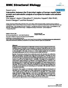
Results Sequence analysis of tetraspanin transmembrane domains: presence of the heptad repeat motif We focused our attention on 28 human tetraspanins identified from the SWISS-PROT and GenBank databases. All tetraspanins have in common four hydrophobic stretches (TM domains) of 20–25 residues, and contain highly conserved residues in the second extracellular loop, in
Page 2 of 20 (page number not for citation purposes)
BMC Structural Biology 2005, 5:11
particular the Cys-Cys-Gly (CCG) motif. Detailed analysis of the large extracellular loop sequences [14], and dendrograms based on full-length alignment can be found in earlier studies [26,27]. The length of each transmembrane domain was established based on previous sequence analysis of tetraspanin sequences [27,28], and on annotations to the database entries. Manual adjustments based on sequence homology and hydrophobicity profiles were done to fully delineate the TM domains. The resulting lengths of TM domains were: TM1 – 23 residues; TM2 – 21 residues; TM3 – 25 residues; TM4 – 25 residues. Two more residues could be added onto the N-terminal part of TM2; however, relatively small sequence conservation of these residues among tetraspanins and occurrence of polar/ charged side chains in some tetraspanins precluded us from doing so for the global alignment. Figures 1 and 2 show a multiple sequence alignment of four TM domains of 28 human tetraspanins. For each position within the domains, consensus residues were determined and classified (with individual color code) in 4 categories: 1) large hydrophobic residues (including Val, Met, Leu, Ile, Phe, Tyr, Trp), 2) small residues (Gly, Ala, Ser and Thr), 3) Cys, and 4) Asn. When more than two types of residues occupied a given position in a TM, a dual-color pattern that reflected the prevalence of the particular residue type was used (Figure 1). Cysteine residues were shown separately due to their importance as palmitoylation target sites. The highly conserved asparagine residue in TM1 was considered separately. No proline residues are found in TM domains 1–3 of human tetraspanins. An inspection of the multiple sequence alignment reveals a repeating heptad amino acid pattern, (abcdefg)n, in TM1, 2 and 3 (Figure 1, 2). Heptad repeats promote helical coiled coil interactions in multiple soluble and membrane-spanning proteins [29-31]. In the heptad repeat, hydrophobic residues in positions a and d are of special importance, as they directly mediate interhelical contacts by creating a tight knobs-into-holes packing in the coiled coil structure [32]. For instance, in the leucine zipper of the yeast transcription factor GCN4, positions a and d contain Val and Leu residues, respectively, with an Asn residue in a single a position forming a hydrogen bond across the GCN4 dimer interface [33]. In TM1 of tetraspanins, highly conserved Asn, Gly and Gly residues (numbers 18, 25 and 32 in the CD9 sequence) appear at d positions of the heptad repeats, and residues 14, 21 and 28 are at a positions (Figure 1). In TM2, residues 67, 74 and 81 (consensus Gly, Gly and Ala, respectively) occupy a positions, whereas residues 63, 70 and 77 are at d positions. Another highly conserved glycine, Gly80, occupies the 3rd g position in TM2. In TM3, the
http://www.biomedcentral.com/1472-6807/5/11
conserved pattern consists of two leucine residues (Leu89 and Leu96) and a glutamate/glutamine residue (Glu/ Gln103) in a positions (Figure 2). Two d positions are also conserved – Phe/Tyr92 and Ile/Val/Leu99. TM4 lacks a conserved heptad pattern and has only a single conserved position, Glu/Gln209 (with four exceptions). These features of TM1-4 of tetraspanins are displayed on helical wheel diagrams (Figure 3). Analysis of TM1 sequences The conserved Asn-Gly-Gly motif, occupying designated d positions of the heptad repeat, is the most prominent structural feature of TM1. We also compared sequences of CD9 orthologs from 10 different organisms (the most available for any tetraspanin) to gain further insight into conservation and variability of the TM1 sequence. As shown in Figure 4, positions a, d and g in TM1 are among the most conserved (0, 1 and 1 substitution, respectively), while interspecies variability tends to occur in other positions: b (5 substitutions), c (4 substitutions), e (4 substitutions) and f (4 substitutions). Thus, the positions typically involved in coiled coil interactions (a and d) are the most conserved.
When residues of TM1 are plotted as a helical wheel, additional structural features are revealed (Figure 3). There are highly conserved aliphatic and aromatic residues in the first three a positions of the heptad motif (Phe15, Trp22 and Leu29 in CD9), as well as in g positions (Leu14, Phe21, Val28 in CD9). The "ridges" formed by these bulky residues are flanking the "groove"-forming Gly residues of the Asn-Gly-Gly position d motif. In contrast, b, c, e and f positions show an overall higher variability among tetraspanins, as also seen in the comparison of CD9 orthologs described above. Analysis of TM2 sequences A landmark feature of TM2 in tetraspanins is the presence of highly conserved glycine residues (Gly67, 74, 77 and 80 in CD9, Figure 1). Other substitutions at these positions are almost exclusively small residues, such as Ala or Ser. In addition, Ala, Ser or Thr occupy position 81. This residue, together with Gly67 and Gly74, forms face a of the helix. Residue Gly77 (position d) is preceded by conserved, chiefly large hydrophobic residues on the same helical face (Leu63 and Met70 in CD9). Extremely conserved Gly80 falls into heptad position g (Figure 3). Among CD9 orthologs, heptad positions a and d are absolutely conserved, whereas other positions have the following number of substitutions: b – 3; c – 2; e – 1; f – 3; g – 1 (Figure 4). Two of the f position residues in TM2 (65 and 79) also show higher variability among different tetraspanins (Figures 1, 3). Cysteine residues are frequently found near the cytoplasmic end of TM2 helix at positions 78 and 79; these cysteines are likely to be palmitoylated.
Page 3 of 20 (page number not for citation purposes)
BMC Structural Biology 2005, 5:11
Net1 CD82 CD37 CD53 Nag2 Net5 BAB55318 Tsn2 Net4 TM4B NM_030927 CD151 CD81 CD9 CO-029 Tsn3 Net2 SAS Net6 CD63 Net7 A15 Tsn6 Ocsp UPIA UPIB RDS ROM
http://www.biomedcentral.com/1472-6807/5/11
TM1
TM2
MMILFNLLIFLCGAALLAVGIWV FLFLFNLIFFILGAVILGFGVWI FLFVFNLFFFVLGSLIFCFGIWI VLFFFNLLFWICGCCILGFGIYL LMFAFNLLFWLGGCGVLGVGIWL MMFLFNLIFWLCGCGLLGVGIWL LMFVFNFFIFLGGACLLAIGIWV LLLGFNLLFWLAGSAVIAFGLWF FIFGFNVIFWFLGITFLGIGLWA LLSLLNGFVAVSGIILVGLGIGG LLFSYNIIFWLAGVVFLGVGLWA LLFTYNCCFWLAGLAVMAVGIWT LLFVFNFVFWLAGGVILGVALWL LLFGFNFIFWLAGIAVLAIGLWL ----- 18------ 25------ 32--SMFTFNFLFWLCGILILALAIWV VLVFLNLIFWGAAGILCYVGAYV LLYALNLLFWLMSISVLAVSAWM ALCALNVVYMLVSLLLIGVAAWG CLCALNLLYTLVSLLLIGIAAWG LLYVLLLAFCACAVGLIAVGVGA SLIIYSTVFWLIGALVLSVGIYA LLIIYSFVFWITGVILLAVGVWG VLLIYTFIFWITGVILLAVGIWG LIFLSNFPFSLLGLLALAIGLWG LLVVGNIIILLSGLSLFAETIWV LLIFGNVIIGCCGIALTAECIFF GLWLMNWFSVLAGIIIFSLGLFL GLWLLSWLLALAGGVILLCSGHL fgabcdefgabcdefgabcdefg
FLIAAGVVVFALGFLGCYGAK VFIGVGAVTMLMGFLGCIGAV VLAISGIFTMGIALLGCVGAL VFVIVGSIIMVVAFLGCMGSI LLIITGAFVMAIGFVGCLGAI LVIAIGTIVMVTGFLGCLGAI ILLAMGGLLFLLGFLGCCGAV VLVGAGALMMAVGFFGCCGAM LFLVVGGVMFILGFAGCIGAL LCLVMGCITVLLGCAGWYGAT LVLMVGVVMFTLGFAGCVGAL ILVVAGTVVMVTGVLGCCATF ILIAVGAVMMFVGFLGCYGAI ILIGAGALMMLVGFLGCCGAV ----- 67------ 74--77-- 80-ILIAVGAIIMILGFLGCCGAI VIIAVGALLFIIGLIGCCATI VMIAVCCFLIIVGMLGYCGTV GVIAVGVFLLLIAVAGLVGAV VVIAVGIFLFLIALVGLIGAV VIIAVGVFLFLVAFVGCCGAC ILILLGVVMFMVSFIGVLASL VLIGTGTTIVVFGLFGCFATC VLIATGTVIILLGTFGCFATC GLALGGLVVSAASLAGCLGAL IAIFCGFSFFMVASFGVGAAL IGIFVGICLFCLSVLGIVGIM SLIGMGVLSCVFNSLAGKICY AALAAGAVALGTGLVGVGASR cdefgabcdefgabcdefgab
Consensus
N
G
G
G
G
G
GA
Consensus color coding: Large hydrophobic (Leu, Ile, Met, Val, Phe, Tyr, Trp) Small (Gly, Ala, Ser, Thr) Cys
Degree of prevalence
Asn
Individual residues color coding: Leu, Ile, Met, Val
G ly
Cys
Phe, Tyr, Trp
Ala, Ser, Thr
Asn
Lys, Arg, His
Asp, Glu, Gln
Figure 1 alignment of the transmembrane domains 1 and 2 of 28 human tetraspanins Sequence Sequence alignment of the transmembrane domains 1 and 2 of 28 human tetraspanins. Residues from select positions of the heptad motifs in TM1 and 2 are highlighted (see text for details). Also highlighted are polar residues and cysteines. Consensus residue types are shown by the color scheme indicated. Boxed residues reflect correlated substitutions for position pairs 22–74 and 25–70 (details are in the text). The numbers refer to CD9 sequence.
Page 4 of 20 (page number not for citation purposes)
BMC Structural Biology 2005, 5:11
http://www.biomedcentral.com/1472-6807/5/11
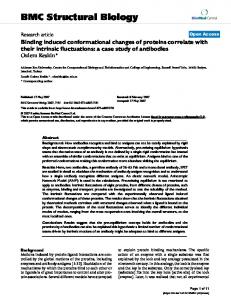
TM3
TM4
Net1 CD82 CD37 CD53 Nag2 Net5 BAB55318 Tsn2 Net4 TM4B
ALVTFFFILLLIFIAEVAAAVVALV LLGLYFAFLLLILIAQVTAGALFYF LLGLYFGMLLLLFATQITLGILIST LLMSFFILLLIILLAEVTLAILLFV LLLTFFLLLLLVFLLEATIAILFFA LLLSFFIVLLVILLAELILLILFFV LLLFFFLFILIIFLAELSAAILAFI VLGSFFTCLLVIFAAEVTTGVFAFI LLKFFSVFLGIIFFLELTAGVLAFV TLLFCILSMVIVLIMEVTAATVVLL NM_030927 LLNFFCGTIVLIFFLELAVAVLAFL CD151 LLRLYFILLLIIFLLEIIAGILAYA CD81 LLGTFFTCLVILFACEVAAGIWGFV CD9 MLGLFFGFLLVIFAIEIAAAIWGYS -89-- 92---96-- 99---103--------CO-029 MLLLFFIGLLLILLLQVATGILGAV Tsn3 GLATFVIILLLVFVTEVVVVVLGYV Net2 LLAWYFGSLLVIFCVELACGVWTYE SAS LLFFYMIILGLVFIFQFVISCSCLA Net6 LLFFYMIILLLVFIVQFSVSCACLA Net7 LLQAFMYILGICLIMELIGGVVALT CD63 LMITFAIFLSLIMLVEVAAAIAGYV A15 MLKLYAMFLSLVFLAELVAGISGFV Tsn6 MLKLYAMFLTLVFLVELVAAIVGFV Ocsp LLRGFSGGILAFLVLEAVAGALVVA UPIA MVLTYLVLMLIVYIFECASCITSYT UPIB ILLAYFILMFIVYAFEVASCITAAT RDS WLKPYLAICVLFNIILFLVALCCFL ROM VLGPLLVAGTAGGGGLLVVALGLAL
AVTVGGVAAGIGGLELAAMIVSMYL LGIILGVGVGVAIIELLGMVLSICL LISIVGICLGVGLLELGFMTLSIFL FLYIGIITICVCVIEVLGMSFALTL LLAVGIFGLCTALVQILGLTFAMTM KHVLGTVGMCILIMQILGMAFSMTL VYLAGALAIGVLAIELFAMIFAMCL LQLIGIVGIGIAGLTIFGMIFSMVL LTIVAGIFIGIALLQIFGICLAQNL SFTLSGSSLGAAVIQRWGSRYVAQA IYIVAGVFIAISLLQIFGIFLARTL LRVIGAVGIGIACVQVFGMIFTCCL IGIAAIVVAVIMIFEMILSMVLCCG IIGAVGIGIAVVMIFGMIFSMILCC -------------- 209---------LIIVIGISFGLAVIEILGLVFSMVL MMHVIWAALAFAAIQLLGMLCACIV LQVLRFLGISIGVTQILAMILTITL LKILGGVGLFFSFTEILGVWLAMRF LRFVGGIGLFFSFTEILGVWLTYRY YTIMAGILLGILLPQFLGVLLTLLY VLVVAAAALGIAFVEVLGIVFACCL MGIIAGVAFGIAFSQLIGMLLACCL MGVVAGISFGVACFQLIGIFLAYCL LAASGGYAIAVVLLQGAELLLAARL YTWGISWFGFAILMWTLPVMLIAMY HAWGVAWFGFAILCWTFWVLLGTMF MNSMGVVTLLIWLFEVTITIGLRYL AGTLGSMLAVTFLLQALVLLGLRYL
gabcdefgabcdefgabcdefgabc C o n se n sus
L
F/Y
L
I/V
E/Q
E/Q
Figure 2 alignment of the transmembrane domains 3 and 4 of 28 human tetraspanins Sequence Sequence alignment of the transmembrane domains 3 and 4 of 28 human tetraspanins. Residues from heptad positions a and d in TM3 are highlighted. Also highlighted are the conserved Glu/Gln residue in TM4, other polar residues and cysteines. The color scheme is as in Figure 1.
Analysis of TM3 and TM4 sequences The TM3 domain provides another example of the heptad repeat pattern. Position a is occupied by two highly conserved leucine and a glutamate/glutamine residue (Leu89,
Leu96 and Glu/Gln103 in CD9). Furthermore, two d positions are conserved – Phe/Tyr92 (aromatic residue) and Ile/Val99 (β-branched aliphatic residue; Figures 2, 3). In addition, residue 100 in position e is generally Phe or Leu.
Page 5 of 20 (page number not for citation purposes)
BMC Structural Biology 2005, 5:11
http://www.biomedcentral.com/1472-6807/5/11
TM1
19
TM2
78 71
26
16
33
23 30
29
15
74
68
67
e b
e
b
a
a
f 13 20 27 34
22
32 25
d
c
82 75
64
81
g
31 24 17
18
77
f
d
63 70
g 66
35
73
28
80
65 72
c
79
62 69 76
21 14
90
TM3
TM4
97 94 101 108
f 91 98 105
213 206 199
104 111 107 100
b
c g
216
a d
207 214 200
93
e
112
217 210 203 196
209
202
195
110
197 204
103 96
109 102 95 88
106
211 89
99 92
198 205 212 219
201 208 215
218
Figurewheel Helical 3 diagrams of transmembrane domains TM1-4 reflecting the consensus residue types Helical wheel diagrams of transmembrane domains TM1-4 reflecting the consensus residue types. The color scheme is as in Figure 1. The numbers refer to CD9 sequence. Heptad positions a through g are indicated for TM1-3. A predicted interaction between positions a and e in TM1 and a and d in TM2 is shown by dotted lines (see Figure 6 and text for details). Arrows reflect the efficiency of intermolecular cross-linking via single cysteines placed in these positions (see Figure 7 and text for details).
Among CD9 orthologs, position a has 1 substitution, positions b, c and f each have 6, positions d and e each have 2, and g has 4. Thus, as for TM1 and TM2, positions a and d are among the most conserved, but overall TM3
has more variable positions than TM1 or TM2 (Figure 3). Less than half of TM3 sequences contain cysteine residues, and those tend to occur at the internal positions of the helix (Figure 2).
Page 6 of 20 (page number not for citation purposes)
BMC Structural Biology 2005, 5:11
http://www.biomedcentral.com/1472-6807/5/11
TM1 18
Human
25
TM2 32
LLFGFNFIFWLAGIAVLAIGLWL
67
74
TM3 81
ILIGAGALMMLVGFLGCCGAV
89
96
103
110
MLGLFFGFLLVIFAIEIAAAIWGYS
Monkey
LLFGFNFIFWLAGIAVLAIGLWL
ILIGAGALMMLVGFLGCCGAV
MLGLFFGFLLVIFAIEIAAAIWGYS
Pig
LLFGFNFIFWLAGIAVLAIGLWL
ILIGAGALMMVVGFLGCCGAV
MLGLFFGFLLVIFAIEIPAAIWGYS
Cow
LLFGFNFIFWLAGIAVLSVGLWL
ILIGAGALMMLVGFLGCCGAV
MLGLFFSFLLVIFAIEVAAAIWGYS
Cat
LLFGFNFIFWLAGIAVLAVGLWL
ILIGAGALMMLVGFLGCCGAV
MLGLFFGFLLVIFAIEIAAAIWGYS
Rat
LLFGFNFIFWLAGIAVLAIGLWL
ILIGAGALMMLVGFLGCCGAV
MLGLFFGFLLVIFAIEIAAAVWGYT
Mouse
LLFGFNFIFWLAGIAVLAIGLWL
ILIGAGALMMLVGFLGCCGAV
MLGLFFGFLLVIFAIEIAAAVWGYT
Chicken
LLFGFNFVFWLAGTAVLAIGLWL
ILIGAGALMMLVGFLGCCGAL
MLGLFFVFLFVIFALEIATAIWGFA
Lamprey
LLFVFNFVFWLAGGAVLGIALWL
VLMGAGALMMLIGFLGCCGAI
MPGSFFVCLLVVFAAEIAAGIWGFL
Zebrafish
SMFLLNSVFWIAGTAVLAVGLWL
ILIAAGALMMVVGFFGCCGAI
MLGLFFFFLLVIFAVEVAAGIWGFS
fgabcdefgabcdefgabcdefg
cdefgabcdefgabcdefgab
gabcdefgabcdefgabcdefgabc
Figure 4 alignment of TM1-3 for ten vertebrate orthologs of CD9 Sequence Sequence alignment of TM1-3 for ten vertebrate orthologs of CD9. Heptad positions a and d in TM1, TM2 and TM3 are highlighted in green. Residues that differ between orthologs are shown in yellow.
TM4 shows less conservation among various tetraspanin family members than the other TM domains (Figures 2, 3). The only highly conserved feature is the glutamate/ glutamine residue in position 209. In addition, one or two cysteine residues can be found at the C-terminal end of TM4 in some tetraspanins (e.g. CD9, CD81, CD151), and many sequences contain additional polar residues (Arg, Lys, His, Asn, Gln). No conserved heptad motif was identified in TM4, as also confirmed by analysis of substitutions in CD9 orthologs (data not shown). Mutational analysis of conserved glycine residues in TM1 and TM2 The conserved nature of the Asn and Gly residues in TM1 and TM2 prompted an analysis of their functional role. To this end, we have probed whether mutations of these residues destabilize the protein molecule. We expressed a construct of the first and second TMs of CD9, connected by the small extracellular loop, and tagged with a C-terminal green fluorescent protein (TM(1+2)-GFP molecule). In human rhabdomyosarcoma RD cells, the wild-type fusion protein localized mostly in a reticular, intracellular pattern, without forming any large aggregates (Figure 5, panel A). Remarkably, when double mutants Gly25Leu + Gly32Leu and Gly67Leu + Gly74Leu were expressed, the protein formed distinct large aggregates in a high proportion of cells (Figure 5, panels C and E). In contrast, double mutant Gly77Leu + Gly80Leu did not form such aggregates (Figure 5, panel G). Results with respective single
mutants were similar to that with double mutants, with the aggregation being somewhat more pronounced for Leu substitutions of Gly67 and Gly74 compared to Gly25 and Gly32 mutations. No aggregation was observed for Asn18Ser and Asn18Tyr mutants (data not shown). Also, nearly identical results were obtained with human HT1080 cells (data not shown). We interpret these results as an indication that aggregating mutants are destabilized or misfolded while non-aggregating mutants retain the wild-type conformation. Intriguingly, mutations to the conserved GG7 motifs caused protein aggregation while the mutation of other glycines had no detectable effect. These results also suggest that wild-type GFP, which has weak tendency to selfassociate, could enhance non-specific interactions of destabilized mutant TM(1+2) CD9 moieties, leading to their aggregation. Consistent with this hypothesis, the aggregation of Gly25Leu + Gly32Leu and Gly67Leu + Gly74Leu double mutants was suppressed when monomeric GFP molecule, Leu221Lys [34] was used (Figure 5, panels D and F). The use of monomeric GFP did not affect intercellular localization of wild-type CD9 TM(1+2) (Figure 5, panel B), or a Gly77Leu + Gly80Leu double mutant (Figure 5, panel H). In summary, Leu substitutions of Gly residues that are part of the Asn-Gly-Gly (NGG7) motif in TM1, or Gly-GlyAla (GGA7) motif in TM2, resulted in destabilization and
Page 7 of 20 (page number not for citation purposes)
BMC Structural Biology 2005, 5:11
http://www.biomedcentral.com/1472-6807/5/11
Figure 5 of wild-type and mutant CD9 TM(1+2)-GFP proteins in human cells Expression Expression of wild-type and mutant CD9 TM(1+2)-GFP proteins in human cells. Human rhabdomyosarcoma RD cells were transfected with constructs encoding CD9 TM(1+2)-GFP fusion proteins that carried mutations indicated. Either wild-type or monomeric (L221K) GFP was used. Images were captures 18–28 hours post-transfection.
Page 8 of 20 (page number not for citation purposes)
BMC Structural Biology 2005, 5:11
http://www.biomedcentral.com/1472-6807/5/11
Figure 6 model of TM1-TM2 interaction in CD9 Structural Structural model of TM1-TM2 interaction in CD9. Shown are space-filling models of CD9 TM1 (panel A), TM2 (panel B), and the two helices together (panel C). Small and large residues of heptad positions a and d, which form the crucial contacts between the helices, are shown in green and red, respectively. Asn18 is shown in blue, Leu14 and Phe17 in yellow, and Gly80 in light green.
aggregation of GFP-fused TM(1+2) proteins, whereas substitutions of Gly77 or Gly80, which are not part of these motifs (Figure 3), failed to show such aggregation. Prediction and modelling of interaction between TM1 and TM2 Consecutive helices in polytopic membrane proteins frequently interact [35]. Sequence analysis of TM1 and TM2 helices of tetraspanins reveals a remarkable complementarity in the distribution of large and small residues at heptad positions a and d along the helical axis (Figure 3), suggesting that these residues may interact. To further elucidate the potential for TM1-TM2 interaction, the putative interface was modeled using a novel algorithm that considers mutational data during each step of a Monte Carlo simulated annealing cycle (see Methods for details). Specifically, Gly25Leu, Gly32Leu, Gly67Leu and Gly74Leu were scored as disruptive mutations, while Asn18Ser, Gly77Leu and Gly80Leu were scored as silent mutations, based on their effects on protein stability (Figure 5 and data not shown).
The resulting model predicts left-handed crossing of TM1 and TM2 helices at an angle of +28°. The key element of the structure is the apposition of bulky and small heptad
position a and d residues, as follows: Gly32-Leu63; Gly67-Leu29; Gly25-Met70; Gly74-Trp22; Asn18-Gly77; Ala81-Phe15 (Figure 6). Our model predicts that each of these residue pairs are in van der Waals contact. Additionally, two potential H-bonds are predicted in this model, indicating close packing: Gly67 Cα to Gly25 carbonyl oxygen, and Trp22 Cα to Met70 carbonyl oxygen. The packing is tighter in the ectodomain-proximal portion of the helices (Figure 6, panel B), as determined by Cα-Cα distances between interacting residue pairs. The key elements of the model are corroborated by the presence of apparently complementary substitutions in TM1 and TM2 sequences of different tetraspanins (Figure 1, boxed residues). For example, Gly74 is predicted to interact with Trp22. In 8 of the 10 tetraspanins that contain a substitution for Gly74, a compensatory substitution occurs at the Trp22 position (Figure 1). Thus, a larger nonglycine side chain at position 74 may necessitate a less bulky non-Trp side chain in position 22. Likewise, the presence of a Cβ at position 25, typically occupied by glycine, necessitates a non-β-branched amino acid at position 70, which is occupied by a β-branched residue in nearly half of all cases. Indeed, we find that in each of 5 cases in which position 25 contains a Cβ, a leucine residue Page 9 of 20 (page number not for citation purposes)
BMC Structural Biology 2005, 5:11
occurs in position 70. This analysis is consistent with our molecular model that suggests Leu70 will pack most favorably against a Cβ at position 25 than a β-branched residue or a methionine. Role of TM1 and TM2 heptad motif residues in CD9 dimerization To probe CD9 dimerization, we used a cysteine-mediated cross-linking approach. We established previously a simple and efficient method for cysteine-mediated cross-linking [25]. After cells are pre-treated with 2-BP for 16–24 hours to expose normally palmitoylated cysteines, the cysteines can be cross-linked using any of the following methods: a) Spontaneous oxidation in Brij97 lysates (a condition that preserves tetraspanin-tetraspanin associations), b) In situ cross-linking, by pre-lysis oxidation of cells with Cu2+-phenanthroline (CuP) to promote disulfide bond formation. c) In situ cross-linking with thiol-reactive cross-linking agents of defined length (e.g. DTME, BMB). The first two approaches produce in essence "zero-length" disulfides, indicative of close proximity of target cysteines and presumably high specificity of interaction. In contrast, chemical cross-linkers with 6–20 Å spacer arm may cross-link with higher efficiency, but not necessarily higher specificity. However, they provide advantages such as variable membrane permeability, and potential linkage cleavability. For tetraspanins such as CD9, membrane-permeable cross-linker DTME (13.3 Ålong, reducible) provides highly specific and efficient cross-linking [25]. Here we have used a cysteine crosslinking strategy, in combination with cysteine-scanning mutagenesis, to map the residues from TM1 and TM2 contributing to the CD9 dimerization interface.
For subsequent cross-linking experiments using CD9 TM(1+2)-GFP protein, the non-dimerizing form of GFP was used. This avoids potential GFP-dependent dimerization and aggregation that can be observed with wild-type GFP, especially when fusions with transmembrane proteins are studied [36]. Importantly, the Leu221Lys mutation in GFP prevented aggregation of mutant forms of CD9 TM(1+2), which was observed with wild-type GFP fusion (Figure 5). The TM(1+2) fragment of CD9 contains three native cysteines – Cys9, Cys78 and Cys79. Singlecysteine mutants of TM(1+2) were constructed, in which a cysteine was placed at various faces of TM1 or TM2 while all of the wild-type cysteines were simultaneously replaced by serines. The mutant proteins were transiently expressed in RD cells (having little endogenous CD9), which were then treated for 16–18 hours with 2-BP. To achieve maximal specificity in cross-linking we used a "zero-length" agent, CuP. First, single-cysteine replacements were constructed for residues Leu14, Phe15, Gly16, Phe17 and Asn18, covering
http://www.biomedcentral.com/1472-6807/5/11
just over one complete helical turn at the beginning of TM1. While residue Asn18 is highly conserved, positions 14, 15 and 17 are occupied by bulky hydrophobic residues in most tetraspanins, whereas position 16 shows less conservation (Figures 1, 4). All of the single-cysteine mutants showed diffused pattern of protein localization, without any signs of aggregation. As shown in Figure 7A, the highest level of intermolecular cross-linking was observed for Leu14Cys and Phe17Cys mutants, a lower level for Phe15Cys and Gly16Cys mutants, and very little cross-linking for Asn18Cys substitution. These results indicate that: a) the first two transmembrane domains of CD9 alone can mediate its dimerization, and b) the g and c residues of TM1 (e.g. Leu14 and Phe17, Figure 3) are likely to be part of the intermolecular interface. Similarly, single-cysteine substitutions were made for residues Gly77, Gly80 and Ala81 in TM2; in addition, proteins carrying a single wild-type cysteine, Cys9, Cys78 or Cys79, were tested. No protein aggregation was observed for any of these single-cysteine mutants. As shown in Figure 7B, the relatively low level of intermolecular crosslinking of wild-type CD9 TM(1+2)-GFP protein was enhanced dramatically in single-cysteine TM2 mutants Gly80Cys and Ala81Cys. The Gly77Cys mutant also had an elevated level of cross-linking. In contrast, any of the three native cysteines (9, 78 and 79) produced level of cross-linking not much greater than the wild-type TM(1+2) protein. Similar results were obtained with cysteine-reactive cross-linker BMB (data not shown). Likewise, comparable results were obtained with singlecysteine mutants of untagged, full-length CD9, using CuP (Figure 7C) as well as DTME cross-linker (data not shown). These cross-linking results for TM1 and TM2 are consistent with our model that places residues Leu14, Phe17 and Gly80 on the same side of the TM1-TM2 pair (Figure 6, panel C). The strong cross-linking with Leu14Cys, Phe17Cys and Gly80Cys places the intermolecular interface toward the c and g phases of the TM1 helix, and the g phase of the TM2 helix, away from its e and f faces containing wild-type cysteines 78 and 79. Critical residues at the TM1-TM2 interface also affect dimerization indirectly. To assess specific CD9 dimerization, we used a Gly80Cys substitution at the intermolecular interface for cross-linking. As shown in Figure 8A, single replacements of conserved heptad residues in positions 18, 25, 32, 67 and 74 (Asn18Ser, Gly25/32/67/ 74→Leu) strongly decreased the cross-linking mediated by Cys80. The effect was most pronounced for mutations of residues, Gly32 and Gly67, located in the tightly packed extracellular end of TM helices (Figure 6). In contrast, mutations of residues closer to the cytoplasmic end
Page 10 of 20 (page number not for citation purposes)
BMC Structural Biology 2005, 5:11
http://www.biomedcentral.com/1472-6807/5/11
A.
Single cysteine Cys: WT
14
15
16
17
18
99 53 33
% dimer:
46.8 48.0 33.7 35.4 46.0 13.7
B.
Single cysteine Cys:
-
WT
9
77
78
79
80
81
82 50 33 27
% dimer: