Synthesis, Spectral and Photodynamic Properties of Lithium Phthalocyanine DANIEL BODA1, RODICA-MARIANA ION2,3* Carol Davila University of Medicine and Pharmacy, 8 Eroilor Sanitari Blv., 050474, Bucharest, Romania 2 ICECHIM, Nanomedicine Research Group, 202 Splaiul Independentei, 060021, Bucharest, Romania 3 Valahia University, 2 Carol I Blv., 130024, Targoviste, Romania
1
The aim of this paper was to synthesize lithium phthalocyanine Li2Pc, to characterize and to test it on culture media from HepG-2 (human hepatocellular carcinoma), by photodynamic test, under irradiation with λ = 632.8 nm, He-Ne laser (30 mW, 25°C, total irradiation time = 30 min, in O2 saturated solution). During irradiation, cells are actively destroyed, especially those loaded with Li2Pc. Some parameters as viability were significantly lowered in culture media from HepG-2 cells in the presence of 25 μg/mL Li2Pc, after irradiation with laser, as compared to control cells (non-irradiated, but treated with Li2Pc). The synthesized phthalocyanine has been characterized from spectral (UV-Vis, FTIR, 1H NMR), photophysical (fluorescence lifetimes and triplet quantum yields) and photochemical point of view, too (singlet oxygen generation). Keywords: lithium phthalocyanine, photodynamic action, photophysics, photochemistry
The chemistry of phthalocyanines (Pcs) gain popularity in the last few decades due to the extensive applications of these compounds [1-4]. Also, the studies on the phthalocyanines represent an active field of research for several decades due to their involvement in various chemical and biological processes [5-7]. Except the phthalocyanine complexes containing non-transition metals or metalloids (e.g. Al(III), Ge(IV) and Zn(II)), which are favoured as good candidates for the use as photosensitizers in for example photodynamic therapy (due to the fact that these metal ions are diamagnetic and are expected to give relatively long-lived excited states), there are new interest on the alkali- metal phthalocyanine complexes, as Li, Na, K [8]. Photodynamic therapy (PDT), well recognized treatment for tumors destruction, utilizes the ability of some selected photosensitizers to exhibit an efficient photodynamic reaction upon activation with tissue penetrating light (around 680 nm) [9]. Phthalocyanines, with an extensively conjugated aromatic chain, exhibit UV-VIS absorption spectra with intense π− π* transitions, usually referred to as Q bands in the range 660-700 nm (ε >105M -1cm -1), have associated higher energy vibrational components in the range 600-660 nm, strongly absorb clinically useful red light. Metalation, which reduces the electron density at the inner nitrogen atoms, is predicted to produce a shift of the λ max to shorter wavelength depending on the electronegativity of the metal [10]. The relationships between the chemical structure and the tumor localizing activity of photosensitizers yield to the conclusion that the optimal tumor localizing efficiency is imparted to be phthalocyanine-type macrocycle, with a strong NIR absorption and high extinction coefficient in the 680-800 nm range, exhibiting water or DMSO solubility, high triplet quantum yield and singlet oxygen quantum yield [11]. The aim of our study was to synthesize a new phthalocyanine structure Li2Pc, to characterize it by specific analitycal techniques (UV-Vis, FTIR, 1H-NMR) and to test it on culture cells HepG-2 (human hepatocellular
carcinoma), by photodynamic action (λ = 632.8 nm, HeNe laser (30 mW, 25 °C, total irradiation time = 30 min, in O2 saturated solution). Experimental part Chemicals 8 g of lithium metal are added to 120 g of phthalonitrile in 1 L of pentanol, and this reaction mixture is heated for 4 h at the boiling temperature. When the reaction mixture has cooled at 80° C, 1 L of benzene are added and the mixture allowed to set for 3 h. A crude dilithium phthalocyanine product is separated by filtration and washed with diethyl ether. The purification is achieved by extraction with acetone, filtering and evaporating the acetone under vacuum. The dilithium phthalocyanine product is stored in dark, avoiding hydrolysis. Anal. calc. For C72H96N8O8Li2: C, 71.15; H, 7.96; N, 9.22. Found: C, 71.27; H, 7.90; N, 9.25%. 1 H NMR (THF-d8): 1.03 (t, 24H, J = 6.9 Hz), 1.47–1.61 (m, 16H), 1.63–1.74 (m, 16H), 1.96–2.08 (m, 16H), 4.46 (t, 16H, J = 6.3 Hz), 8.88 (s, 8H). UV–Vis in THF; λmax (nm), ε (L mol_1 cm_1): 660 (2.55 · 105); 632 (3.6 · 104); 599 (3.92 · 104); 357 (1.0 · 105); 282 (6.0 · 104). Instrumentation 1 H-NMR spectra were obtained in deuterated chloroform (CDCl3) using Varian Mercury NMR spectrometer operated at 400 MHz. Chemical shifts (δ) are reported in parts per million (ppm) relative to the residual CHCl3 peak (7.26 ppm). Coupling constants (J) are reported in hertz (Hz). Absorption spectra UV-Vis were registered by means of a SPECORD M400, Carl Zeiss Jena spectrophotometer with double beam and equipped with a microprocessor. Quartz cuvettes with 0.5-2 cm optical path lengths. Molar extinction coefficients at a given wavelength, ε, were obtained using the Beer-Lambert law, over the concentration range 10-410-7 mol dm-3• FTIR absorption measurements were performed using Perkin Elmer Spectrum GX (Perkin Elmer Inc., USA) with
* email:
[email protected] REV. CHIM. (Bucharest) ♦ 65 ♦ No. 11 ♦ 2014
http://www.revistadechimie.ro
1271
TDS detector (deuterated triglycine sulfate) in KBr pellettes, resolution of 4 cm-1, with 32 scans, operated both in transmission and in ATR device (total reflection attenuated). Singlet oxygen generation by photodegradation of DPBF Measurements were carried out in a quartz cell (1cm x 1cm) at 25oC. Oxygen-saturated solutions (V=0.2 ml) containing sensitizer and 1,3-diphenylisobenzofuran (DPBF) (c=0.858 x 10⁻⁴ M) in DMSO were used. The photolysis cell was placed into the light beam of an UV-Vis spectrophotometer (Carl Zeiss Jena as described above), with DMSO as reference. The decreasing DPBF concentration was followed by a wave program measuring the absorbance at 420 nm (ε =23300 M -1.cm -1) as a function of irradiation time (irradiation cycles, 20 x 20 s) [12]. The quantum yields Φ(1O2) were taken from the intercept of the Stern-Volmer plots according to the following equation: 1/Φ[DPBF] = 1/Φ(1O2) + 1/Φ (1O2) • kd/ka •1/[DPBF]
(1)
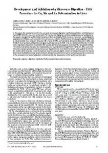
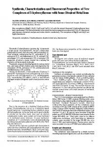
The singlet oxygen trap, 1,3-diphenylisobenzofuran (DPBF), obtained from Fluka (Purum grade) was used as received. Cell activation Cell suspensions incubated for 24 h in Li2Pc compound were subject to irradiation after washing them 3 times in culture medium to remove excess photosensitizer and resuspending at 2.0 × 10⁵ cells• mL-1 in RPMI1640 culture medium without phenol red or FCS. The cell suspensions were irradiated at different laser wave lengths and irradiation times; the best cellular destruction was obtained using the following parameters: λ = 632.8 nm, He-Ne laser (30 mW, 25 °C), total irradiation time = 30 min, in O 2 saturated solution. After irradiation, cell suspensions were washed again twice in RPMI1640 culture medium without phenol red with FCS for removal of cell debris generated during the irradiation. Culturing and treatment of HepG-2 cells HepG-2 human adenocarcinoma cell line was routinely grown as monolayer culture in a combination of E-199 and Iscove ,s modified Dulbecco , s medium (IMDM) supplemented with 5 to 10% calf serum, penicillin (100 U/ mL) and streptomycin (100 mg/mL). The culture was maintained at 37oC, 5% CO2, 95% humidity. For routine passages, adherent cells were detached using a 0.05% trypsin (Gibco) - 0.02% ethylendiamino tetraacetic (EDTA) mixture. The cells were seeded in 96-well plates (Cellstar) at a concentration of 2 x 10⁴ cells/well. Some hours later, after the attachment of the cells, culture medium containing Li2Pc was added – final concentration of the Li2Pc – 25 μg/mL. Samples of cells grown in not modified medium served as a control. After 24 h incubation irradiation was performed with a laser (λ = 632 nm, 30 mW). Experiments without light irradiation were also performed. Cell culture was collected 24 h after the irradiation. Results and discussions Phthalocyanines are azaporphyrins consisting of four benzoindole nuclei connected by nitrogen bridges in a 16membered ring of alternating carbon and nitrogen atoms around a central metal atom, which forms stable chelates with metal cations (dia- or paramagnetic ion). Dilithium phthalocyanine (fig. 1) has become one of the most 1272
Fig. 1. The structure of Li2Pc
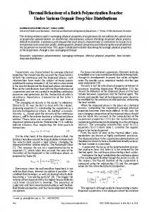
exciting compounds of phthalocyanine research. It was firstly synthesized by Barrett et al. in 1938 and has been a fundamental basis for the future production of this phthalocyanine [13]. Lithium phthalocyanines (unsubstituted) exhibit good absorption properties, and a long-time use as dyes with a good stability. It is well known that these compounds absorb in the UV and visible regions of the spectrum (fig. 2), with absorptions (λmax) at 320-370 nm (B-band) and 650-700 nm (Q-band). The Q-band is generally more intense having molar absorptivities (ε) with magnitudes of 104 – 106 M-1cm-1. The phthalocyanins strongly absorb in this region, due to disruption in the phthalocyanine π-system caused by the nitrogen atoms being in the meso-positions (higher electronegativity than carbon attracts π-electron density) and the peripheral benzene rings that extend the π-system [14]. Therefore, the red portion of the spectrum is strongly absorbed by phthalocyanins (Pcs) and their metallocomplexes (MPcs) while the blue/violet portion (320-370 nm; B-band) is less well absorbed.
Fig.2. The absorption spectrum of Li2Pc and H2Pc
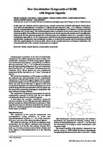
From FTIR spectrum it is visible the broad band around 3500 cm -1 (CH-groups), 1600 cm -1 (-C=C- stretching vibrations of phenyl rings); 1000 cm-1 (vibration of the porphyrin ring or pyrole units); 800 cm-1 (vibration of pyrrole ring) and 450 cm-1 (Li-N), (fig. 3). Photochemical and photophysical parameters Photochemical studies include singlet oxygen quantum yields and photodegradation. In the present paper we will show only singlet oxygen quantum yield. Photophysical studies include fluorescence lifetimes, and triplet quantum yields and lifetimes [15]. During photosensitization, the phthalocyanine molecule is first excited to the singlet state and through intersystem crossing forms the triplet state, and then transfers the energy to ground state oxygen, O2(3Σg), generating excited singlet state oxygen, O2(1Δg), the main cytotoxic species,
http://www.revistadechimie.ro
REV. CHIM. (Bucharest) ♦ 65♦ No. 11 ♦ 2014
2
2
2
2
2
1 2
1 1 1
1
1
Fig. 3. The FTIR spectra of H2Pc and Li2Pc
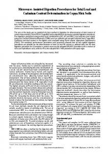
specifically to Type II mechanism [16-18]. The singlet oxygen quantum yields are expected to be comparable to triplet state yields if quenching of the latter by triplet oxygen is efficient [19]. Singlet oxygen quantum yields (ΦΔ) for the MPc complexes may be conveniently determined using a singlet oxygen quencher such as 1,3-diphenyl-isobenzofuran (DPBF) [20]. Both fluorescence and triplet quantum yields parameters may be determined by the comparative methods, using well known references such as chlorophyll. Triplet lifetimes of ZnPc in DMSO is relatively long (350 μs)[21], For Li2Pc, there were obtained the following parameters: ΦF = 0.489 (fluorescence quantum yield determined with standard chlorophyll a in ether (ΦF = 0.32) [22]); ΦT = 0.6 (triplet quantum yield); ΦΔ = 0.38 (singlet oxygen quantum yield). All these values indicated us that Li2Pc is a relatively good sensitizer, and its singlet oxygen efficiency will be tested during photodynamic tests on HepG-2 cells, as is shown below. In vitro cytotoxicity studies Viability and cell proliferation capacity of HepG-2 cells have been determined in the presence of Li2Pc in the concentration range 20-25 μg/L, for 2 and 24 h incubation. The results obtained indicate the fact that for HepG-2 cells following 24h of incubation with Li2Pc at the concentration 25 μg/mL tends to exert a slight effect on the proliferative capacity (fig. 4). Cell viability was assessed as cell membrane integrity using Cytotox96 Non-Radioactive Cytotoxicity Assay kit (Promega) [23,24], a test that quantifies lactate dehydrogenase (LDH) release by damaged cells. Trypan Blue exclusion test was also performed in order to establish the percentage of dead cells. Cell viability tested with the Trypan Blue exclusion test matched perfectly the pattern of cell membrane integrity (LDH release). The results are expressed as indexes compared to control (LDH index). Some decreases in viability were observed in culture medium from HepG-2 tumour cells treated with photosensitiser (25 μg/mL, 30 mW). The obtained results are in good concordance with similar other results obtained by our group [25,26]. The study concerning zinc phtalocyanine properties was presented in [27].
REV. CHIM. (Bucharest) ♦ 65 ♦ No. 11 ♦ 2014
Fig.4. Effect of PcLi2 (25 μg/mL) on viability of HepG-2 cells
Conclusions Lithium phthalocyanine Li2Pc, shown in this paper, was characterized from spectral (UV-Vis, FTIR, 1H NMR), photophysical (fluorescence lifetimes and triplet quantum yields) and photochemical point of view, too (singlet oxygen generation). It has been tested on culture media from HepG-2 (human hepatocellular carcinoma), by photodynamic test, under irradiation with λ = 632.8 nm, He-Ne laser (30 mW, 25 °C, total irradiation time = 30 min, in O2 saturated solution). During irradiation, cells are actively destroyed. Some parameters as viability were significantly lowered in culture media from HepG-2 cells in the presence of 25 μg/Ll Li2Pc after irradiation with laser, as compared to control cells (non-irradiated, but treated with Li2Pc). References 1. ION, R.M., Photosensitizers in Medicine, Environment and Security, 1st Ed., XVI, Springer Netherlands, Dordrecht, Heidelberg, London, New York, Nyokong, Tebello; Ahsen, Vefa (Eds.), 2012, p.315-350. 2. ION, R.M., GRIGORESCU, M., STIRBET, A., Rev. Chim.(Bucharest), 48, no. 12, 1997, p.923 3. ION, R.M., SCARLAT, F., Rev. Chim.(Bucharest), 55, no. 11, 2004, p. 900 4. ION, R.M., APOSTOL, S., Rev. Chim.(Bucharest), 56, no. 6, 2005, p.607 5. ION, R.M., BODA, D., Rev. Chim.(Bucharest), 59, no..2, 2008, p.205 6. TAMPA, M., MATEI, C., POPESCU, S., GEORGESCU, S.-R., NEAGU, M., CONSTANTIN, C., ION, R.M., Rev. Chim.(Bucharest), 64, no 6, 2013, p. 639 7. ION, R. M., PETRE, G., Rev. Chim.(Bucharest), 47, no. 2, 1996, p. 113 8. LAGORIO, M. G., DICELIO, L. E., San ROMAN, E. A., J.Photochem. Photobiol. A: Chem., 3, 1989, p.615. 9. AGIRTAS, S., ION, R.M., BEKAROGLU, O., Mat.Sci. Eng.C:Biomimetic Materials Senzors Systems, 7, nr.2, 2000, p. 105 10.ECKUT, H., SCHIER, A., Angew. Chem. Intern., Ed. English 2, 1981, p.280. 11.ROSENTHAL, I., Photochem. Photobiol., 53, 1991, p.859 12.MATEI, C., TAMPA, M., ION, R.M., NEAGU, M., CONSTANTIN, C., Digest J. Nanomat. Biostruct., 7, nr. 4, 2012, p. 1535 13.BARRETT, P.A., FRYE, D. A., LINSTEAD, R. P. J. Chem. Soc., 1938, p.1157. 14.ALLEN, C., SHARMAN, W., VAN LIER, J., J. Porph. Phthal., 5, 2001, p.161 15.LEZNOF, C.C, LEVER, A.B.P., Phthalocyanines, Properties and Applications. VCH Publishers, New York., 4, 1989, p.25 16.NYOKONG, T., http://eprints.ru.ac.za/979/1/
http://www.revistadechimie.ro
1273
17.FRACKOWIAK, D., WIKTOROWICZ, K., PL ANNER, A ., WASZKOWIAK, A., ION, R.M., Int.J.Photoenergy, 2, 2002, p. 52 18.FRACKOWIAK, D., PLANNER, A., WASZKOWIAK, A., BOGUTA, A., MANIKOWSKI, H., ION, R.M., WIKTOROWICZ, K., J.Photochem. Photobiol., A:Chem., 141,2001, p. 101; 19.ION, R.M., Curr. Topics BioPhys., 24, 2000, p. 21. 20.FOLEY, S., JONES, G., LIUZZI, R., MCGARVEY, D.J., PERRY, M.H., TRUSCOTT, T.G., J.Chem.Soc. Perkin Trans., 2, nr. 9, 1997, p. 1725 21.SPILLER, W., KLIESCH, H., WOHRLE, D., HACKBARTH, S., RODER, B., SCHNURPFEIL, G. J.Porph. Phthal., 2, nr.2, 1998, p. 145. 22.BISHOP, S.M., BEEBY, A., PARKER, A.W., FOLEY, M.S.C., PHILIPS, D., J. Photochem. Photobiol. A: Chem., 90, 1995, p. 39.
23.OGUNSIPE, A., NYOKONG, T., J. Photochem. Photobiol. A: Chem., 173, nr. 2, 2005, p. 211 24.KORZENIEWSKI, C., CALLEWAERT, DM., J.Immunol. Meth., 64, 1983, p. 313 25.FRACKOWIAK, D., WASZKOWIAK, A., ION, R.M., WIKTOROWICZ, K., COFTA, I., MANIKOWSKI, H., Acta Biochim.Pol., 48, nr. 1, 2001, p.257. 26.ION, R.M., SORESCU, A.A., NUTA, A., RADITOIU, V., Proc. ARSAAdvanced Research in Scientific Areas, EDIS Publ., Zilina, 2, 2013, p.294. 27. YOUSSEF, T.E., Al-TURAIF, H., BALEANU, D., Rev. Chim. (Bucharest), 65, no. 11, 2014, p. 1268 Manuscript received: 21.01.2014
1274
http://www.revistadechimie.ro
REV. CHIM. (Bucharest) ♦ 65♦ No. 11 ♦ 2014