Clinical Care/Education/Nutrition/Psychosocial Research B R I E F
R E P O R T
Bone Age Corresponds With Chronological Age at Type 1 Diabetes Onset in Youth ANISSA MESSAAOUI, MD HARRY DORCHY, MD, PHD
OBJECTIVE — To our knowledge, only two controversial articles have reported the study of bone age at diagnosis in diabetic children. The aim of this study was to compare chronological age with bone age and to evaluate the impact of A1C on bone age in children at diagnosis of type 1 diabetes. RESEARCH DESIGN AND METHODS — In 496 diabetic children, height was measured at diagnosis and height SD score was calculated using the British 1990 growth reference. Bone age was determined according to the Greulich and Pyle method, and A1C levels were measured. RESULTS — Participants’ height was normal for age and sex. No significant differences were found between chronological age and bone age, and there was no correlation between ⌬ (bone age ⫺ chronological age) and A1C. CONCLUSIONS — This study showed that height and bone maturation among diabetic children are normal for age and sex and independent of A1C at diagnosis of type 1 diabetes. Diabetes Care 32:802–803, 2009
A
dequate insulin secretion is needed to promote growth (1). Many controversies remain about height and bone maturation in diabetic children. Since 1959, the Greulich and Pyle atlas (2) has been used to assess bone age (3). There are only few data on skeletal maturation in diabetic children at diagnosis, and these are controversial (4,5). The aim of this study was to compare chronological age with bone age and to evaluate the impact of A1C on bone age at diagnosis of type 1 diabetes.
ing height was measured and transformed into an SD score (SDS) according to the British 1990 growth reference (6). A1C levels were measured by chromatography (normal A1C ⬍7.7%) before March 1990 in 10 patients (3 girls and 7 boys) and by high-performance liquid chromatography (normal A1C ⬍6.2%) after March 1990 (204 girls and 282 boys). Radiographies of left hands and wrists were made at diagnosis of type 1 diabetes. Six different trained radiologists analyzed the X rays and evaluated the bone age according to the Greulich and Pyle method (2). Linear regression analysis was used to test for the correlations between chronological age and bone age and between ⌬ (bone age ⫺ chronological age) and A1C. P ⬍ 0.05 was considered to be significant.
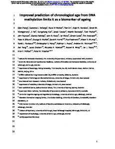
and 8.8 ⫾ 4.3 years, respectively, for the entire population. Chronological age and bone age were 8.12 ⫾ 4.1 and 8.4 ⫾ 4.4 years, respectively, for girls and 9.0 ⫾ 3.9 and 9.1 ⫾ 4.2 years, respectively, for boys. Chronological age and bone age were 7.0 ⫾ 3.2 and 7.2 ⫾ 3.5 years, respectively, for children ⬍12 years of age and 13.8 ⫾ 1.3 and 14.0 ⫾ 1.6 years, respectively, for children ⱖ12 years of age. There was a strongly significant correlation between chronological age and bone age for the entire population of 496 patients (r ⫽ 0.967; P ⬍ 0.001), for girls (r ⫽ 0.981; P ⬍ 0.001), for boys (r ⫽ 0.957; P ⬍ 0.001), for those ⬍12 years of age (r ⫽ 0.956; P ⬍ 0.001), and for those ⱖ12 years of age (r ⫽ 0.653; P ⬍ 0.001) (Fig. 1). Bone age corresponded with chronological age: ⌬ (bone age ⫺ chronological age) was median 0.0 years (25th, 75th percentiles ⫺1.0, 1.0) for the entire population, 0.1 years (⫺0.9, 1.1) for girls, and 0.0 years (⫺1.1, 1.1) for boys. The ⌬ (bone age ⫺ chronological age) was 0.0 years (⫺0.9, 0.9) for those ⬍12 years of age and 0.1 years (⫺1.4, 1.6) for those ⱖ12 years of age. There was no correlation between ⌬ (bone age ⫺ chronological age) and A1C (P ⫽ NS).
The costs of publication of this article were defrayed in part by the payment of page charges. This article must therefore be hereby marked “advertisement” in accordance with 18 U.S.C. Section 1734 solely to indicate this fact.
CONCLUSIONS — Many studies have been published about insulin deficiency and growth in patients with type 1 diabetes (1). The presence of growth hormone excess has been argued in the pathogenesis of type 1 diabetes since Young (1937) showed the diabetogenic effect of pituitary extracts (7). Adequate insulin secretion or insulin replacement in diabetic children is needed to promote growth. The stunted growth in Mauriac syndrome was seen in poorly controlled diabetic children (8). Many controversies about height and skeletal maturation remain. At diagnosis, diabetic children have been reported as being taller than (4,9), shorter than (9,10), or similar to (9,11) control subjects. Bone age has been reported as being advanced (4,12) or delayed (5) in children and adolescents with type 1 diabetes. To our knowledge, only two controversial studies reported on bone age at diagnosis in diabetic children: Edelsten et al. (4) reported advanced
802
DIABETES CARE, VOLUME 32, NUMBER 5, MAY 2009
RESEARCH DESIGN AND METHODS — A total of 663 subjects less than 18 years of age developed type 1 diabetes before the age of 18 years, from 1986 to 2008, and attended the Diabetology Clinic of the University Children’s Hospital Queen Fabiola. At diagnosis, bone age was determined in 496 patients (207 [42%] girls and 289 [58%] boys), of which 376 (76%) were ⬍12 years of age and 120 (24%) ⱖ12 years of age. Stand-
RESULTS — At diagnosis, mean ⫾ SD height SDS was 0.35 ⫾ 0.95 in girls and 0.37 ⫾ 1.07 in boys (P ⫽ NS). Chronological age and bone age were 8.7 ⫾ 4.0
● ● ● ● ● ● ● ● ● ● ● ● ● ● ● ● ● ● ● ● ● ● ● ● ● ● ● ● ● ● ● ● ● ● ● ● ● ● ● ● ● ● ● ● ● ● ● ● ●
From the Diabetology Clinic, University Children’s Hospital Queen Fabiola, Brussels, Belgium. Corresponding author: Harry Dorchy,
[email protected]. Received 27 December 2008 and accepted 10 February 2009. Published ahead of print at http://care.diabetesjournals.org on 19 February 2009. DOI: 10.2337/dc08-2317. © 2009 by the American Diabetes Association. Readers may use this article as long as the work is properly cited, the use is educational and not for profit, and the work is not altered. See http://creativecommons. org/licenses/by-nc-nd/3.0/ for details.
Messaaoui and Dorchy
Figure 1—Bone age (BA) versus chronological age (CA) in the 496 patients at diagnosis of type 1 diabetes.
bone aging in 38 girls and 39 boys, and Holl et al. (5) reported retarded bone aging in 201 girls and 188 boys. We reported bone age at diagnosis of type 1 diabetes in 207 girls and 289 boys. We did not find any significant differences between chronological age and bone age in either the entire population of 496 patients or in the subgroups (by age and sex). We used the Greulich and Pyle atlas (2) to determine the skeletal maturation. Published in 1959, this method is still applicable (3). It has been shown that the use of the Greulich and Pyle atlas is simple and reproductive and that the intra- and interobserver errors are small (13). In the Greulich and Pyle atlas, SDs for skeletal age are reported and depend on chronological age and sex (2). In each of our subgroups, the difference between chronological age and bone age was in the range of the Greulich and Pyle atlas SDs. The correlation between bone age and chronological age was less strong in the subgroup of subjects ⱖ12 years of age. This can be explained by the precision of the Greulich and Pyle method: the older the child, the greater the SD for skeletal age (2). We evaluated the impact of A1C at diagnosis on bone age and did not find any correlation. These findings are com-
DIABETES CARE, VOLUME 32, NUMBER 5, MAY 2009
patible with the fact that the median duration of typical symptoms of type 1 diabetes before diagnosis (polyuria, polydipsia, weight loss, and tiredness) has been evaluated as being 3 weeks (25th and 75th percentiles, 1 and 8 weeks, respectively) in children (14). The short exposure to important insulin deficiency does not impair the mechanisms by which the growth hormone–IGF-I axis allows normal growth (1). On the contrary, it has been clearly demonstrated that height and growth gains after diagnosis are lower with poor glycemic control (1,11). In conclusion, this study showed that bone maturation is normal for age and sex and independent of A1C at diagnosis of type 1 diabetes. However, careful monitoring of height and weight in diabetic patients is necessary, and good metabolic control must be maintained to allow normal growth and development in diabetic patients. Acknowledgments — No potential conflicts of interest relevant to this article were reported. References 1. Chiarelli F, Giannini C, Mohn A. Growth, growth factors and diabetes. Eur J Pediatr
2004;151:U109 –U117 2. Greulich W, Pyle S. Radiographic Atlas of Skeletal Development of the Hand and Wrist. 2nd ed. Stanford, CA, Stanford Univ. Press, 1959 3. Van Rijn R, Lequin M, Robben S, Hop W, Van Kuijk C. Is the Greulich and Pyle atlas still valid for Dutch Caucasian children today? Pediatr Radiol 2001;31:748 –752 4. Edelsten A, Hughes I, Oakes S, Gordon I, Savage D. Height and skeletal maturity in children with newly-diagnosed juvenileonset diabetes. Arch Dis Child 1981;56: 40 – 44 5. Holl R, Heinze E, Seifert M, Grabert M, Teller W. Longitudinal analysis of somatic development in paediatric patients with IDDM: genetic influences on height and weight. Diabetologia 1994;37:925–929 6. Freeman J, Cole T, Chinn S, Jones P, White E, Preece M. Cross sectional stature and weight reference curves for the UK, 1990. Arch Dis Child 1995;73:17–24 7. Theodoridis C, Chance G, Brown G, Williams J. Plasma insulin and growth hormone levels in untreated diabetic children. Arch Dis Child 1970;45:70 –72 8. Dorchy H, van Vliet G, Toussaint D, Ketelbant-Balasse P, Loeb H. Mauriac syndrome: three cases with retinal angiofluorescein study. Diabete Metab 1979;3: 195–200 9. Japan and Pittsburgh Childhood Diabetes Research Groups. Height at onset of insulin-dependent mellitus in high- and lowrisk countries. Diabetes Res Clin Pract 1989;6:173–176 10. Ernould C, Devroede M, Dorchy H, Loeb H. Observation of growth in diabetic Belgian children and adolescents. Pediatr Adolesc Endocr 1977;2:70 – 83 11. Thon A, Heinze E, Feilen K, Holl R, Schmidt H, Koletzko S, Wendel U, Nothjunge J. Development of height and weight in children with diabetes mellitus: report on two prospective multicentre studies, one cross-sectional, one longitudinal. Eur J Pediatr 1992;151:258 –262 12. Evans N, Robinson V, Lister J. Growth and bone age of juvenile diabetics. Arch Dis Child 1972;47:589 –593 13. Lynnerup N, Belard E, Buch-Olsen K, Sejrsen B, Damgaard-Pedersen K. Intra- and interobserver error of the Greulich-Pyle method as used on a Danish forensic sample. Forensic Sci Int 2008;179:242.e1– 242.e6 14. Dorchy H, Gorus F, Vandewalle C, Weets I, Decochez K, ver Elst K, Roggemans M-P, Belgian Diabetes Registry. Inaugural manifestations of type I diabetes mellitus in children, adolescents, and adults younger than 40 years. Ann Pediatr (Paris) 1998;45:543–548
803